© Daffodil International University
i
Study on Electroencephalography (EEG) Past, Present
& Future
A Thesis submitted in partial fulfillment of the requirements for the Award of Degree of Bachelor of Science in Electrical and Electronic Engineering
By
Mehedi Chowdhury ID: 131-33-1238
Supervised by
Md. Mahmudur Rahman Assistant Professor
Department of Electrical and Electronics Engineering Faculty of Engineering
Daffodil International University
D
EPARTMENT OFE
LECTRICAL ANDE
LECTRONICE
NGINEERINGF
ACULTY OFE
NGINEERINGDAFFODIL INTERNATIONAL UNIVERSITY
August-2018
© Daffodil International University
ii
Certification
This is to certify that this thesis entitled “Electroencephalography (EEG) Past, Present & Future”
is done by the following student under my direct supervision and this work has been carried out by him in the laboratories of the Department of Electrical and Electronic Engineering under the Faculty of Engineering of Daffodil International University in partial fulfillment of the requirements for the degree of Bachelor of Science in Electrical and Electronic Engineering. The presentation of the work was held on.
Signature of the Candidate
____________________________
Name: Mehedi Chowdhury ID: 131-33-1238
Countersigned
_____________ _______________
Md. Mahmudur Rahman Assistant Professor
Department of Electrical and Electronic Engineering Faculty of Engineering
Daffodil International University
© Daffodil International University
iii
Dedicated To…
My beloved Parents And
All of My Teachers
© Daffodil International University
iv
ABSTRACT
The field of electroencephalography (EEG) has witnessed a dramatic development during the last decade.Electroencephalography (EEG) has been in continuous development over at least 70 years and is firmly established as a tool in the management of epilepsy.The electroencephalogram that had been principally used as a ‘post-hoc’ diagnostic procedure is now fully used as an ‘on-line’
monitor of neural function with its excellent temporal resolution.
For a while, the technique fell into disregard because of difficulties with interpretation, specificity and sensitivity. Whilst clinicians have to be aware of these problems, they have been largely addressed by recent computer digitization of signals, which permits longer standard recordings and monitoring linked to a simultaneous video.Neurophysiological monitoring in the operating room, neurological intensive care unit (ICU) and during endovascular procedures allows early identification of impending neurological deficits before irreversible neurological impairment.
These techniques are not only an essential component of a specialist epilepsy service, where inpatient video-EEG telemetry is vital both for diagnosis and assessment before neurosurgical treatment, but also in general and acute medical settings, particularly for the management of status epilepticus. Further developments in computing will extend the use of EEG in all of these roles and long-term monitoring for diagnosis and management of coma will become more widely available.The advent of digital EEG with digital storage and the ability to manipulate data with digital reformatting, filter and sensitivity changes has allowed us to maximize the information and reduce artifacts. These changes have revolutionized the way in which EEG is performed and interpreted.
Keywords: EEG; Epilepsy; Technology; Telemetry; Clinical applications; Monitoring.
© Daffodil International University
v
TABLE OF CONTENTS
CANDIDATE DECLARATION i
CERTIFICATE OF APPROVAL ii
DEDICATION iii
ABSTRACT iv TABLE OF CONTENTS v-vii LIST OF FIGURES viii
Chapter 1
Introduction to Brain Signal 1-2Chapter 2
Introduction to Electroencephalography (EEG) 3-9 2.1 What is Electroencephalography (EEG) 32.2 EEG generation 4-5 2.3 Types of EEG 5-8 2.3.1 Normal EEG 5
2.3.2 Sleep EEG 6
2.3.3 Common Physiological Artifacts 6
2.3.4 The Posterior Dominant Rhythm 7
2.3.5 Provocation techniques 7
2.3.6 The Developmental EEG: Premature, Neonatal and Children 8
2.4 Benefits of using Electroencephalography (EEG) 9
Chapter 3
History of Electroencephalography (EEG)10-19 3.1 Invention of Electroencephalography 10
3.2 Progress of Electroencephalography 10-13 3.2.1 Richard Caton’s work 10
3.2.2 Carlo & Emil Du’s work 11
3.2.3 First Electroencephalographic 11
© Daffodil International University
vi 3.2.4 Limitations 12
3.2.5 Works done by Other Scientists 12-13
3.4 A Brief History of EEG 13-19
Chapter 4
Electroencephalography (EEG) Signal Processing 20-32 4.1 Fundamentals of Electroencephalography (EEG) Signal Processing 20 4.1.1 EEE signal modeling 20-21 4.2 Various Bands 21-23 4.2.1. Delta band 21 4.2.2. Theta band 21-22 4.2.3. Alpha band 22 4.2.4. Beta band 23 4.2.5. Gamma band 23 4.2.6 Other Waves 23-24 4.3 EEG Recording and Measurement 24-25 4.4Conventional Electrode Positioning 25-27 4.5 Number and distribution of electrodes 27 4.6 Clean EEG data and artefacts 28-32 4.6.1 Physiological artifacts 28-31 4.6.2 External sources of artifacts 31-32Chapter 5
Application of Electroencephalography 33-40 5.1 Medical or Clinical Application 33-37 5.1.1 Dementia 33-35 5.1.2 Additional work done by EEG 355.1.3 Epileptic Seizure and Nonepileptic Attacks 35-36 5.1.4 Psychiatric Disorders 36
5.1.5 Physiologic 36-37 5.2 External Effects 37 5.3 Applications of EEG monitoring 38-39
© Daffodil International University
vii 5.3.1 Long-term Video-EEG Monitoring 38 5.3.2 Pitfalls in Video-EEG Monitoring 38-39 5.3.3 Long-term Video-EEG Monitoring in a Preoperative Evaluation 39
5.4 Various Application 40
Chapter 6
Advantage & Disadvantage of Electroencephalography (EEG)41-43 7.1 Advantage of Electroencephalography (EEG) 41 7.2 Disadvantages Electroencephalography (EEG) 41-42 7.3 Limitations 42 7.4 Abnormal activity 42-43
Chapter 7
Discussion & Conclusion 44 8.1 Discussion & Conclusion 44REFERENCES 45-54
© Daffodil International University
viii
LIST OF FIGURES
Fig. 1.1 Activities inside human brain 2
Fig. 2.1 Epileptic spike and wave discharges monitored with EEG 3 Fig: 2.2 REM sleep is characterized by a more typically wake-appearing, desynchronized,
mixed-frequency background, which may contain alpha frequencies, characteristic centrally dominant sharply contoured sawtooth waves, and rapid eye movement artifacts in lateral frontal electrode sites. Copyright 2013. Mayo Foundation for Medical Education and Research. All rights reserved. Figure courtesy of Erik K. St. Louis, MD. 8
Fig: 3.1 The first human EEG recording obtained by Hans Berger 11
Fig: 3.2 Hans Berger (1873-1941) 11
Fig: 3.3 Frist String Galvanometer with recording apparatus 12 Fig: 3.4 Normal EEG recording from an adult using a longitudinal temporal and transverse
bipolar montage. 14
Fig: 3.5 Scalp EEG recording of ictal onset in a patient with absence epilepsy using a
longitudinal bipolar montage. 14 Fig: 3.6 recording of ictal onset in a patient with right mesial temporal epilepsy due to hippocampal sclerosis using subdural electrodes inserted to cover the inferior surface of the temporal lobe cortex. Each strip of electrodes has four contacts, with no. 1 being most
Medial. 16 Fig: 4.1 Frequency variance per second 22
Fig: 4.2 Placement of Electrodes 26
Fig: 4.3 Muscle activity of electric current 29
Fig: 4.4 Eye movement 30
Fig: 4.5 Blinking effect 30
Fig: 4.6 Movement of electrodes 31
Fig: 4.7 Noise variation over frequency 32
Fig: 5.1 An EEG recording setup 33
© Daffodil International University
1
Chapter 1
Introduction to Brain Signal
Take a minute and put your hand onto your head and think about what you are touching. Within a centimeter of your fingers, there is a piece of the most mysterious and rarely understood matter in the universe. And that is our brain.
Inside each of our brains, there are roughly 100 billion highly specialized cells called neurons.
They make about 500 trillion connections called synapsis. These unique cells transmit important information alarming us the sense and interacts with the world around us. If you want to take a closer look, you will see that this information is transmitted between neurons using chemicals called neurotransmitters where tiny structures called synaptic vesicles fuse with the membrane of one neuron and release chemicals signals into the gap. The second neuron can receive them.
Scientists already knew some about how this neurotransmission process works. But now after over 10 years of collaborated research of Stanford University and SLAC national accelerator laboratory along with the ultra-bright X-rays, scientists now have a better idea of exactly how these tiny vesicles might fuse with the membrane of one neuron to transmit their signals. The key to this fusion is the collaboration between special proteins called snares and synaptotagmin-1. They are then triggered by calcium to cause the vesicle of fuse with the membrane of the neuron. When a synaptic vesicle comes close enough to the membrane, the proteins connect with the two and enter a pre-fusion state. Next when the neuron fires, calcium arrives and triggers the proteins which bend the neural membrane towards the vesicle membrane and draw the two together. This finally triggers fusion allowing the neurotransmitters to leave the neuron. This experiment represents the first time when scientists have seen how synaptotagmin-1 interacts with the snares of the atomic scale and scientists are more confident that this protein resembles before calcium arrives allowing the fusion process and resulting neurotransmission to happen very quickly getting information from point A to point B in less than a millisecond. The end result is that our nervous system can work at an incredible speeds enabling us to sense, react to and interact with the world around us.
© Daffodil International University
2 Fig: 1.1 Activities inside human brain
And now the scientists have been able to use the bright X-ray SSRL, the LCLS and the ANL light source to see how this particular process works. It opens the door to better understand our nervous system and ways that even our brains can think of.
© Daffodil International University
3
Chapter 2
Introduction to Electroencephalography (EEG)
2.1 What is Electroencephalography (EEG)
Electroencephalography (EEG) is the recording of electrical activity along the scalp.
EEG measures voltage fluctuations resulting from activation of neurons of the brain.
We can use EEG techniques to detect brainwaves.
During the EEG test, small electrodes like cup or disc type are placed on the scalp.
They pick the brain’s electrical signal and send them to a machine called Electroencephalogram.
Fig: 2.1 Epileptic spike and wave discharges monitored with EEG
© Daffodil International University
4
2.2 EEE generation
Inside each of our brains, there are roughly 100 billion highly specialized cells called neurons.
They make about 500 trillion connections called synapsis. These unique cells transmit important information alarming us the sense and interacts with the world around us.
If you want to take a closer look, you will see that this information is transmitted between neurons using chemicals called neurotransmitters where tiny structures called synaptic vesicles fuse with the membrane of one neuron and release chemicals signals into the gap. The second neuron can receive them.
Scientists already knew some about how this neurotransmission process works. But now after over 10 years of collaborated research of Stanford University and SLAC national accelerator laboratory along with the ultra-bright X-rays, scientists now have a better idea of exactly how these tiny vesicles might fuse with the membrane of one neuron to transmit their signals.
The key to this fusion is the collaboration between special proteins called snares and synaptotagmin-1. They are then triggered by calcium to cause the vesicle of fuse with the membrane of the neuron. When a synaptic vesicle comes close enough to the membrane, the proteins connect with the two and enter a pre-fusion state.
Next when the neuron fires, calcium arrives and triggers the proteins which bend the neural membrane towards the vesicle membrane and draw the two together. This finally triggers fusion allowing the neurotransmitters to leave the neuron.
This experiment represents the first time when scientists have seen how synaptotagmin-1 interacts with the snares of the atomic scale and scientists are more confident that this protein resembles before calcium arrives allowing the fusion process and resulting neurotransmission to happen very quickly getting information from point A to point B in less than a millisecond.
The end result is that our nervous system can work at an incredible speeds enabling us to sense, react to and interact with the world around us. The key to this fusion is the collaboration between special proteins called snares and synaptotagmin-1. They are then triggered by calcium to cause the vesicle of fuse with the membrane of the neuron.
© Daffodil International University
5 When a synaptic vesicle comes close enough to the membrane, the proteins connect with the two and enter a pre-fusion state.
Next when the neuron fires, calcium arrives and triggers the proteins which bend the neural membrane towards the vesicle membrane and draw the two together. This finally triggers fusion allowing the neurotransmitters to leave the neuron.
This experiment represents the first time when scientists have seen how synaptotagmin-1 interacts with the snares of the atomic scale and scientists are more confident that this protein resembles before calcium arrives allowing the fusion process and resulting neurotransmission to happen very quickly getting information from point A to point B in less than a millisecond.
2.3 Types of EEG
There are several different types of EEG and can be listed as the following:
2.3.1 Normal EEG
This experiment represents the first time when scientists have seen how synaptotagmin-1 interacts with the snares of the atomic scale and scientists are more confident that this protein resembles before calcium arrives allowing the fusion process and resulting neurotransmission to happen very quickly getting information from point A to point B in less than a millisecond.
Scientists already knew some about how this neurotransmission process works. But now after over 10 years of collaborated research of Stanford University and SLAC national accelerator laboratory along with the ultra-bright X-rays, scientists now have a better idea of exactly how these tiny vesicles might fuse with the membrane of one neuron to transmit their signals.
This experiment represents the first time when scientists have seen how synaptotagmin-1 interacts with the snares of the atomic scale and scientists are more confident that this protein resembles before calcium arrives allowing the fusion process.
© Daffodil International University
6 2.3.2 Sleep EEG
If you want to take a closer look, you will see that this information is transmitted between neurons using chemicals called neurotransmitters where tiny structures called synaptic vesicles fuse with the membrane of one neuron and release chemicals signals into the gap. The second neuron can receive them.
Scientists already knew some about how this neurotransmission process works. But now after over 10 years of collaborated research of Stanford University and SLAC national accelerator laboratory along with the ultra-bright X-rays, scientists now have a better idea of exactly how these tiny vesicles might fuse with the membrane of one neuron to transmit their signals.
2.3.3 Common Physiological Artifacts
Scientists already knew some about how this neurotransmission process works. But now after over 10 years of collaborated research of Stanford University and SLAC national accelerator laboratory along with the ultra-bright X-rays, scientists now have a better idea of exactly how these tiny vesicles might fuse with the membrane of one neuron to transmit their signals. The key to this fusion is the collaboration between special proteins called snares and synaptotagmin-1. They are then triggered by calcium to cause the vesicle of fuse with the membrane of the neuron.
When a synaptic vesicle comes close enough to the membrane, the proteins connect with the two and enter a pre-fusion state. Next when the neuron fires, calcium arrives and triggers the proteins which bend the neural membrane towards the vesicle membrane and draw the two together. This finally triggers fusion allowing the neurotransmitters to leave the neuron. This experiment represents the first time when scientists have seen how synaptotagmin-1 interacts with the snares of the atomic scale and scientists are more confident that this protein resembles before calcium arrives allowing the fusion process and resulting neurotransmission to happen very quickly getting information from point A to point B in less than a millisecond.
© Daffodil International University
7 5.3.4 The Posterior Dominant Rhythm
The key to this fusion is the collaboration between special proteins called snares and synaptotagmin-1. They are then triggered by calcium to cause the vesicle of fuse with the membrane of the neuron. When a synaptic vesicle comes close enough to the membrane, the proteins connect with the two and enter a pre-fusion state. Next when the neuron fires, calcium arrives and triggers the proteins which bend the neural membrane towards the vesicle membrane and draw the two together. This finally triggers fusion allowing the neurotransmitters to leave the neuron. This experiment represents the first time when scientists have seen how synaptotagmin-1 interacts with the snares of the atomic scale and scientists are more confident that this protein resembles before calcium arrives allowing the fusion process and resulting neurotransmission to happen very quickly getting information from point A to point B in less than a millisecond. The end result is that our nervous system can work at an incredible speeds enabling us to sense, react to and interact with the world around us.
5.3.5 Provocation Techniques
When a synaptic vesicle comes close enough to the membrane, the proteins connect with the two and enter a pre-fusion state. Next when the neuron fires, calcium arrives and triggers the proteins which bend the neural membrane towards the vesicle membrane and draw the two together. This finally triggers fusion allowing the neurotransmitters to leave the neuron. This experiment represents the first time when scientists have seen how synaptotagmin-1 interacts with the snares of the atomic scale and scientists are more confident that this protein resembles before calcium arrives allowing the fusion process and resulting neurotransmission to happen very quickly getting information from point A to point B in less than a millisecond. The key to this fusion is the collaboration between special proteins called snares and synaptotagmin-1. They are then triggered by calcium to cause the vesicle of fuse with the membrane of the neuron. When a synaptic vesicle comes close enough to the membrane, the proteins connect with the two and enter a pre-fusion state. Next when the neuron fires, calcium arrives and triggers the proteins which bend the neural membrane towards the vesicle membrane and draw the two together. This finally triggers fusion allowing the neurotransmitters to leave the neuron.
© Daffodil International University
8
2.4 The Developmental EEG: Premature, Neonatal and Children
This finally triggers fusion allowing the neurotransmitters to leave the neuron. This experiment represents the first time when scientists have seen how synaptotagmin-1 interacts with the snares of the atomic scale and scientists are more confident that this protein resembles before calcium arrives allowing the fusion process and resulting neurotransmission to happen very quickly getting information from point A to point B in less than a millisecond.Figure 2.2 REM sleep is characterized by a more typically wake-appearing, desynchronized, mixed-frequency background, which may contain alpha frequencies, characteristic centrally dominant sharply contoured sawtooth waves, and rapid eye movement artifacts in lateral frontal electrode sites. Copyright 2013. Mayo Foundation for Medical Education and Research. All rights reserved. Figure courtesy of Erik K. St. Louis, MD.
If you want to take a closer look, you will see that this information is transmitted between neurons using chemicals called neurotransmitters where tiny structures called synaptic vesicles fuse with the membrane of one neuron and release chemicals signals into the gap. The second neuron can receive them.
© Daffodil International University
9
2.5 Benefits of using Electroencephalography (EEG)
Electroencephalography has several benefits. The main benefit of Electroencephalography is its high time conductivity. It can take hundreds to thousands of shots of electrical activity across within a second. It is very useful but it has some disadvantages too.
© Daffodil International University
10
Chapter 3
History of Electroencephalography (EEG)
3.1 Invention of Electroencephalography
Inside each of our brains, there are roughly 100 billion highly specialized cells called neurons.
They make about 500 trillion connections called synapsis. These unique cells transmit important information alarming us the sense and interacts with the world around us. If you want to take a closer look, you will see that this information is transmitted between neurons using chemicals called neurotransmitters where tiny structures called synaptic vesicles fuse with the membrane of one neuron and release chemicals signals into the gap. The second neuron can receive them.
Scientists already knew some about how this neurotransmission process works. But now after over 10 years of collaborated research of Stanford University and SLAC national accelerator laboratory along with the ultra-bright X-rays, scientists now have a better idea of exactly how these tiny vesicles might fuse with the membrane of one neuron to transmit their signals. The key to this fusion is the collaboration between special proteins called snares and synaptotagmin-1. They are then triggered by calcium to cause the vesicle of fuse with the membrane of the neuron. When a synaptic vesicle comes close enough to the membrane, the proteins connect with the two and enter a pre-fusion state. Next when the neuron fires, calcium arrives and triggers the proteins which bend the neural membrane towards the vesicle membrane and draw the two together. This finally triggers fusion allowing the neurotransmitters to leave the neuron.
3.2. Progress of Electroencephalography
3.2.1 Richard Caton’s work
Richard Caton (1842– 1926) invented it in 1875.
© Daffodil International University
11 3.2.2 Carlo & Emil Du’s work
Carlo Matteucci 1811 and Emil Du Bois-Reymond 1818 invented it first.
3.2.3 First Electroencephalographic
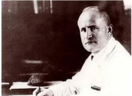
Hans Berger 1873 discover EEG signals first.
Fig: 3.1 The first human EEG recording obtained by Hans Berger in 1924.
Fig: 3.2 Hans Berger(1873-1941)
This experiment represents the first time when scientists have seen how synaptotagmin-1 interacts with the snares of the atomic scale and scientists are more confident that this protein resembles before calcium arrives allowing the fusion process and resulting neurotransmission to happen very quickly getting information from point A to point B in less than a millisecond.
© Daffodil International University
12 3.2.4 Limitations
This experiment represents the first time when scientists have seen how synaptotagmin-1 interacts with the snares of the atomic scale and scientists are more confident that this protein resembles before calcium arrives allowing the fusion process and resulting neurotransmission to happen very quickly getting information from point A to point B in less than a millisecond.
Fig: 3.3 Frist String Galvanometer with recording apparatus 3.2.5 Works by other scientists
Take a minute and put your hand onto your head and think about what you are touching. Within a centimeter of your fingers, there is a piece of the most mysterious and rarely understood matter in the universe. And that is our brain.
Inside each of our brains, there are roughly 100 billion highly specialized cells called neurons.
They make about 500 trillion connections called synapsis. These unique cells transmit important information alarming us the sense and interacts with the world around us. If you want to take a closer look, you will see that this information is transmitted between neurons using chemicals called neurotransmitters where tiny structures called synaptic vesicles fuse with the membrane of one neuron and release chemicals signals into the gap. The second neuron can receive them.
© Daffodil International University
13 Scientists already knew some about how this neurotransmission process works. But now after over 10 years of collaborated research of Stanford University and SLAC national accelerator laboratory along with the ultra-bright X-rays, scientists now have a better idea of exactly how these tiny vesicles might fuse with the membrane of one neuron to transmit their signals. The key to this fusion is the collaboration between special proteins called snares and synaptotagmin-1. They are then triggered by calcium to cause the vesicle of fuse with the membrane of the neuron. When a synaptic vesicle comes close enough to the membrane, the proteins connect with the two and enter a pre-fusion state.
3.3 Brief History of EEG
Take a minute and put your hand onto your head and think about what you are touching. Within a centimeter of your fingers, there is a piece of the most mysterious and rarely understood matter in the universe. And that is our brain.
Inside each of our brains, there are roughly 100 billion highly specialized cells called neurons.
They make about 500 trillion connections called synapsis. These unique cells transmit important information alarming us the sense and interacts with the world around us. If you want to take a closer look, you will see that this information is transmitted between neurons using chemicals called neurotransmitters where tiny structures called synaptic vesicles fuse with the membrane of one neuron and release chemicals signals into the gap. The second neuron can receive them.
© Daffodil International University
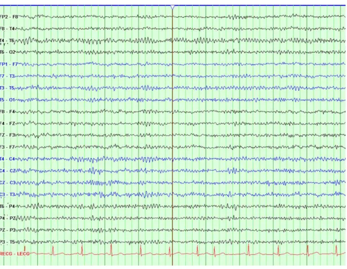
14 Fig: 3.4 Normal EEG recording from an adult using a longitudinal temporal and transverse bipolar montage.
Fig: 3.5 Scalp EEG recording of ictal onset in a patient with absence epilepsy using a longitudinal bipolar montage.
© Daffodil International University
15 Take a minute and put your hand onto your head and think about what you are touching. Within a centimeter of your fingers, there is a piece of the most mysterious and rarely understood matter in the universe. And that is our brain.
Inside each of our brains, there are roughly 100 billion highly specialized cells called neurons.
They make about 500 trillion connections called synapsis. These unique cells transmit important information alarming us the sense and interacts with the world around us.
If you want to take a closer look, you will see that this information is transmitted between neurons using chemicals called neurotransmitters where tiny structures called synaptic vesicles fuse with the membrane of one neuron and release chemicals signals into the gap. The second neuron can receive them.
Scientists already knew some about how this neurotransmission process works. But now after over 10 years of collaborated research of Stanford University and SLAC national accelerator laboratory along with the ultra-bright X-rays, scientists now have a better idea of exactly how these tiny vesicles might fuse with the membrane of one neuron to transmit their signals.
The key to this fusion is the collaboration between special proteins called snares and synaptotagmin-1. They are then triggered by calcium to cause the vesicle of fuse with the membrane of the neuron.
When a synaptic vesicle comes close enough to the membrane, the proteins connect with the two and enter a pre-fusion state. Next when the neuron fires, calcium arrives and triggers the proteins which bend the neural membrane towards the vesicle membrane and draw the two together.
This finally triggers fusion allowing the neurotransmitters to leave the neuron. This experiment represents the first time when scientists have seen how synaptotagmin-1 interacts with the snares of the atomic scale and scientists are more confident that this protein resembles before calcium arrives allowing the fusion process and resulting neurotransmission to happen very quickly getting information from point A to point B in less than a millisecond.
The end result is that our nervous system can work at an incredible speeds enabling us to sense, react to and interact with the world around us.
© Daffodil International University
16 Fig: 3.6 Recording of ictal onset in a patient with right mesial temporal epilepsy due to hippocampal sclerosis using subdural electrodes inserted to cover the inferior surface of the temporal lobe cortex.
Take a minute and put your hand onto your head and think about what you are touching. Within a centimeter of your fingers, there is a piece of the most mysterious and rarely understood matter in the universe. And that is our brain.
Inside each of our brains, there are roughly 100 billion highly specialized cells called neurons.
They make about 500 trillion connections called synapsis.
These unique cells transmit important information alarming us the sense and interacts with the world around us. If you want to take a closer look, you will see that this information is transmitted between neurons using chemicals called neurotransmitters where tiny structures called synaptic vesicles fuse with the membrane of one neuron and release chemicals signals into the gap. The second neuron can receive them.
© Daffodil International University
17 Scientists already knew some about how this neurotransmission process works. But now after over 10 years of collaborated research of Stanford University and SLAC national accelerator laboratory along with the ultra-bright X-rays, scientists now have a better idea of exactly how these tiny vesicles might fuse with the membrane of one neuron to transmit their signals. The key to this fusion is the collaboration between special proteins called snares and synaptotagmin-1.
They are then triggered by calcium to cause the vesicle of fuse with the membrane of the neuron.
When a synaptic vesicle comes close enough to the membrane, the proteins connect with the two and enter a pre-fusion state. Next when the neuron fires, calcium arrives and triggers the proteins which bend the neural membrane towards the vesicle membrane and draw the two together. This finally triggers fusion allowing the neurotransmitters to leave the neuron. This experiment represents the first time when scientists have seen how synaptotagmin-1 interacts with the snares of the atomic scale and scientists are more confident that this protein resembles before calcium arrives allowing the fusion process and resulting neurotransmission to happen very quickly getting information from point A to point B in less than a millisecond.
The end result is that our nervous system can work at an incredible speeds enabling us to sense, react to and interact with the world around us.
Take a minute and put your hand onto your head and think about what you are touching. Within a centimeter of your fingers, there is a piece of the most mysterious and rarely understood matter in the universe. And that is our brain.
Inside each of our brains, there are roughly 100 billion highly specialized cells called neurons.
They make about 500 trillion connections called synapsis.
These unique cells transmit important information alarming us the sense and interacts with the world around us. If you want to take a closer look, you will see that this information is transmitted between neurons using chemicals called neurotransmitters where tiny structures called synaptic vesicles fuse with the membrane of one neuron and release chemicals signals into the gap. The second neuron can receive them.
Scientists already knew some about how this neurotransmission process works. But now after over 10 years of collaborated research of Stanford University and SLAC national accelerator laboratory
© Daffodil International University
18 along with the ultra-bright X-rays, scientists now have a better idea of exactly how these tiny vesicles might fuse with the membrane of one neuron to transmit their signals. The key to this fusion is the collaboration between special proteins called snares and synaptotagmin-1. They are then triggered by calcium to cause the vesicle of fuse with the membrane of the neuron. When a synaptic vesicle comes close enough to the membrane, the proteins connect with the two and enter a pre-fusion state. Next when the neuron fires, calcium arrives and triggers the proteins which bend the neural membrane towards the vesicle membrane and draw the two together. This finally triggers fusion allowing the neurotransmitters to leave the neuron. This experiment represents the first time when scientists have seen how synaptotagmin-1 interacts with the snares of the atomic scale and scientists are more confident that this protein resembles before calcium arrives allowing the fusion process and resulting neurotransmission to happen very quickly getting information from point A to point B in less than a millisecond. The end result is that our nervous system can work at an incredible speeds enabling us to sense, react to and interact with the world around us.
Take a minute and put your hand onto your head and think about what you are touching. Within a centimeter of your fingers, there is a piece of the most mysterious and rarely understood matter in the universe. And that is our brain.
Inside each of our brains, there are roughly 100 billion highly specialized cells called neurons.
They make about 500 trillion connections called synapsis. These unique cells transmit important information alarming us the sense and interacts with the world around us. If you want to take a closer look, you will see that this information is transmitted between neurons using chemicals called neurotransmitters where tiny structures called synaptic vesicles fuse with the membrane of one neuron and release chemicals signals into the gap. The second neuron can receive them.
Scientists already knew some about how this neurotransmission process works. But now after over 10 years of collaborated research of Stanford University and SLAC national accelerator laboratory along with the ultra-bright X-rays, scientists now have a better idea of exactly how these tiny vesicles might fuse with the membrane of one neuron to transmit their signals. The key to this fusion is the collaboration between special proteins called snares and synaptotagmin-1. They are then triggered by calcium to cause the vesicle of fuse with the membrane of the neuron. When a synaptic vesicle comes close enough to the membrane, the proteins connect with the two and enter a pre-fusion state. Next when the neuron fires, calcium arrives and triggers the proteins which bend
© Daffodil International University
19 the neural membrane towards the vesicle membrane and draw the two together. This finally triggers fusion allowing the neurotransmitters to leave the neuron.
This experiment represents the first time when scientists have seen how synaptotagmin-1 interacts with the snares of the atomic scale and scientists are more confident that this protein resembles before calcium arrives allowing the fusion process and resulting neurotransmission to happen very quickly getting information from point A to point B in less than a millisecond. The end result is that our nervous system can work at an incredible speeds enabling us to sense, react to and interact with the world around us.
© Daffodil International University
20
Chapter 4
Electroencephalography (EEG) Signal Processing
4.1. Fundamentals of Electroencephalography (EEG) Signal Processing
These unique cells transmit important information alarming us the sense and interacts with the world around us. If you want to take a closer look, you will see that this information is transmitted between neurons using chemicals called neurotransmitters.
Inside each of our brains, there are roughly 100 billion highly specialized cells called neurons.
They make about 500 trillion connections called synapsis. These unique cells transmit important information alarming us the sense and interacts with the world around us. If you want to take a closer look, you will see that this information is transmitted between neurons using chemicals called neurotransmitters where tiny structures called synaptic vesicles fuse with the membrane of one neuron and release chemicals signals into the gap. The second neuron can receive them.
Scientists already knew some about how this neurotransmission process works. But now after over 10 years of collaborated research of Stanford University and SLAC national accelerator laboratory along with the ultra-bright X-rays, scientists now have a better idea of exactly how these tiny vesicles might fuse with the membrane of one neuron to transmit their signals. The key to this fusion is the collaboration between special proteins called snares and synaptotagmin-1. They are then triggered by calcium to cause the vesicle of fuse with the membrane of the neuron. When a synaptic vesicle comes close enough to the membrane, the proteins connect with the two and enter a pre-fusion state.
4.1.1 EEE signal modeling
Inside each of our brains, there are roughly 100 billion highly specialized cells called neurons.
They make about 500 trillion connections called synapsis. These unique cells transmit important information alarming us the sense and interacts with the world around us. If you want to take a closer look, you will see that this information is transmitted between neurons using chemicals
© Daffodil International University
21 called neurotransmitters where tiny structures called synaptic vesicles fuse with the membrane of one neuron and release chemicals signals into the gap. The second neuron can receive them.
Scientists already knew some about how this neurotransmission process works. But now after over 10 years of collaborated research of Stanford University and SLAC national accelerator laboratory along with the ultra-bright X-rays, scientists now have a better idea of exactly how these tiny vesicles might fuse with the membrane of one neuron to transmit their signals.
4.2 Various Bands
Inside each of our brains, there are roughly 100 billion highly specialized cells called neurons.
They make about 500 trillion connections called synapsis. These unique cells transmit important information alarming us the sense and interacts with the world around us. If you want to take a closer look, you will see that this information is transmitted between neurons using chemicals called neurotransmitters where tiny structures called synaptic vesicles fuse with the membrane of one neuron and release chemicals signals into the gap. The second neuron can receive them.
Scientists already knew some about how this neurotransmission process works. But now after over 10 years of collaborated research of Stanford University and SLAC national accelerator laboratory along with the ultra-bright X-rays, scientists now have a better idea of exactly how these tiny vesicles might fuse with the membrane of one neuron to transmit their signals
4.2.1 Delta band (1-4 Hz)
Delta waves lie within the range of 0.5– 4 Hz. These waves are primarily associated with deep sleep and may be present in the waking state. It is very easy to confuse artefact signals caused by the large muscles of the neck and jaw with the genuine delta response.
4.2.2. Theta band (4-8 Hz)
Theta waves lie within the range of 4– 7.5 Hz. The term theta might be chosen to allude to its presumed thalamic origin. Theta waves appear as consciousness slips towards drowsiness. Theta waves have been associated with access to unconscious material, creative inspiration and deep meditation.
© Daffodil International University
22
Fig: 4.1 Frequency variance per second
4.2.3. Alpha band (8 - 12 Hz)
These unique cells transmit important information alarming us the sense and interacts with the world around us. If you want to take a closer look, you will see that this information is transmitted between neurons using chemicals called neurotransmitters where tiny structures called synaptic vesicles fuse with the membrane of one neuron and release chemicals signals into the gap.
© Daffodil International University
23 4.2.4. Beta band (12- 25 Hz)
Scientists already knew some about how this neurotransmission process works. But now after over 10 years of collaborated research of Stanford University and SLAC national accelerator laboratory along with the ultra-bright X-rays, scientists now have a better idea of exactly how these tiny vesicles might fuse with the membrane of one neuron to transmit their signals
4.2.5 Gamma band (above 25 Hz)
These unique cells transmit important information alarming us the sense and interacts with the world around us. If you want to take a closer look, you will see that this information is transmitted between neurons using chemicals called neurotransmitters where tiny structures called synaptic vesicles fuse with the membrane of one neuron and release chemicals signals into the gap.
4.2.6 Other Waves
Within a centimeter of your fingers, there is a piece of the most mysterious and rarely understood matter in the universe. And that is our brain.
Inside each of our brains, there are roughly 100 billion highly specialized cells called neurons.
They make about 500 trillion connections called synapsis.
These unique cells transmit important information alarming us the sense and interacts with the world around us. If you want to take a closer look, you will see that this information is transmitted between neurons using chemicals called neurotransmitters where tiny structures called synaptic vesicles fuse with the membrane of one neuron and release chemicals signals into the gap.
The second neuron can receive them.
Scientists already knew some about how this neurotransmission process works. But now after over 10 years of collaborated research of Stanford University and SLAC national accelerator laboratory along with the ultra-bright X-rays, scientists now have a better idea of exactly how these tiny vesicles might fuse with the membrane of one neuron to transmit their signals.
The key to this fusion is the collaboration between special proteins called snares and synaptotagmin-1. They are then triggered by calcium to cause the vesicle of fuse with the
© Daffodil International University
24 membrane of the neuron. When a synaptic vesicle comes close enough to the membrane, the proteins connect with the two and enter a pre-fusion state.
Next when the neuron fires, calcium arrives and triggers the proteins which bend the neural membrane towards the vesicle membrane and draw the two together. This finally triggers fusion allowing the neurotransmitters to leave the neuron.
This experiment represents the first time when scientists have seen how synaptotagmin-1 interacts with the snares of the atomic scale and scientists are more confident that this protein resembles before calcium arrives allowing the fusion process and resulting neurotransmission to happen very quickly getting information from point A to point B in less than a millisecond. The end result is that our nervous system can work at an incredible speeds enabling us to sense, react to and interact with the world around us.
4.3 EEG Recording and Measurement
Scientists already knew some about how this neurotransmission process works. But now after over 10 years of collaborated research of Stanford University and SLAC national accelerator laboratory along with the ultra-bright X-rays, scientists now have a better idea of exactly how these tiny vesicles might fuse with the membrane of one neuron to transmit their signals. The key to this fusion is the collaboration between special proteins called snares and synaptotagmin-1. They are then triggered by calcium to cause the vesicle of fuse with the membrane of the neuron. When a synaptic vesicle comes close enough to the membrane, the proteins connect with the two and enter a pre-fusion state. Next when the neuron fires, calcium arrives and triggers the proteins which bend the neural membrane towards the vesicle membrane and draw the two together. This finally triggers fusion allowing the neurotransmitters to leave the neuron.
This experiment represents the first time when scientists have seen how synaptotagmin-1 interacts with the snares of the atomic scale and scientists are more confident that this protein resembles before calcium arrives allowing the fusion process and resulting neurotransmission to happen very quickly getting information from point A to point B in less than a millisecond. The end result is that our nervous system can work at an incredible speeds enabling us to sense, react to and interact with the world around us.
© Daffodil International University
25 These unique cells transmit important information alarming us the sense and interacts with the world around us. If you want to take a closer look, you will see that this information is transmitted between neurons using chemicals called neurotransmitters where tiny structures called synaptic vesicles fuse with the membrane of one neuron and release chemicals signals into the gap.
4.4 Conventional Electrode Positioning
The key to this fusion is the collaboration between special proteins called snares and synaptotagmin-1. They are then triggered by calcium to cause the vesicle of fuse with the membrane of the neuron. When a synaptic vesicle comes close enough to the membrane, the proteins connect with the two and enter a pre-fusion state. Next when the neuron fires, calcium arrives and triggers the proteins which bend the neural membrane towards the vesicle membrane and draw the two together. This finally triggers fusion allowing the neurotransmitters to leave the neuron. This experiment represents the first time when scientists have seen how synaptotagmin-1 interacts with the snares of the atomic scale and scientists are more confident that this protein resembles before calcium arrives allowing the fusion process and resulting neurotransmission to happen very quickly getting information from point A to point B in less than a millisecond. The end result is that our nervous system can work at an incredible speeds enabling us to sense, react to and interact with the world around us.
Points to note in the 10-20 system:
Nasion (Nz)
The noise between the eyes at the top of the nose.
Inion (Iz)
The bump at the back of the head.
© Daffodil International University
26
Fig: 4.2 Placement of Electrodes
The second neuron can receive them. Scientists already knew some about how this neurotransmission process works. But now after over 10 years of collaborated research of Stanford University and SLAC national accelerator laboratory along with the ultra-bright X-rays, scientists now have a better idea of exactly how these tiny vesicles might fuse with the membrane of one neuron to transmit their signals. The key to this fusion is the collaboration between special proteins called snares and synaptotagmin-1. They are then triggered by calcium to cause the vesicle of fuse with the membrane of the neuron. When a synaptic vesicle comes close enough to the membrane, the proteins connect with the two and enter a pre-fusion state. Next when the neuron fires, calcium arrives and triggers the proteins which bend the neural membrane towards the vesicle membrane and draw the two together. This finally triggers fusion allowing the neurotransmitters to leave the
© Daffodil International University
27 neuron. This experiment represents the first time when scientists have seen how synaptotagmin-1 interacts with the snares of the atomic scale and scientists are more confident that this protein resembles before calcium arrives allowing the fusion process and resulting neurotransmission to happen very quickly getting information from point A to point B in less than a millisecond. The end result is that our nervous system can work at an incredible speeds enabling us to sense, react to and interact with the world around us.
4.5 Number and distribution of electrodes
These unique cells transmit important information alarming us the sense and interacts with the world around us. If you want to take a closer look, you will see that this information is transmitted between neurons using chemicals called neurotransmitters where tiny structures called synaptic vesicles fuse with the membrane of one neuron and release chemicals signals into the gap.
The second neuron can receive them. Scientists already knew some about how this neurotransmission process works. But now after over 10 years of collaborated research of Stanford University and SLAC national accelerator laboratory along with the ultra-bright X-rays, scientists now have a better idea of exactly how these tiny vesicles might fuse with the membrane of one neuron to transmit their signals. The key to this fusion is the collaboration between special proteins called snares and synaptotagmin-1. They are then triggered by calcium to cause the vesicle of fuse with the membrane of the neuron. When a synaptic vesicle comes close enough to the membrane, the proteins connect with the two and enter a pre-fusion state. Next when the neuron fires, calcium arrives and triggers the proteins which bend the neural membrane towards the vesicle membrane and draw the two together. This finally triggers fusion allowing the neurotransmitters to leave the Neuron.
4.6 Clean Electroencephalography data and artefacts
The second neuron can receive them. Scientists already knew some about how this neurotransmission process works. But now after over 10 years of collaborated research of Stanford University and SLAC national accelerator laboratory along with the ultra-bright X-rays, scientists now have a better idea of exactly how these tiny vesicles might fuse with the membrane of one
© Daffodil International University
28 neuron to transmit their signals. The key to this fusion is the collaboration between special proteins called snares and synaptotagmin-1. They are then triggered by calcium to cause the vesicle of fuse with the membrane of the neuron. When a synaptic vesicle comes close enough to the membrane, the proteins connect with the two and enter a pre-fusion state. Next when the neuron fires, calcium arrives and triggers the proteins which bend the neural membrane towards the vesicle membrane and draw the two together. This finally triggers fusion allowing the neurotransmitters to leave the Shell.
4.6.1 Physiological artefacts
The second neuron can receive them. Scientists already knew some about how this neurotransmission process works. But now after over 10 years of collaborated research of Stanford University and SLAC national accelerator laboratory along with the ultra-bright X-rays, scientists now have a better idea of exactly how these tiny vesicles might fuse with the membrane of one neuron to transmit their signals. The key to this fusion is the collaboration between special proteins
© Daffodil International University
29 called snares and synaptotagmin-1. They are then triggered by calcium to cause the vesicle.
Fig: 4.3 Muscle activity of electric current
Eye movements
They are then triggered by calcium to cause the vesicle of fuse with the membrane of the neuron. When a synaptic vesicle comes close enough to the membrane, the proteins connect with the two and enter a pre-fusion state. Next when the neuron fires, calcium arrives and triggers the proteins which bend the neural membrane towards the vesicle membrane and draw the two together. This finally triggers fusion allowing the neurotransmitters to leave the neuron. This experiment represents the first time when scientists have seen how synaptotagmin-1 interacts with the snares of the atomic scale and scientists are more confident that this protein resembles before
© Daffodil International University
30 calcium arrives allowing the fusion process and resulting neurotransmission to happen very quickly getting information from point A to point B in less than a millisecond.
Fig: 4.4 Eye movement
Blinking:
Fig: 4.5 Blinking effect
© Daffodil International University
31 The second neuron can receive them. Scientists already knew some about how this
neurotransmission process works. But now after over 10 years of collaborated research of
Stanford University and SLAC national accelerator laboratory along with the ultra-bright X-rays, scientists now have a better idea of exactly how these tiny vesicles might fuse with the
membrane of one neuron to transmit their signals.
The key to this fusion is the collaboration between special proteins called snares and synaptotagmin-1. They are then triggered by calcium to cause the vesicle of fuse with the membrane of the neuron. When a synaptic vesicle comes close enough to the membrane, the proteins connect with the two and enter a pre-fusion state. Next when the neuron fires, calcium arrives and triggers the proteins which bend the neural membrane towards the vesicle membrane and draw the two together.
4.6.2 External sources of artefacts
Movement of an electrode can cause many artefacts (Fig 4.6).
Fig: 4.6 Movement of electrodes
Line noise
Line noise (50 Hz in the United States, 60 Hz in the Europe) probably have strong artefacts on the electrode listing - it is quite common in the raw Electroencephalography data.
© Daffodil International University
32 Fig: 4.7 Noise variation over frequency
© Daffodil International University
33
Chapter 5
Application of Electroencephalography (EEG)
5.1 Medical or Clinical Applications
It has severe medical applications.
Fig: 5.1 An EEG recording setup
The key to this fusion is the collaboration between special proteins called snares and synaptotagmin-1. They are then triggered by calcium to cause the vesicle of fuse with the membrane of the neuron. When a synaptic vesicle comes close enough to the membrane, the proteins connect with the two and enter a pre-fusion state.
5.1.1 Dementia
Dementia is a syndrome that consists of a decline in intellectual and cognitive abilities. This consequently affects the normal social activities, mode, and the relationship and interaction with other people. EEG is often used to study the effect of dementia. In most cases, such as in primary degenerative dementia, e.g. Alzheimer’s, and psychiatric disorder, e.g. depression with cognitive impairment, the EEG can be used to detect the abnormality.
© Daffodil International University
34 Dementia is classified into cortical and subcortical forms. The most important cortical
dementia is Alzheimer’s disease (AD), which accounts for approx- imately 50 % of the cases. Other known cortical abnormalities are Pick’s disease and Creutzfeldt– Jakob diseases (CJD). They are characterized clinically by findings such as aphasia, apraxia, and agnosia.
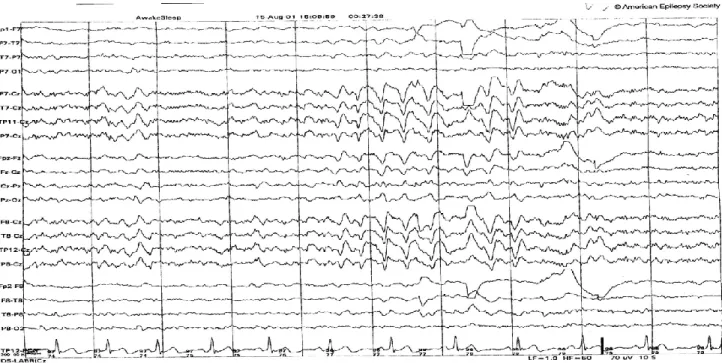
CJD can often be diagnosed using the EEG signals. Figure shows a set of EEG signals from a CJD patient. On the other hand, the most common subcortical diseases are Parkinson’s disease, Huntington’s disease, lacunar state, normal pressure hydrocephalus, and progressive supranuclear palsy. These diseases are characterized by forgetfulness, slowing of thought processes, apathy, and depression. Generally, subcortical dementias introduce less
abnormality to the EEG patterns than the cortical ones.
In AD the EEG posterior rhythm (alpha rhythm) slows down and the delta and theta wave activities increase. On the other hand, beta wave activity may decrease. In severe cases epileptiform discharges and triphasic waves can appear. In such cases, cognitive impairment often results. The spectral power also changes; the power increases in delta and theta bands and decreases in beta and alpha bands and also in mean frequency.
The EEG wave morphology is almost the same for AD and Pick’s disease. Pick’s disease involves the frontal and temporal lobes. An accurate analysis followed by an efficient classification of the cases may discriminate these two diseases. CJD is a mixed cortical and subcortical dementia. This causes slowing of the delta and theta wave activities and, after approximately three months of the onset of the disease, periodic sharp wave complexes are generated that occur almost every second, together with a decrease in the background activity [54]. Parkinson’s disease is a subcortical dementia, which causes slowing down of the background activity and an increase of the theta and delta wave activities. Some works have been undertaken using spectral analysis to confirm the above changes [55]. Some other disorders such as depression have a lesser effect on the EEGs and more accurate analysis of the EEGs has to be performed to detect the signal abnormalities for these brain disorders.
The key to this fusion is the collaboration between special proteins called snares and synaptotagmin-1. They are then triggered by calcium to cause the vesicle of fuse with the membrane of the neuron. When a synaptic vesicle comes close enough to the membrane, the
© Daffodil International University
35 proteins connect with the two and enter a pre-fusion state. Next when the neuron fires, calcium arrives and triggers the proteins which bend the neural membrane towards the vesicle membrane and draw the two together.
5.1.2 Additional work done by EEG
The EEG wave morphology is almost the same for AD and Pick’s disease. Pick’s disease involves the frontal and temporal lobes. An accurate analysis followed by an efficient classification of the cases may discriminate these two diseases. CJD is a mixed cortical and subcortical dementia. This causes slowing of the delta and theta wave activities and, after approximately three months of the onset of the disease, periodic sharp wave complexes are generated that occur almost every second, together with a decrease in the background activity [54]. Parkinson’s disease is a subcortical dementia, which causes slowing down of the
background activity and an increase of the theta and delta wave activities. Some works have been undertaken using spectral analysis to confirm the above changes [55].
They are characterized clinically by findings such as aphasia, apraxia, and agnosia. CJD can often be diagnosed using the EEG signals. Figure shows a set of EEG signals from a CJD patient. On the other hand, the most common subcortical diseases are Parkinson’s disease, Huntington’s disease, lacunar state, normal pressure hydrocephalus, and progressive supranuclear palsy. These diseases are characterized by forgetfulness, slowing of thought
processes, apathy, and depression. Generally, subcortical dementias introduce less abnormality to the EEG patterns than the cortical ones.
5.1.3 Epileptic Seizure and Nonepileptic Attacks
They are characterized clinically by findings such as aphasia, apraxia, and agnosia. CJD can often be diagnosed using the EEG signals. Figure shows a set of EEG signals from a CJD patient. On the other hand, the most common subcortical diseases are Parkinson’s disease, Huntington’s disease, lacunar state, normal pressure hydrocephalus, and progressive supranuclear palsy. These diseases are characterized by forgetfulness, slowing of thought
processes, apathy, and depression. Generally, subcortical dementias introduce less abnormality to the EEG patterns than the cortical ones.
© Daffodil International University
36 Inside each of our brains, there are roughly 100 billion highly specialized cells called neurons.
They make about 500 trillion connections called synapsis. These unique cells transmit important information alarming us the sense and interacts with the world around us. If you want to take a closer look, you will see that this information is transmitted between neurons using chemicals called neurotransmitters where tiny structures called synaptic vesicles fuse with the membrane of one neuron and release chemicals signals into the gap. The second neuron can receive them.
Scientists already knew some about how this neurotransmission process works. But now after over 10 years of collaborated research of Stanford University and SLAC national accelerator laboratory along with the ultra-bright X-rays, scientists now have a better idea of exactly how these tiny vesicles might fuse with the membrane of one neuron to transmit their signal.
5.1.4 Psychiatric Disorders
Inside each of our brains, there are roughly 100 billion highly specialized cells called neurons.
They make about 500 trillion connections called synapsis. These unique cells transmit important information alarming us the sense and interacts with the world around us. If you want to take a closer look, you will see that this information is transmitted between neurons using chemicals called neurotransmitters where tiny structures called synaptic vesicles fuse with the membrane of one neuron and release chemicals signals into the gap. The second neuron can receive them.
Scientists already knew some about how this neurotransmission process works. But now after over 10 years of collaborated research of Stanford University and SLAC national accelerator laboratory along with the ultra-bright X-rays, scientists now have a better idea of exactly how these tiny vesicles might fuse with the membrane of one neuron to transmit their signal
5.1.5 Physiologic
Inside each of our brains, there are roughly 100 billion highly specialized cells called neurons.
They make about 500 trillion connections called synapsis. These unique cells transmit important information alarming us the sense and interacts with the world around us.
© Daffodil International University
37 If you want to take a closer look, you will see that this information is transmitted between
neurons using chemicals called neurotransmitters where tiny structures called synaptic vesicles fuse with the membrane of one neuron and release chemicals signals into the gap.
The second neuron can receive them.
Scientists already knew some about how this neurotransmission process works. But now after over 10 years of collaborated research of Stanford University and SLAC national accelerator laboratory along with the ultra-bright X-rays, scientists now have a better idea of exactly how these tiny vesicles might fuse with the membrane of one neuron to transmit their signal
5.2 External effects
Inside each of our brains, there are roughly 100 billion highly specialized cells called neurons.
They make about 500 trillion connections called synapsis. These unique cells transmit important information alarming us the sense and interacts with the world around us. If you want to take a closer look, you will see that this information is transmitted between neurons using chemicals called neurotransmitters where tiny structures called synaptic vesicles fuse with the membrane of one neuron and release chemicals signals into the gap. The second neuron can receive them.
Scientists already knew some about how this neurotransmission process works. But now after over 10 years of collaborated research of Stanford University and SLAC national accelerator laboratory along with the ultra-bright X-rays, scientists now have a better idea of exactly how these tiny vesicles might fuse with the membrane of one neuron to transmit their signals. The key to this fusion is the collaboration between special proteins called snares and synaptotagmin-1. They are then triggered by calcium to cause the vesicle of fuse with the membrane of the neuron. When a synaptic vesicle comes close enough to the membrane, the proteins connect with the two and enter a pre-fusion state. Next when the neuron fires, calcium arrives and triggers the proteins which bend the neural membrane towards the vesicle membrane and draw the two together. This finally triggers fusion allowing the neurotransmitters to leave the neuron. This experiment represents the first time when scientists have seen how synaptotagmin-1 interacts with the snares of the atomic scale and scientists are more confident that this protein resembles before calcium arrives allowing the fusion process and resulting neurotransmission to happen very quickly getting information from point A to point B in less than a millisecond. The end result is that our nervous system can work at an incredible speeds enabling us to sense, react to and interact with the world around us.
© Daffodil International University
38
5.3 Applications of EEG monitoring
. The key to this fusion is the collaboration between special proteins called snares and synaptotagmin-1. They are then triggered by calcium to cause the vesicle of fuse with the membrane of the neuron. When a synaptic vesicle comes close enough to the membrane, the proteins connect with the two and enter a pre-fusion state. Next when the neuron fires, calcium arrives and triggers the proteins which bend the neural membrane towards the vesicle membrane and draw the two together. This finally triggers fusion allowing the neurotransmitters to leave the neuron. This experiment represents the first time when scientists have seen how synaptotagmin-1 interacts with the snares of the atomic scale and scientists are more confident that this protein resembles before calcium arrives.
5.3.1 Long-term Video-EEG Monitoring
Dementia is a syndrome that consists of a decline in intellectual and cognitive abilities. This consequently affects the normal social activities, mode, and the relationship and interaction with other people. EEG is often used to study the effect of dementia. In most cases, such as in primary degenerative de