Diagnostic Biomarkers of Parkinson’s Disease: The Present and the Future
By Labiba Ferdous
16146043
A thesis submitted to the Department of Pharmacy in partial fulfillment of the requirements for the degree of
Bachelor of Pharmacy (Hons.)
Department of Pharmacy Brac University
January 2021
© 2021. Brac University All rights reserved.
i
Declaration
It is hereby declared that
1. The thesis submitted is my own original work while completing degree at BRAC University.
2. The thesis does not contain material previously published or written by a third party, except where this is appropriately cited through full and accurate referencing.
3. The thesis does not contain material which has been accepted, or submitted, for any other degree or diploma at a university or other institution.
4. I have acknowledged all main sources of help.
Student’s Full Name & Signature:
Labiba Ferdous 16146043
ii
Approval
The thesis titled “Diagnostic Biomarkers of Parkinson’s Disease: The Present and the Future”
submitted by Labiba Ferdous (16146043) of Spring, 2016 has been accepted as satisfactory in partial fulfillment of the requirement for the degree of Bachelor of Pharmacy (Hons.) on 14.01.2021.
Examining Committee:
Supervisor:
(Member)
______________________________
Ms. Marzia Alam
Lecturer, Department of Pharmacy Brac University
Program Coordinator:
(Member)
_______________________________
Dr. Hasina Yasmin
Professor, Department of Pharmacy Brac University
Departmental Head:
(Chair)
_______________________________
Dr. Eva Rahman Kabir
Chairperson, Department of Pharmacy Brac University
iii
Ethics Statement
The study does not involve any kind of animal and human trial.
iv
Abstract
Parkinson’s disease (PD) is a progressive nervous system disorder that is familiar as a common neurodegenerative disorder. PD requires precise biomarkers for prodromal and preclinical diagnosis, to provide quality treatment during the initial phase of PD. Such biomarkers may be efficient enough to monitor the progression of disease. The purpose of this review is to highlight recent developments in the discovery of biomarkers of PD based on biochemical (urate, protein DJ-1, 𝛼-synuclein, peptides, IGF-1, homocysteine etc.) and genetic factors. Significant progress has been made in research on biomarkers for PD and a growing number of candidate biomarkers have been identified for PD, but they do not have satisfactory specificity when used individually.
Therefore, a comprehensive composition of multimodal biomarkers can improve diagnostic accuracy and simplify personalized medicine implementation.
Keywords
Parkinson's disease; biochemical; non-invasive diagnosis;genetic; biomarkers
v
Dedication
Dedicated to my parents.
vi
Acknowledgment
First and foremost, I would like to express my deepest gratitude towards Almighty Allah for giving me the blessing, the strength, the patience and chance to complete my project work properly.
I am really fortunate that I had the kind and supportive supervision of Ms. Marzia Alam (Lecturer of Pharmacy Department of Brac University). Her constant encouragement and careful monitoring motivated to do my work properly. She was very helpful to me whenever I faced any problem in my work and helped me to resolve any kind of inaccuracy in my work. Moreover, I would like to express my special thanks to Dr. Eva Rahman Kabir, Chairperson of the Department of Pharmacy Brac University who gave me the opportunity to conduct my project work.
In the end, I acknowledge my sincere gratitude to my parents, friends and seniors for their devotion, support and faith in my ability throughout the whole journey.
vii
Table of Contents
Declaration... i
Approval ... ii
Ethics Statement... iii
Abstract ... iv
Dedication ... v
Acknowledgment ... vi
List of Tables ... x
List of Figures ... xi
List of Acronyms ... xii
Glossary ... xiv
Chapter 1 ... 1
1.1 Introduction ...1
1.2 Rationale of the Study ...2
1.3Aim of the Review ...3
1.4 Objective of the Study ...3
Chapter 2 ... 4
Methodology ...4
Chapter 3 ... 5
3.1 What is Parkinson’s Disease? ...5
3.2 History of PD ...5
3.3 Causes of Parkinson’s Disease ...6
viii
3.4 Symptoms of Parkinson’s Disease ...7
3.5 Stages of Parkinson’s disease ...9
3.6 Treatment of Parkinson’s Disease ... 10
3.7 Pathogenesis of Parkinson’s Disease ... 11
3.8 The Necessity of Various Types of PD Biomarkers ... 11
3.9 Types of Biomarkers and their Purpose ... 12
Chapter 4 ... 14
Diagnostic Biochemical Biomarkers ... 14
4.1 Oxidative stress related biomarkers ... 14
4.1.1 Urate ... 14
4.1.2 Protein DJ-1 ... 15
4.1.3 Coenzyme Q10 ... 17
4.1.4 Homocysteine (Hcy) ... 17
4.1.5 8-OHdG (8-Hydroxydeoxyguanosine) ... 18
4.1.6 Advanced Oxidized Protein Products (AOPP) ... 19
4.2 Biomarkers Associated with Abnormal Protein Accumulation and Aggregation ... 20
4.2.1 𝛼-synuclein ... 20
4.2.2 β-Glucocerebrosidase and Ubiquitin C-terminal hydrolase-L1(UCH-L1) ... 22
4.2.3 Amyloid beta 42(Aβ42) ... 23
4.2.4 Tau protein ... 24
4.2.5 Neurofilament light chain protein (NFL) ... 25
4.3 Neurotrophins related biomarkers ... 25
4.3.1 Brain-derived neurotrophic factor (BDNF) ... 25
4.3.2 Insulin-like growth factor 1 (IGF-1) ... 26
4.4 Biomarkers Associated with Neuroinflammatory Reaction ... 27
Chapter 5 ... 28
Genetic Biomarkers ... 28
Chapter 6 ... 31
Other biomarkers ... 31
5.1 MicroRNA (MiRNA) ... 31
5.2 Peptides ... 32
ix
Chapter 7 ... 35
Discussion ... 35
Chapter 8 ... 37
Prospect ... 37
Chapter 9 ... 39
Conclusion ... 39
Future Directions: ... 39
References ... 40
x
List of Tables
1. Table 1: Name of gene, Protein encoded, Role of gene as a diagnostic tool of PD
xi
List of Figures
1. Figure 1: Difference between Parkinson’s and Non-Parkinson’s brain (Embogama, 2016).
xii
List of Acronyms
PD Parkinson’s Disease
PDD Parkinson’s Disease with Dementia DaTSCAN Dopamine transporter scan
APD Atypical Parkinsonism Disease
MSA Multiple System Atrophy
PSP Progressive Supranuclear Palsy
DLB Dementia with Lewy-bodies
CBD Cortico-basal Degeneration
PDI Peripheral Decarboxylase Inhibitor
ROS Reactive Oxygen Species
WHO World Health Organization
DATATOP Deprenyl and Tocopherol Antioxidant Therapy of Parkinsonism PRECEPT Parkinson's Research Examination of the CEP-1347 Trial
UPSIT University of Pennsylvania Smell Identification Test RBDSQ Rapid eye movement sleep Behavior Disorder Screening
Questionnaire
4-HNE 4-hydroxy-2-nonenal
UPDRS The Unified Parkinson’s Disease Rating Scale
CSF Cerebrospinal Fluid
MCI Mild Cognitive Impairment
AD Alzheimer’s Disease
CAA Cerebral Amyloid Angiopathy
xiii
RT-QuIC Real-Time Quaking-Induced Conversion H&Y stage Hoehn-Yahr stage
ELISA Enzyme-Linked Immunosorbent Assay
CSFR1 Colony-stimulating factor receptor 1
EPHA4 Ephrin type-A receptor 4
TIMP1 Tissue metalloproteinase inhibitor-1
MMPs Matrix Metalloproteinases
GWAS Genome-Wide Association Studies
SPP1 Secreted Phosphoprotein 1
LRP1 Lipoprotein Receptor-related Protein 1
DATATOP Deprenyl and Tocopherol Antioxidant Therapy of Parkinsonism and PRECEPT Parkinson's Research Examination of the CEP-1347 Trial
MPTP-induced 1-methyl-4-phenyl-1,2,3,6-tetrahydropyridine-induced UCH-L1 Ubiquitin C-terminal hydrolase-L1
Hcy Homocysteine
8-OHdG 8-Hydroxydeoxyguanosine
AOPP Advanced Oxidized Protein Products
Aβ42 Amyloid beta 42
NFL Neurofilament Light chain protein IGF-1 Insulin-like growth Factor 1 BDNF Brain-derived Neurotrophic Factor
MicroRNA MiRNA
xiv
Glossary
Lewy bodies: Lewy bodies are the inclusion bodies, abnormal aggregations of protein that develop inside nerve cells affected by Parkinson’s disease.
NMDA Receptor: The NMDA Receptor is a glutamate and ion channel protein receptor that is activated when glycine and glutamate bind to it.
SNCA Gene: The SNCA gene provides instructions for making a small protein called alpha-synuclein.
Transcriptomics Transcriptomics is the study of the transcriptome, the complete set of RNA transcripts that are produced by the genome, under specific circumstances or in a specific cell, using high-throughput methods, such as microarray analysis.
Juvenile-onset PD Juvenile-onset PD is a rare disorder that presents before the age of 21 years.
Familial PD Parkinson’ disease with a family history is called familial PD.
xv
Sporadic PD Sporadic PD can be explained as a disease occurring randomly in a population with no known cause.
Autosomal dominant PD
If the LRRK2 or SNCA genes are involved, Parkinson's is likely inherited from just one parent. That's called an autosomal dominant pattern, which is when a person only need one copy of a gene to be altered for the disorder to happen.
1
Chapter 1
1. 1 Introduction
Living with Parkinson’s disease is challenging because it causes gradual changes in motor function and many other non-motor symptoms, such as depression, sleep disorders, pain and cognitive dysfunction which a person cannot ignore (Living with Parkinson’s Disease | American Parkinson Disease Assoc., 2016). So, this disease significantly impacts not only the quality of life of the person suffering from PD but also their family. Many people develop dementia as the condition worsens over time, which can cause profound memory loss and make it difficult maintain relationships (Jennifer G. Goldman, 2012). Also, they constantly need caregiver because they face difficulty to in physical movement and forget things easily. Moreover, as PD affects the muscles, the speech of a person may become weaker and harder to comprehend. Changes in the ability to think can make communication more challenging over time, which can make it difficult to engage in social activities (The MNT Editorial Team, 2018). These are some of the complications of life with PD. Therefore, early diagnosis and appropriate treatment can be helpful. Thus, it is important to discover effective and comprehensive biomarkers of PD, which may help in early diagnosis and treatment and may give a person hope to live a better life.
PD is usually seen in the population of the age 60 years and above, about one percent of whom are affected (Lisak et al., 2010).Males with a round 3:2 ratio are more often affected than females (Kalia & Lang, 2015).
2
Parkinson’s disease is an incurable disease but patients can be treated to improve the symptoms (Samii et al., 2004) Various studies indicate that, with increased age and dementia as the highest predictors of increased mortality, the mean period before death varies from 6.9 to 14.3 years (Sveinbjornsdottir, 2016).
1.2 Rationale of the Study
There is neuroimaging and genetic biomarkers which are currently available for commercial usage (He et al., 2018) but misdiagnosis frequently takes place in the early stage of this disease. Like DaTSCAN (Dopamine transporter scan), which tests the density of sites of the nigrostriatal dopamine transporter, can help the clinicians make the right diagnosis. However, it’s clinical usefulness has been questioned, as most evaluations contrasted DaTSCAN with clinical tests, which were found to be inaccurate in 6% to 25% of cases.In other words, most early Parkinson's disease patients had an usual DaTSCAN, and there was no strong proof that DaTSCAN is accurate in early Parkinson's disease diagnosis (Galbraith, 2019). Moreover, previously in most of the cases, the disease was confirmed at the autopsy of the patient. Thus, there is an obvious necessity to invent a specific biomarker which will facilitate clinicians among patients suspected of a variety of Parkinson's disease to assess a lot of timely and accurate medical diagnosis. It is also important to discover biomarkers that will assist clinicians in predicting disease progression (Mosley et al., 2012). For the following reasons, the discovery of highly accurate and sensitive markers for
preclinical PD or early PD is a high research priority since:
(1) Before the classic symptoms develop, it will enable the identification of at-risk people, a
3
stage in which neuroprotective therapies are supposed to have their greatest impact;
(2) It will help to compare PD with the other causes of Parkinsonism.
(3) It will accelerate research into PD therapeutics aimed at etiopathogenesis and pathogenesis.
(Wu et al., 2011).
1.3Aim of the Review
The target of this review is to compile all of the possible genetic and biochemical biomarkers of Parkinson’s disease which are expected to contribute towards the early diagnosis of the disease and to reveal how their pathophysiology is interconnected with Parkinson’s disease.
1.4 Objective of the Study
The objectives of this review are,
● To compile information about the emerging and existing biomarkers of Parkinson’s disease
● To collect information of early diagnosis of Parkinson’s Disease
● To identify relationships among different biomarkers
● To identify the effective use of combined biomarkers
4
Chapter 2
Methodology
The information compiled for this review was collected through a three-staged process: 1) collection of possibly relevant articles using several sources, 2) selection of definitely relevant articles, and 3) analysis of selected studies. A systematic review of articles was performed to summarize these recent advances. The information was collected from relevant articles Pub-med, ScienceDirect. Basically, those articles were chosen on the basis of updated information about biomarkers for initial diagnosis of PD. Also “recent, advanced, biomarkers, Parkinson’s disease, diagnosis, early etc.” these keywords were used to gather information for this review paper. Then the references were searched via Mendeley library and inserted them into the review paper. Also, a bibliography of all the references that have already been cited was created.
5
Chapter 3
3.1 What is Parkinson’s Disease?
According to WHO, Parkinson's disease is the second most prevalent progressive disorder of the nervous system after Alzheimer's disease (He et al., 2018). It is a neurodegenerative disease in which the patient's condition is incurable due to continuous death of neurons of substantia nigra that impedes patient's movement and mental function. Substantia nigra contains a large number of neurons which releases dopamine with which substantia nigra communicates with movement controlling parts of the brain like basal ganglia and frontal lobe. So, when neuronal death occurs in the substantia nigra they cannot communicate and function effectively, which results in dyskinesia, tremor, amnesia in the people who have PD. (Berman & Bayati, 2018).
3.2 History of PD
In 1817, in "An Essay on the Shaking Palsy," a British physician James Parkinson described Parkinson's disease as a neurological syndrome(A.J., 2007) This disease is mentioned under the name Kampavata in the old Indian medical system of Ayurveda and Parkinson's disease was identified by doctor Galen in AD 1755 as "Shaking palsy" in the western medical system (Dr. Marc Halpern, 2015).Public attention campaigns encompass World Parkinson's Day (on James Parkinson's birthday, 11 April) and the use of a red tulip as a symbol of the disease (Findley, 2007).
6
3.3 Causes of Parkinson’s Disease
The key source of dopamine for the mammalian CNS is dopaminergic neurons (Chinta &
Andersen, 2005). Researchers have found that dopamine is a neurotransmitter which is related to movement, memory, motivation, feelings and other functions of the human body (Emamzadeh & Surguchov, 2018). Progressive dopaminergic neuron loss in the substantia nigra of the brain is one of the main pathological changes observed in PD patients. Another remarkable pathological change observed is the collection of intraneuronal inclusions of alpha- synuclein called "Lewy bodies" (He et al., 2018).
It was a common hypothesis among researchers in the early years that PD is caused mainly by environmental factors such as exposure to pesticides, previous head injury, rural living, beta- blocker use, agricultural occupation and drinking well water. But present studies are suggesting that PD develops from the presence of both environmental and genetic factors (Kalia & Lang, 2015). That is why the cause of this disease is a mystery (Kalia & Lang, 2015). If a person has a family history, he is genetically predisposed to having PD. Also, the people who get exposed to certain pesticides including paraquat and rotenone have a high risk to get affected by this disease. Moreover, having a history of head injury increases the risk of PD later in life (Kalia
& Lang, 2015).
7
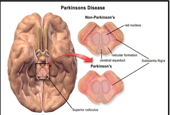
Figure 1: Difference between Parkinson’s and Non-Parkinson’s brain (Embogama, 2016).
3.4 Symptoms of Parkinson’s Disease
There are two types of symptoms in PD and they are- i. Motor symptoms
ii. Non-motor symptoms
In case of motor symptoms, they come out slowly which is the reason this disease usually begins years before diagnosis (Kalia & Lang, 2015). The main motor symptoms of PD are-
● Tremor
● Rigidity
● Bradykinesia
● Postural instability (Martínez-Martin et al., 2015)
8
PD carries identical clinical features with atypical Parkinsonism (APD) disease like-Dementia with lewy bodies (DLB), MSA (Multiple system atrophy), CBD (Corticobasal degeneration), Progressive supranuclear palsy (PSP), etc. these common clinical conditions making PD’s diagnosis complex. This is the reason why the world needs reliable biomarkers for early and exact diagnosis to detect or predict PD (Santaella et al., 2020).
The non-motor symptoms of PD include anosmia (loss of smelling sensation) and ageusia (loss of sense of flavor), depression, anxiety, changes in sleep cycle, gastrointestinal complications, swallowing problems, sexual dysfunction, pain, fatigue, hallucinations and psychosis, impulse control disorders (compulsive gambling, shopping or eating etc.) (Leeman & Potenza, 2011), cognitive impairment (slowed thinking, difficulties in word finding, difficult in learning and remembering information) and dementia (Butt et al., 2018) . PD with dementia (PDD) occurs in some regions, such as subcortical and cortical areas, due to neuropathological changes. A study carried out by Summerfield and his group of researchers compared PD patients within a certain age range with and without dementia with healthy control subjects. In this study PD with dementia patients revealed that the amount of gray matter decreases compared to healthy controls in many brain regions such as bilateral hippocampus, bilateral putamen, para hippocampal area, left anterior cingulate gyrus, etc. In case of PD the most affected areas are the hippocampus, thalamus, and anterior cingulate (Summerfield et al., 2005).
9
3.5 Stages of Parkinson’s disease
There are several forms of PD is identified, familial PD, sporadic PD, juvenile on-set PD and autosomal dominant PD are most common forms of such disease. Parkinson’s disease can occur in families as a result of their parents transmitting defective genes to a child therefore the family history of Parkinson's disease is called familial PD (Poewe et al., 2017). Besides, in a population where patients are tested with PD without a known cause, sporadic PD can be explained as a disease that occurs randomly (Modi et al., 2016). Moreover, in children and teenagers, Parkinson's- like symptoms can develop. This type of the condition is referred to as juvenile Parkinsonism, and is often associated with particular genetic mutations at high risk of PD (Mahmoudi et al., 2015).
Lastly, Parkinson’s is probably inherited from just one parent if the LRRK2 or SNCA genes are engaged. This is considered as an autosomal dominant pattern, which is when only one copy of a gene is required for a person to be altered for the disorder to occur (Sundal et al., 2012).
Parkinson’s disease has three stages- i. Pre-clinical
ii. Prodromal iii. Clinical
Preclinical PD may be defined as preceding the onset of the PD motor function,despite the fact that the presence of few non motor feature can be there or subclinical abnormalities might also additionally exist that may be detected with the aid of neuroimaging or with the aid of biological test results (Wu et al., 2011) (Jankovic, 2008). Prodromal PD can be described as a stage where
10
some variety of non-motor or subtle motor symptoms can be present but do not meet the current diagnostic standard (Mahlknecht et al., 2015).
Lastly, the clinical PD is a stage where classical motor symptoms are present and the diagnosis is based on those motor features (He et al., 2018). Mainly there are four clinical features, in short, these features can be known as TRAP (T=Tremor at rest, R=Rigidity, A= Akinesia or bradykinesia and P=Postural instability). In addition, among the characteristic features of Parkinsonism, flexed posture and freezing (motor blocks) were included, with PD as the most common type (Jankovic, 2008). There are several known forms of autosomal recessive Parkinsonism. Typically, the phenotype is defined by parkinsonism with levodopa-responsive without atypical features in three forms due to mutations in DJ-1 (PARK7), PINK1 (PARK6) or parkin (PARK2) (Bonifati, 2012).
3.6 Treatment of Parkinson’s Disease
Anti-Parkinson medicationLevodopacoupled with Carbidopa a peripheral decarboxylase inhibitor (PDI) can be first line treatment of symptomatic treatment for Parkinson disease. In addition, Istradefylline (Trade name: Nourianz) is the latest medication for PD which has been recently approved by FDA.
11
3.7 Pathogenesis of Parkinson’s Disease
The antioxidant activity of enzymes in PD patients, is low and there is a high level of oxidative stress. For this reason in the neurodegenerative mechanism of PD, oxidative damage is considered as a prominent contributor (Burkhardt & Weber, 1994; Fahn & Cohen, 1992).
In PD neuronal cells of the patients become more vulnerable to damage from ROS and reactive nitrogen species. This occurs due to glutathione peroxidase and catalase reduction in PD brain (Fahn & Cohen, 1992). In such patients, high oxidative stress, protein accumulation, DNA damage, mitochondrial dysfunction, and lipid peroxidation are prevalent in their brain tissues (Danielson &
Andersen, 2008; Swerdlow et al., 1996; Yoritaka et al., 1996). In the pre-clinical stage of the disease, due to the selective neurodegeneration, the dopaminergic neurons of pars compacta region of substantia nigra become more sensitive to ROS and reactive nitrogen species at the beginning than other neurons. There is no proper explanation for the vulnerability of dopaminergic neurons to oxidative damage (Fahn & Cohen, 1992). Nonetheless, one of the demonstrations proposes that dopamine can impart neurotoxicity on its own. After all, in the presence of molecular oxygen, it can produce ROS and is also capable to auto-oxidating into neuromelanin. Later, it facilitates the formation of oxyradicals (Fahn & Cohen, 1992).
.
3.8 The Necessity of Various Types of PD Biomarkers
Some PD biomarkers include (1)prodromal, preclinical or premotor stage biomarkers; (2) motor stage biomarkers; (3) biomarkers of risk or susceptibility (DeKosky & Marek, 2003), (Chen-
12
Plotkin et al., 2018). According to these classes, biomarkers could be based on clinical, biochemical, imaging, genetic or proteomic or various combinations of them (Sharma et al., 2013).
It may be difficult to invent an effective diagnosis of PD because its phenotype resembles several common clinical characteristics of APD, especially in the pre-clinical stage of PD. And when diagnosis was made by movement disorder experts, misdiagnosis rates in clinicopathological series of PD and APD patients were as high as 24 percent (Tolosa et al., 2006). The trials of ELLDOPA, CALM-PD and REAL-PET showed no evidence of striatal dopaminergic defects in certain subjects reported as having early PD mainly based on medical signs (Fahn, 2005). Thus, there is an obvious need to find particular biomarkers that may assist clinicians to set up a greater well timed and accurate differential analysis among sufferers who carry the symptoms of Parkinson’s disease (Devos et al., 2013) .
3.9 Types of Biomarkers and their Purpose
Premotor biomarkers might be diagnostic biomarkers and might identify PD earlier, before advanced disease progression has happened and at a period when neuroprotective treatments could pause or delay the neuronal loss. Risk biomarkers are used to indicate groups with a higher degree of probability of growing clinical PD (He et al., 2018). Progression of disease can be obtained by biomarkers of the motor stage and alongside this will help us with identification of the several available therapies and their effectiveness for improving the disease condition, in times when there are visible motor symptoms as a result of the SN neurons distortions.
13
Recently, several groups of studies showed numerous examples of the use of a combination of multimodal biomarkers in patients with PD. Furthermore, Nalls demonstrated a feasible, harmless predictive model for selecting patients with preclinical and prodromal PD in a population-based modeling experiment. The UPSIT value was accountable for 63.1 percent of the established variance, 13.6 percent for genetic score , 11.4 percent for family history,6.0 percent for gender and 5.9 percent for age, as contrasted to the defined β coefficients of such regression model (Nalls,2015). Moreover, in order to help predict progression of the disease in PD patients, a study has demonstrated statistical methods using genetic data, clinical expertise, imaging measures and molecular biomarkers. Finally, in both the early and late stages of the disease, 12 important predictors have been identified that have strong predictive accuracy. Most notably, the key purpose of these predictors is to classify patients at risk of progressive motor disease development (Latourelle et al., 2017). Furthermore, in 2017, a study showed that the mean DAT imaging caudate uptake, UPSIT value, CSF Aβ42 and RBDSQ summation would predict cognitive deficits with an AUC of 0.80.Many clinical evidence and biomarker variables were used in this experiment as predictors for identifying individuals at risk of cognitive degression (Schrag et al., 2017).
14
Chapter 4
Diagnostic Biochemical Biomarkers
One of the most sensitive and specific biomarkers that play a key role in identifying prodromal PD is biochemical biomarkers. Several studies have shown that both body fluids and tissues can be investigated for biochemical biomarkers. Currently, an impressive number of experiments have revealed that mitochondrial dysfunction, oxidative stress, the formation of Lewy bodies, neuroinflammation and various mechanisms associated with PD growth have been shown.
Therefore, the biochemical biomarkers will be reviewed from the above aspects (He et al., 2018).
4.1 Oxidative stress related biomarkers 4.1.1 Urate
With the removal ofperoxynitrite, hydroxyl radicals, hydroxyl peroxide andsinglet oxygen, urate plays an important antioxidant role. Urates can inhibit the human brain's oxidative damage (Hwang, 2013) , higher urate levels can be correlated with a diminished risk of PD. Many experiments have supported the probability of urate of being defensive against PD. An in vitro study was conducted by Sara Cipriani and her colleagues where they had shown that in PD models, spontaneous degression of cultured substantia nigral neurons can be prevented by urate and also prevent dopaminergic neurons death due to oxidative and mitochondrial toxins. (Cipriani et al., 2012), (Cipriani et al., 2012b). Meantime an in vivo study conducted on PD mouse models by Xiqun Chen and team found that in the advanced phenotype and histopathologic findings, the
15
concentration of urate was elevated in the central nervous system (X. Chen et al., 2013) (Gong et al., 2012). Moreover,Parkinson's Research Examination of the CEP-1347 Trial (PRECEPT) and Deprenyl and Tocopherol Antioxidant Therapy of Parkinsonism (DATATOP) indicated that delayed disease development rates are linked to higher levels of serum urate (Ascherio et al., 2009).
Also, CSF urate concentration was first identified in these studies as a predictor of the rate of clinical decrease in PD. A Phase II clinical trial that tested elevating urate levels as a treatment strategy for disease modification in the PRECEPT trial simplified these inventions. In the PRECEPT trials secondary end point showed that the UPDRS score of PD individuals with lower levels of blood urate was considerably in excess of that of group of controls (Schwarzschild et al., 2008). Concentration ofCSF urate is responsible for 10 percent peripheral blood concentration, and is primarily dependent on two important factors, peripheral blood urate concentration and blood brain barrier integrity (Ascherio et al., 2009).It also serves as a robust biomarker of pd by exploring an integration of data that observed urate present in the CSF and serum that acts like a protective characteristic of PD. In addition, the DATATOP analysis also showed that urate levels were lower in patients with cognitive impairment than in patients with PD-free cognitive impairment (D. et al., 2011). There is also a negative relationship in a study between urate concentrations and PD cognitive impairment intensity (Y. Wang et al., 2012). Urate can thus be a successful predictor for PD diagnosis and disease growth detection.
4.1.2 Protein DJ-1
Protein DJ-1 is referred to as a multifunctional inclusive cysteine protease because it participates in several cellular metabolic processes like RNA binding and protease, mitochondrial and
16
molecular chaperones regulation. In addition, it also plays a neuroprotective role during neurodegeneration in preventing oxidative stress. (Saito, 2014). Oxidative stress-related diseases like PD can occur due to diminished function of DJ-1. Therefore, inherited recessive PD is connected with the DJ-1 gene PARK7 mutations and causes familial PD (Abou-Sleiman et al., 2003).
Waragai et al. conducted a study in which it was recommended a direct link between the levels of DJ-1 and the disease stage. (Waragai et al., 2007). A quantitative analysis of CSF DJ-1 levels without blood contamination found that CSF DJ-1 levels decreased relative to normal controls in PD patients, AD patients, MSA patients (Herbert et al., 2014).
Besides, extensive experiments have suggested that the oxidative response of Cys-106 plays an important role in pathogenesis of PD (Ariga et al., 2013; Hao et al., 2010; Saito & Noguchi, 2016) because if exposed to oxidative stress, the residue of cysteine at position 106 is specifically oxidized and the antioxidative activity of Cys-106 helps to establish the roles of DJ-106 (Saito, 2014). In addition, it has been seen in a study that in the erythrocytes of untreated PD patients, levels of DJ-1 oxidation in Cys-106 are noticeably high in comparison with controls which are non-PD and medicated patients (Saito et al., 2009).
Lin et al. have also shown that 4-hydroxy-2-nonenal (4-HNE) modified blood DJ-1 levels are remarkably reduced in the advanced stage of PD, which indicate that 4-HNE modified DJ-1 might play the role of diagnostic biomarker of PD. Therefore, the oxidized DJ-1 promises to be an
17
effective PDD biomarker. (Lin et al., 2012). However, the levels of DJ-1 and seriousness of PD are not associated with each other (Herbert et al., 2014). Even then, DJ-1 can be a potential PD biomarker.
4.1.3 Coenzyme Q10
Coenzyme Q10 simplifies the function of the mitochondrial electron transport chain as an antioxidant (Kikusato et al., 2016). Yen et al. performed in vitro and in vivo studies indicating that the defective expression in PD patients of mitochondrial complex 1 contributes to neuronal toxicity disruption and redox balance. CoQ10 is efficient enough to prevent neurodegeneration against mitochondrial deficiency as a lipophilic antioxidant. Researchers found that in human plasma at the initial oxidation point, ubiquinol-10 was oxidized into ubiquinone-10, so the percentage of the total oxidized form of coenzyme Q10 (% CoQ-10) will be a valid biomarker of oxidative stress (Yen, 2014). Moreover, Sohmiya discovered that total CoQ10 plasma levels were substantially reduced and a strong increase was observed in the %CoQ10 plasma in patients with PD relative to usual controls. Especially, H&Y scores evaluated that %CoQ-10 is likely to rise with disease progression (Sohmiya et al., 2004). Most importantly, in the diagnosis of PD, CoQ10 levels and CoQ-10 percentages are indicated to have significance as biomarkers. Furthermore, CoQ10 supplementation could be a viable way to treat and prevent PD (Sohmiya et al., 2004).
4.1.4 Homocysteine (Hcy)
Hcy is generated by the methylation process as a natural amino acid in the body (Leng et al., 2018).
In vivo and in vitro experiments conducted by Kocer et al. found thatthese generated Hcy have
18
toxic effects on dopaminergic neurons (Kocer et al., 2016). The elevated Hcy level of hyperhomocysteinemia is a significant risk factor in the general population for cerebrovascular diseases(Leng et al., 2018). Hyperhomocysteinemia destroys neuronal DNA, activates NMDA receptors and leads to dopaminergic neuron death acceleration, which suggests it is neurotoxic to the substantia nigra as well. In addition, as the amount of Hcy increases, it can lead to oxidative stress sharpness in the dopaminergic neurons, resulting in PD-like pathology caused by MPTP (1- methyl-4-phenyl-1,2,3,6-tetrahydropyridine-induced) (Obeid et al., 2009). Hence, dyskinesia can also be an indication of neurodegeneration that is associated with hyperhomocysteinemia.
(Zoccolella et al., 2006). A research conducted by Irizarry et al. has shown that the Hcy levels are comparatively higher than normal controls in patients with PD, MCI (mild cognitive impairment), AD (Alzheimer’s Disease) and cerebral amyloid angiopathy (CAA), with PD patients in particular having the highest rates compared to non-PD patients. (Irizarry et al., 2005). A study assumed that risk factors in PD were supposed to be higher CSF and plasma concentrations of Hcy (Rozycka et al., 2014). Moreover, it is known in PD patients, elevated levels of homocysteine are associated with decreased cognition and higher plasma Aβ levels (Zoccolella et al., Irizarry et al., 2005;
2006). Even though, Hy levels, vitamin B12 and folate are helpful in reversing the hyperhomocysteinemia associated with levodopa (Postuma, 2006).
4.1.5 8-OHdG (8-Hydroxydeoxyguanosine)
8-OHdG is known to be one of the potential biomarkers of oxidative stress damage to DNA and is the primary result of DNA oxidation reactions with hydroxyl radicals and guanine residues. Many studies have shown that 8-OHdG levels in the substantia nigra of PD patients are significantly
19
increased (Alam et al., 1997). As a result, several studies have shown that the amount of CSF and serum 8-OHdG of the PD patient is considerably higher in comparison to the controls (García- Moreno, 2013;Gmitterova, 2009). Besides, 8-OHdG concentrations are linked to the duration of illness and also to the %CoQ-10 (Isobe, 2010). 8-OHdG is well known to be excreted in the urine and so elevated urinary 8-OHdG concentrations in PD patients are also much higher than in control subjects. In addition, hallucinations with a correlation coefficient of 0.85777 are predominantly correlated with urinary 8-OHdG levels (Hirayama et al., 2011). Thus, it can be said that the urinary 8-OHdG can be a credible biomarker for the diagnosis of PD.
4.1.6 Advanced Oxidized Protein Products (AOPP)
AOPP is recognized for halogenative stress as a credible biomarker for PD. In a quantitative analysis of AOPP concentrations with ELISA, researchers found that PD patient’s AOPP concentration level is remarkably higher than the control subjects. Moreover, the serum AOPP levels in PD patients depend on the Hoehn-Yahr stage (H&Y stage) or levodopa dose or disease duration because it is shown that in PD patients, serum AOPP levels are inversely proportional to the mentioned parameters. That is why it is claimed that AOPP can act a crucial role in disease progression control and PD diagnosis (García-Moreno et al., 2013).
20
4.2 Biomarkers Associated with Abnormal Protein Accumulation and Aggregation
4.2.1 𝛼-synuclein
𝛼-synuclein is known as the essential component of the Lewy bodies cytoplasmic inclusions. Also, it is we know that the existence of Lewy bodies in surviving neurons is an irregular protein accumulation with alpha-synuclein that is a pathological feature of PD. (Bandopadhyay, 2016). 𝛼- synuclein also plays an effective role in the pathogenesis of PD by the phosphorylation, misfolding and abnormal accumulation. The idea of prion-like transmission of misfolded alpha-synuclein is Braak's theory of expanding neuropathology (George, 2013). Some histology-based studies have tested CSF alpha-synuclein levels with the assistance of ELISA and have reported that the level of alpha-synuclein CSF in patients with PD is substantially lower than that of controls without PD (Mollenhauer, 2011; Parnetti, 2014). Moreover, in a study by Tokuda et al. found thhigh degree of adverse relationship between the levels of CSF alpha-synuclein and Hoenh-Year stage at there was a high degree of negative relation between CSF alpha-synuclein levels and Hoenh-Year stage, indicating the seriousness of the disease (Tokuda Takahiko et al., 2006).
(RT-QuIC) Real-time quaking-induced conversion has given an innovative technique that can detect irregular alpha-synuclein CSF in PD patients with 100% specificity and 95% sensitivity (Fairfoul, 2016).Renowned scientist Groveman and coworkers showed that the RT-QuIC method can be adequate to identify prodromal PD due to its high diagnostic accuracy and quick detection capability (Groveman et al., 2018). On the other hand, many studies suggested that the 𝛼-synuclein
21
fibrosis tends to produce the most neurotoxic species which is called the soluble oligomeric 𝛼- synuclein (Gallea & Celej, 2014). As a result, Takuda et al. have mentioned that PD patients have significantly elevated oligomeric forms of alpha-synuclein (T. Tokuda, 2010). PD can also be differentiated from control groups by measuring the oligomeric alpha-synuclein (o-alpha- syn)/total alpha-synuclein (t-alpha-syn) ratio with 90.6 percent specificity and 89.3 percent sensitivity with 0.948 ofAUC (T. Tokuda et al., 2010).Nonetheless, compared to controls which are non-PD multiple studies conducted by various groups on alpha-synuclein blood levels in patients of PD (Prakash & Tan, 2010; Duran, 2010). Recently, in PD patients, the RBC oligomeric alpha-synuclein/total protein ratio has been reported to be significantly higher than in normal controls, which could be an efficient way to differentiate patients of PD withsensitivity of 79.0 percent and 64.7 percent specificity from normal controls (X. Wang et al., 2015).
Phosphorylated alpha-synuclein could be a useful biomarker for PD patients since a study found that phosphorylated alpha-synuclein levels were significantly higher than control levels in patients of PD (Foulds, 2013).So, the basic neuropathology of such disease would be more accurately reflected by phosphorylated al-synuclein. It can also inhibit the fibrillation of alpha-synuclein and this will show that phosphorylation of alpha-synuclein occurs relatively late in the disease's development. More effective biomarkers of progression of the disease in PD may also be more effective in future medical trials, particularly for substances modifying diseases, delaying progression of disease (Foulds, 2013). In addition, in order to diagnose PD, it can be indicated that alpha-synuclein truncation levels can be used. Also, assembling these measures with other markers would produce more reliable results (He et al., 2018).Besides that, pathogenic forms of alpha- synuclein can develop in patients of PD in the Lewy body submandibular gland precursor. Lately,
22
in order to detect Lewy bodies, in a study salivary gland tissue needle core biopsies is used in which 75 percent of PD patients were tested positive (Adler et al., 2014). Consequently, the irregular evolution of alpha-synuclein may also be known as the latest successful preclinical biomarker of PD in submandibular glands.
4.2.2 β-Glucocerebrosidase and Ubiquitin C-terminal hydrolase- L1(UCH-L1)
On one hand, β-Glucocerebrosidase is a lysosomal hydrolase encoded by the gene GBA1. It plays a crucial role in the deterioration of alpha-synuclein (Sidransky et al., 2009). Especially, the potential risk factor for PD is known to be β-Glucocerebrosidase because alpha-synuclein aggregation contributes significantly to PD pathogenesis. In CSF patients with PD, the activity of β-glucocerebrosidase has been decreased in different studies, most notably in the preclinical stage of PD (García-Moreno et al., 2013). Furthermore, a study conducted by Parnetti and associates indicated that in order to improve the diagnostic accuracy of 82 percent sensitivity along with 71 percent specificity of PD, the mixed estimation of the β-glucocerebrosidase activity and o-alpha- syn/t-alpha-syn ratio will be crucial (Parnetti et al., 2014).
On the other hand, UCH-L1 was discovered by Jackson et al. as one of the proteins which are brain-specific associated with PD (Thompson & Jackson, 1981). UCH-L1 has a vital role in directing brain protein metabolism by pairing with the proteasome pathway to eliminate the unnecessary, altered, oxidized neuronal cytosol proteins (Betarbet, 2005; Bishop, 2016). Hence,
23
a reduced rate of alpha-synuclein degradation will result from the dysfunction or amount of UCH- L1. In order that it will eventually lead the neurodegeneration and neurocyte death. A study mentioned that the PD patients' CSF concentrations of UCH-L1 were found to be lower relative to usual controls (Jiménez-Jiménez et al., 2014). Moreover, lowest levels of CSF UCH-L1 were observed in PD patients suffering from other neurodegenerative diseases, differentiating this group from those suffering from other neurodegenerative diseases, such as MSA and PSP. As an outcome, UCH-L1 with a moderate specificity of 67% and satisfactory sensitivity of 89% may be a promising diagnostic biomarker for PD. Furthermore, as a combination, CSF alpha-synuclein and CSF UCH-L1 levels have a positive association and may become a more precise diagnostic technique for prodromal PD (Mondello et al., 2014).
4.2.3 Amyloid beta 42(Aβ42)
Aβ42 is primarily derived from the amyloid precursor protein hydrolysis. A study by Clinton et al.
indicates that by promoting polymerization of alpha-synuclein, Aβ42 may increase the productivity of Lewy body diseases (Schulte & Gasser, 2011). Particularly, various experiments showed that the PD patients have lower CSF Aβ42 levels compared to control subjects (Siderowf et al., 2010; Montine et al., 2010; Parnetti et al., 2013).
Apparently in PD patients progressing to PD with dementia, lower levels of CSF Aβ42 have been found at 18-month follow-up compared to baseline levels and this outcome could be associated with neocortical regionsLewy body pathology and diffuse amyloid plaque pathogenesis of PDD patients (Andersson et al., 2011; Mollenhauer et al., 2006; Montine et al., 2010). This presented
24
credible information for the use of amyloid beta 42 in the measures of cognitive capacities in patients of PD with a sensitivity of above 85 percent. Therefore, One of the initial biomarkers that can help evaluate the progression of patients with PD from CI-PD to PDD is decreased CSF Aβ42 levels(Alves et al., 2014).
4.2.4 Tau protein
Tau protein is a protein associated with microtubules that can be found primarily in healthy neuron axons in the CNS and peripheral nervous system (PNS). A significant number of studies have reported that the concentrations of tau protein and phosphorylated tau protein (p-tau) in CSF of PD patients are lower than those of AD,MSA,PDD and DBL patients (Hall et al., 2012). Also, with a sensitivity of 82 percent and a specificity of 81 percent, the combined CSF p-tau and t-tau levels can distinguish PD patients from MSA (Herbert et al., 2014). Zhang et al verified an inverse connection between the p-tau/t-tau ratio in UPDRS and CSF (Unified Parkinson's Disease Rating Scale) or UPDRS motor score (UPDRS III) (P. Zhang et al., 2010). Moreover, PD patients with higher levels of CSF t-tau and p-tau are found to be at high risk of visuospatial dysfunction and memory failure. In PD patients, Tau pathology generally tends to be associated with cognitive impairment. It may be used in such patients as an essential indicator of cognitive decline (Andersson et al., 2011). Besides, Montine and colleagues suggested that the number of cases with an elevated t-tau/Aβ index rose from 15 percent in patients with PD and 29 percent in patients with CI-PD to 45 percent in patients withPDD. Therefore, the combination of tau protein and Aβ can give more significantly prognostic value which may predict cognitive function in PD patients (Montine et al., 2010).
25
4.2.5 Neurofilament light chain protein (NFL)
In the PNS and CNS, neurofilaments are recognized as the main structural components of axons.
Also, in the transport of nerve impulses and the preservation of neuronal morphology integrity, NFL plays a crucial role like one of the three subunits. Abnormal neurofilament phosphorylation has been observed in PD patients (Vågberg et al., 2015). A study conducted by Hansson et al.
mentioned that there was no increase in NFL levels in patients with CSF or serum PD because of less extreme axonal degeneration (Hansson et al., 2017). Therefore, it can be a useful biomarker of disease progression for Parkinson’s disease.
4.3 Neurotrophins related biomarkers
4.3.1 Brain-derived neurotrophic factor (BDNF)
A study conducted by Scalzo et al mentioned that BDNF is considered one of the most widespread neurotrophic variables that may control synaptic plasticity and cell survival and inhibit apoptosis- mediated cell death (Scalzo et al., 2010). The neurodegenerative process occurs because of insufficient supply of neurotrophic factors. Usually, BDNF is synthesized by neurons, but microglial cells generate BDNF when damaged neurons are involved. Several studies indicate that in PD patients, BDNF expression in substantia nigra is diminished, specifically in the region called ventral lateral of the brain (Howells e, 2000) and it performs an important role in the survival of dopaminergic neurons (Scalzo et al., 2010). Most importantly, BDNF can be helpful for early diagnosis of PD because of its lower levels in the early stage of the disease. Also, an effective treatment for PD is indicated by up-regulation of BDNF concentrations. Nevertheless, a study
26
reported a conflicting outcome that increased BDNF levels were also associated with more acute symptoms over a longer period of illness (Scalzo et al., 2010). Another study found that BDNF amounts in the PD patient’s CSF are far higher than normal controls because of excessive generation of glial cells arising from brain injury (Mashayekhi & Salehi, 2009). So, the combined concentrations of the BDNF in CSF and serums perform the role of essential early diagnostic marker for PD.
4.3.2 Insulin-like growth factor 1 (IGF-1)
IGF-1 exerts neuroprotective effect by enhancing survival of neurons and inhibiting neuronal apoptosis (Fernandez & Torres-Alemán, 2012). Typically, neurons play an important role in IGF- 1 synthesis, but IGF-1 is generated in case of damaged brain reactive microglia. Various studies showed several correlations between IGF-1 and PD, such as (1) the survival of dopaminergic and substantia nigra neurons is developed by IGF-1; (2) IGF-1 defends dopaminergic neurons against programmed apoptosis of cells (Bernhard et al., 2016; Mashayekhi et al., 2010). Moreover, many studies agreed with the results which showed that PD patients carry an increased level of CSF IGF (Mashayekhi et al., 2010). In the meantime, Godau et al. published that in PD patients the concentration of serum IGF-1 is higher than control subjects (Godau et al., 2010, 2011). The longitudinal MODEP study suggests that patients with PD may have considerably increased serum concentrations of IGF-1 compared to stable controls for more than 3.5 years. However, patients with PD at the early stage or preclinical stage (3.5 years period of disease) do not display an effective rise of IGF-1 in serum (Bernhard et al., 2016). Therefore, in advanced disease stages, serum IGF-1 is considered a standard biomarker. As a matter of fact, in multiple brain regions
27
crucial to cognitive function, different IGF-1 receptors are identified, with the maximum concentrations in the hippocampus and frontal cortex. As a consequence, reduced IGF-1 serum levels are linked with poor functional capacity and verbal memory in patients with PD. For the initial diagnosis of PDD, IGF-1 is seen as a beneficial biomarker (Picillo et al., 2017).
4.4 Biomarkers Associated with Neuroinflammatory Reaction
A research conducted by Mosley RL and team mentioned that the neuroinflammatory reaction is effective in decreasing dopaminergic neurons in patients with PD. Researchers have reported that microglial cells of nigrostriatal region are abnormally activated in both PD patients and animal models of PD (Mosley et al., 2012). Besides, TNF-alpha, IL-6, IL-1β inflammatory cytokines are secreted by abnormally activated microglia that invade dopaminergic neurons and result to neuron degeneration (Cebrián et al., 2014; More et al., 2013). The enhancement of such inflammatory factors can also be a possible biomarker of PD progression for early detection and recognition. In addition, it is confirmed that increased CSF IL-8 levels are predicted to be associated with a decreased (MoCA) Montreal Cognitive Assessment records and increased risk of cognitive damage in PD patients, based on another study (Z. Liu et al., 2016; Yu et al., 2014). So, to evaluate the intensity of cognitive impairment in PD patients, IL-8 and CRP can be effective neuroinflammatory biomarkers. In this condition, an effective treatment strategy for PD can be given by medicines targeting inflammatory mediators (He et al., 2018).
28
Chapter 5
Genetic Biomarkers
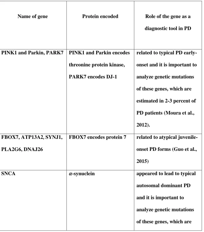
Genetic factors are predicted to have a significant impact on sensitivity to PD. Recently, there have been at least 20 genes approved to be associated with familial forms of PD, while more than 23 PD genetic risk sites have been identified in genome-wide association studies (GWAS) (Deng et al., 2018). Accordingly, of these eight genes, PINK1, DJ-1 and Parkin are related to normal early- onset PD and atypical types of juvenile-onset PD are related with SYNJ1, ATP13A2, FBOX7, DNAJC6, PLA2G6. It is probable that autosomal-recessive genetic forms are likely to be associated with these eight genes (Guo et al., 2015). The other three genes, VPS35, SNCA and LRRK2, appear to contribute to conventional autosomal dominant PD (Moura et al., 2012; Shen et al., 2016). Also,the study of mutations of genes in LRRK2, PINK1, DJ-1, Parkin, GBA, and SNCA is of great importance to 2 to 3 percent of patients with PD. In a study Siddiqui et al.
mentioned that the alpha-synuclein, which is the main Lewy body part, is encoded by SNCA (Siddiqui et al., 2016). Moreover, according to reports, 5-10% of PD (PDD) patients show GBA mutations. Therefore, GBA has, so far, been the most successful for PD related genetic risk factor.
Patients may have genetic factors in the etiology of PD for many years before the onset of clinical features. Therefore, Yung et al. mentioned that in peripheral blood the positive genescan probably become eligible biomarkers for the diagnosis of Parkinsonian syndromes along with PD because
the disease pathophysiology is linked to the proteins (Yang et al., 2015).
29
Table 1: Name of gene, Protein encoded, Role of the gene as a diagnostic tool in PD
Name of gene Protein encoded Role of the gene as a
diagnostic tool in PD
PINK1 and Parkin, PARK7 PINK1 and Parkin encodes threonine protein kinase, PARK7 encodes DJ-1
related to typical PD early- onset and it is important to analyze genetic mutations of these genes, which are estimated in 2-3 percent of PD patients (Moura et al., 2012).
FBOX7, ATP13A2, SYNJ1, PLA2G6, DNAJ26
FBOX7 encodes protein 7 related to atypical juvenile- onset PD forms (Guo et al., 2015)
SNCA 𝛼-synuclein appeared to lead to typical
autosomal dominant PD and it is important to analyze genetic mutations of these genes, which are
30
estimated in 2-3 percent of PD patients (Guo et al., 2015).
LRRK2 Dardarin appeared to lead to typical
autosomal dominant PD andit is important to analyze genetic mutations of these genes, which are estimated in 2-3 percent of PD patients(Siddiqui et al., 2016) .
VPS35 796-amino acid protein appeared to lead to typical
autosomal dominant PD (Moura et al., 2012).
GBA Lysosomal membrane
protein
it has been reported that 5- 10% of patients have GBA mutations, therefore GBA gene has been the most important PD genetic risk factor so far (Yang et al., 2015).
31
Chapter 6
Other biomarkers
5.1 MicroRNA (MiRNA)
MiRNA is a series of small, non-coding single-stranded molecules that can control the appearance of their target genes through translational inhibition or degradation of messenger RNA (Khodadadian et al., 2018). The pathogenesis of PD can occur due to these MiRNAs because most of the PD related genes are regulated by them. Moreover, the development of PD and the specific phase of disease usually based on variations in the expression of MiRNAs, resulting in the heterogeneity of the miRNAs. In fact, up- or down-regulation of inflammatory reaction (Zhou et al., 2016; Nair & Ge, 2016), downregulation of proteinDJ-1 (Z. Zhang & Cheng, 2014; Xiong et al., 2014), pathogenic upregulation of LRRK2 protein(Rassu et al., 2017), dysregulation of IGF (W. Kim et al., 2014) ,overexpression of alpha-synuclein also the death of dopaminergic neurons can all be associated with miRNA dysfunction (Z. Zhang & Cheng, 2014).
Circulating miRNAs carry some significant features. They are likely to be highly stable and quantifiable, tissue-specific, abundant, and several years before the beginning of PD are up- or down-regulated, and these characteristics portray miRNAs as a modern candidate of confined biomarkers to identify initial-stage PD and observe disease progression. A study conducted by Martins and collaborators found that 18 miRNAs in PD patients were under-expressed when a miRNA expression profiling study was performed. It was also found that miR-30c, miR-30b, miR- 26a were possibly associated with sensitivity of PD (Martins et al., 2011). Also, four beneficial
32
candidate biomarkers(miR-1826, miR-505, miR-450b-3p, miR-626) were found in a study(Khoo et al., 2012). Furthermore, a study that showed that miR-331-5p is upregulated in PD patients was conducted by Cardo et al. In addition, three upregulated miRNAs (miR-324-3p,miR-24, miR-223) and two downregulated miRNAs (miR-148b and miR-30c) were seen by Vallelunga. Later on, by performing an RNA-seq approach, Ding et al. observed 5 novel miRNAs where four miRNAs were downregulated in patients with PD and miR-195 was upregulated (Leggio et al., 2017).
Lately, miR-4639-5p was effectively upregulated in patients with PD. Especially, this miRNA can be established as a reliable, effectivepreclinical PD diagnosis biomarker without relying on age of disease onset, gender, L-DOPA treatment and severity of PD motor feature (Y. Chen et al., 2017).
Not only is miRNA profiling promising to be a modern method for diagnosing PD, but it also intends to include new treatments based on miRNA for PD care.
5.2 Peptides
In recent years, it has been identified by using proteomic technologies that some peptides have correlation with various disease stages and severity but most of these candidate peptides have not gone through any quantitative analysis. Besides, a group of researchers were devoted to the establishment of novel biomarkers using mass spectrometry (MS) and accurate inclusion mass screening (AIMS)with high sensitivity, accuracy and specificity (Jaffe et al., 2008; Whiteaker et al., 2011). They discovered that there were substantial variations between PD and controls in 17 peptides in CSF, and most of these 17 peptides had nothing to do with subjects' age and gender.
Among them, secreted phosphoprotein 1 (SPP1) with an AUC of 0.791 and low-density lipoprotein receptor-related protein 1 (LRP1) with an AUC of 0.706 are the best performing individual
33
peptides. Moreover, Colony-stimulating factor receptor 1 (CSFR1), LRP1, SPPI, Tissue metalloproteinase inhibitor-1 (TIMP1) and Ephrin type-A receptor 4 (EPHA4) these 5 peptides can combined elevate AUC to 0.873 in recognition of patients with PD from healthy individuals also can distinguish PD patients very well from patients with AD with a value of 0.990 in AUC.
This group of peptides, therefore, can be an effective biomarker that can deliver adequate PD specificity and diagnostic sensitivity, also correlate with the severity of the disease. Carecchio and Comi discovered in 2011 that in neurons, SPPI, the glycosylated phosphoprotein tends to perform the part of a double-edged sword in case of neurodegenerative disorders. Also it can be neurotoxic in some cases and induce cell death, but in others it has a strong neuroprotective effect (Comi &
Carecchio, 2011).
Furthermore, LRP1 acts as a receptor involved in a signaling pathway that can transport of Aβ along with other brain ligands, regulate the clearance and maintain neuronal integrity and brain lipid homeostasis (Kanekiyo et al., 2013; Q. Liu et al., 2010). A study done by Jian et al. mentioned that stimulating factor 1 and IL-344 for the colony, CSFR1 acts as a receptor that can give brain damage survival signals and neuroprotective signals. Neurodegeneration and excitotoxin-induced cell death can also be exacerbated by lack of CSFR1 (Luo et al., 2013). On the other hand, EPHA4 is a member of the subclass of Ephrin receptor tyrosine kinases that, through its ephrin ligands, can control synapse formation and neuronal plasticity, during neural development can lead axons (Klein, 2009).
Moreover, TIMP1 functions as an inhibitory molecule, and a broad class of matrix
34
metalloproteinases (MMPs) are inhibited by the primary activity of TIMP1.First of all, in response to neuronal cell stress, MMP-3 plays a crucial role in activating neuroinflammation in the pathophysiology of PD. The negative charge of the alpha-synuclein C-terminal portion can also be eliminated, allowing the C-terminally truncated protein extremely hydrophobic and even more cytotoxic for aggregation (E. M. Kim & Hwang, 2011).
Additionally, researcher Kim and Hwang demonstrated that DJ-1 is capable to providing protection against mitochondrial, proteasomal and oxidative stresses, which is cleaved by MMP- 3. Consequently, TIMPI has a neuroprotective role in various ways (E. M. Kim & Hwang, 2011).
35
Chapter 7 Discussion
Parkinson’s Disease is a neurodegenerative disease that is associated with different kinds of biomarkers. As the average lifespan of the population is increasing, the prevalence of such diseases is increasing, since with increasing age is an inherent risk factor for development of PD (He et al., 2018). Therefore, the proper diagnosis plays a major role in the treatment of the disease. There are many biomarkers which are associated with Parkinson’s Disease in different ways. But in some cases, those biomarkers are not very specific in every case and can be associated with other neurodegenerative diseases as well.The most widely used and most effective biomarkers of PD can be the neuroimaging biomarkers. Under neuroimaging biomarkers, transcranial sonography carries most of the ideal features that a biomarker should have, like it is cheap, painless, safe and non-invasive. Also, MRI is potential enough to become a biomarker for preclinical PD. Secondly, one of the most effective biomarkers of PD can be the biochemical biomarkers especially CSF (Cerebrospinal Fluid) 𝛼-synuclein, amyloid beta 1-42 in CSF and advanced oxidized protein products (AOPP) can predict the development of PD and such biomarkers are the focus of the current review.As PD is mainly related to the neurons, the CSF biomarkers are the closest ones to the disease site. Moreover, there are several other biomarkers that include peptides and microRNA.
For preclinical PD diagnosis, MiR-4639-5p may be a stable potential biomarker because it does not depend on gender, age of onset of the disease, severity of motor symptoms of PD, and treatment with L-DOPA. On the other hand, in separating PD patients from healthy people, peptides can be used and PD can also be differentiated from other neurodegenerative diseases, since these particular peptides can signify various stages of disease and PD severity. Besides, molecular imaging technology carries dopaminergic imaging and non-dopaminergic imaging, can be used to
36
detect PD patients. Additionally, a significant move in the illustration of the chain molecular cases directing neurodegeneration in PDD was the discovery of the SNCA gene encoding alpha- synuclein (Gasser, 2010). Moreover, evaluation of the role of common genetic variability as a risk factor for such disease has been effectively applied to quantitative characteristics such as PD age (Latourelle et al., 2009).Since genetics are known to have a major effect on PD susceptibility, it is likely that genetic biomarkers will become candidate biomarkers to diagnose Parkinsonian and PD syndromes (Deng et al., 2018).
There are many biomarkers like DAT imaging, levodopa challenge, substantia nigra hyperechogenicity etc. will become candidate biomarkers to diagnosis of PD but those are still being questioned as their exact relation with the Parkinson’s Disease is still not clear. So, there should be more confirmatory studies to establish the biomarkers and to make the diagnostic procedures more efficient (Cova & Priori, 2018).
37
Chapter 8 Prospect
The advanced approaches to identifying our PD biomarkers have been analyzed by a large number of study groups. They've developed a lot of candidate biomarkers lately. Transcriptomics, proteomics, and metabolomics have also been used in PD biomarker research in recent years (Caudle et al., 2010). Also, a scientific study showed that gene microarrays' high productivity capabilities, the relative appearance of PD patient blood components and it is possible to use normal controls to better understand the role of particular proteins in PD pathogenesis and to detect effective biomarkers related to PD (Caudle, 2010). Besides, by quantifying their comparative differences in illustration from one phase of disease to another, they can offer a highly efficient platform and generate comprehensive protein identification data. A Japanese study mentioned that Proteomics would provide a more thorough understanding of the underlying pathogenesis of PD by using different body tissues to discover specific protein markers, such as CSF or tissue, serum and plasma (Misato et al. 2014). Lastly, in order to provide a way to separate patients with PD from general controls, irrespective of status of medication, metabolomics concentrates on metabolite quantification and detection. Besides, electrochemical coulometric arrays were used in metabolomic analysis that have the capability to discover metabolic statuses that can be used as potential biomarkers. Before that, a study identified using this technique that can distinguish controls from PD patients (Amara & Standaert, 2013). Therefore, transcriptomics, proteomics and metabolomics are promising to advance in the discovery of effective PD biomarkers.
So far, numerous favorable biomarkers have been suggested but the perfect biomarkers are still
38
evasive(He et al., 2018). At different stages of the disease process, different important role has been performed by biomarkers. Additionally, the different biomarkers profiles differ widely from one person to another. One biomarker will be not sufficient enough for early diagnosis and predict disease track with appropriate sensitivity or specificity (He et al., 2018) . Recently, several groups of studies showed numerous examples of the use of a combination of multimodal biomarkers in patients with PD. Fereshtehnejad et al. created a bunch analysis with detailed data of these different biomarkers for predicting progress with adequate reliability. It is an advanced approach which can be utilized to distinct PD subtypes (Fereshtehnejad et al., 2017).
39
Chapter 9
Conclusion
Effective progress has been made in the study of biomarkers for PD, and a growing number of candidate biomarkers have