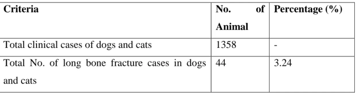
Intramedullary pinning (IMP) is one of the most commonly used techniques and the best choice for the treatment of long bone fractures (Kaur et al., 2015). Therefore; the aim of this study was to investigate the incidence of long bone fractures along with treatment of femur fractures in dogs and cats in SAQTVH, CVASU, Chattogram. The attachment of the adductor muscle to the caudal part of the diaphysis is also an important source of periosteal vessels, which becomes important in the healing of diaphyseal fractures (Kaderly et al., 1982).
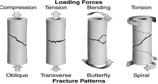
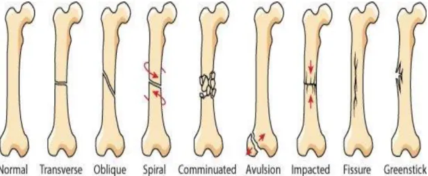
A load-deformation curve allows the detection of the mechanical behavior (e.g., stiffness, strength, energy absorption capacity) of the entire structure, such as the whole bone or the implant-bone composite (Nordin et al., 1980; Schwarz, 1991). The location of the fracture in a bone (epiphysis, metaphysis, or diaphysis) and the pattern of the fracture (e.g., spiral, transverse, oblique, comminuted) are detected by the (1) type of bone (cortical vs. cancellous), (2) apparent density or porosity of the bone, (3) rate at which the bone is loaded (fast versus slow), (4) orientation of the bone microstructure. Intramedullary pins are satisfactory for shaft fractures of the femur in small dogs and cats (Hill, 1977; Phillips, 1979).
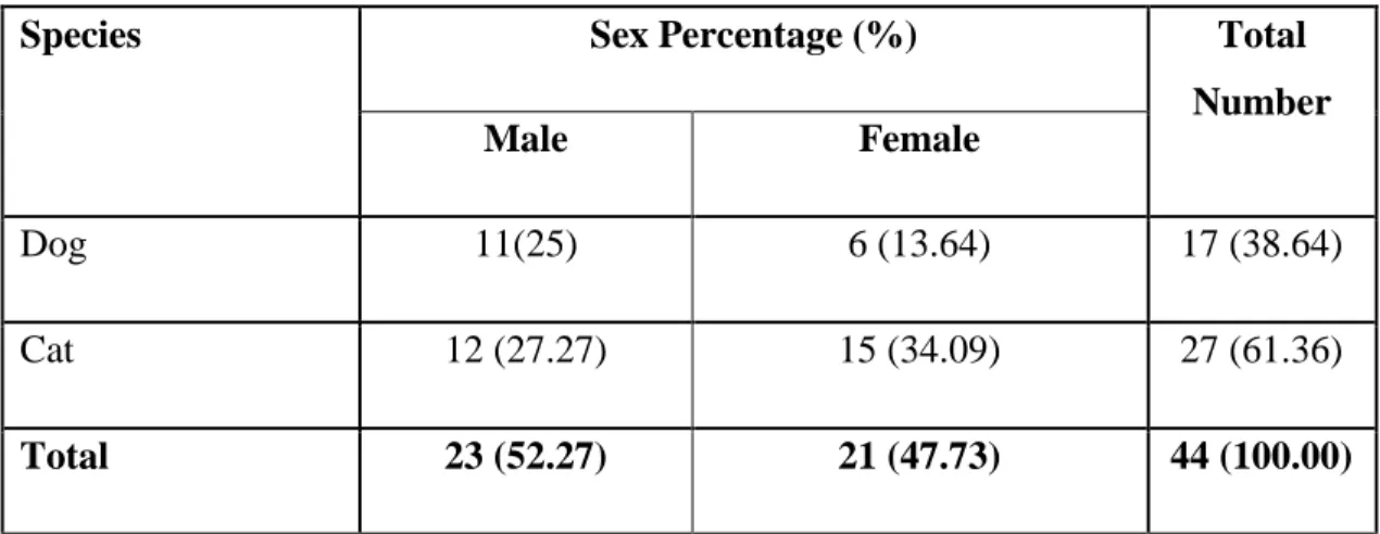
Signals including special breed, age, sex and body weight of the animal were also recorded.
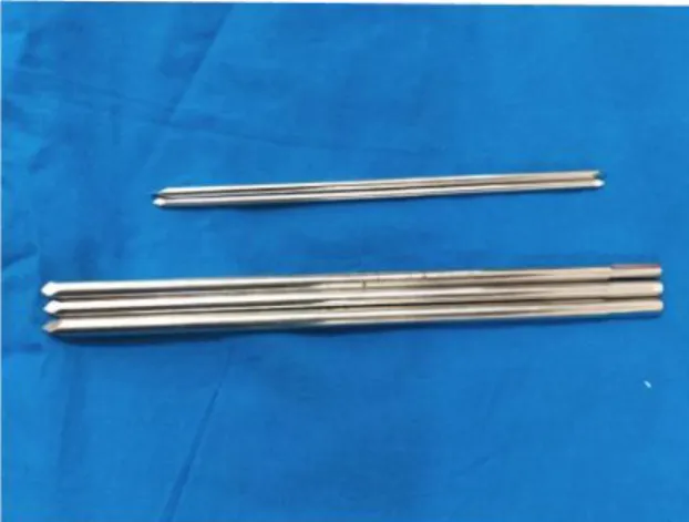
Intramedullary pinning (IMP) technique 1. Instruments and implants used for the study
- Orthopedic implants
- Imaging tools
- Computed radiography (CR)
- Patient preparation
- Premedication
- Induction and maintenance of anesthesia
- Surgical procedure
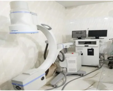
In addition, a portable X-ray machine (Figure 6) was used to generate X-ray and focused on those cassettes. Two different sized cassettes were used; i) 35 cm X 43 cm and ii) 24 cm X 30 cm. Premedication was performed by Atropine sulfate (Inj. Tropin Vet®, ACME Pharmaceutical, BD) at a dose of 0.04mg/kg subcutaneously. Xylazine Hydrochloride at the dose of 1mg/kg (Inj. Xylaxin®, Indian Immunologicals Limited, India) intramuscularly.
Intravenous fluid administration was instituted through the IV cannula at a rate of 10 ml/kg/hour. Ketalar®, Popular Pharmaceutical, BD) was given at 8 mg/kg body weight intravenously. Sedil®, Square Pharmaceuticals Ltd. BD) was given at 0.5 mg/kg body weight intravenously.
In the case of cats, only ketamine hydrochloride was used at a rate of 10 mg/kg body weight intravenously.
IMP Procedure
- Removal of implants
- Parameters studied for fracture management 1. Lameness grade
- Functional limb outcome
- Radiographic assessment
- Post fracture management complications
- Data analysis
Ceftriaxone (Trizon vet®, Acme Laboratories Ltd., BD) at a dose of 50 mg/kg body weight was administered intramuscularly at 24-hour intervals to all dogs and cats until postoperative day 7. Meloxicam (Mel Vet®, Acme Laboratories Ltd., BD) @ 0.5 mg/kg SC until day 3 after surgery and Diphenhydramine HCl (Inj.Phenadryl®, Acme Pharmaceuticals Ltd. BD) @ 1.5 mg/kg IM were administered until the 7th day after surgery or Claudication grade was assigned before and after fracture treatment (days 1, 15, 30, and 45) based on the severity of clinical signs to assess response to treatment.
Limb functional outcome was assessed on day 45 of post-fracture management and categorized as excellent, good, fair and poor (Clark, 1986). Radiographs were taken before and after fracture management (days 1, 15, 30, and 45) to assess response to treatment. Complications of post-fracture management were studied and recorded, if possible Joint stiffness / Muscle atrophy / Pin loosening / Pin migration / Seroma formation / Bone infection / Skin infection / Internal rotation of the affected limb / External rotation of the affected limb / Other.
The data obtained regarding the incidence from the study were imported into Microsoft Excel- 2010 and transferred to the statistical software STATA-11 for calculating the percentage of different variables.
CHAPTER- 4 RESULTS
Incidence of long bone fracture
- Overall incidence of long bone fracture
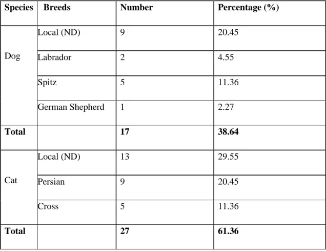
- Incidence of long bone fracture according to species
- Incidence of long bone fracture according to sex
- Incidence of long bone fracture on the basis of closed and open fracture
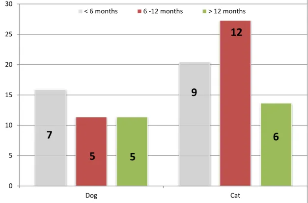
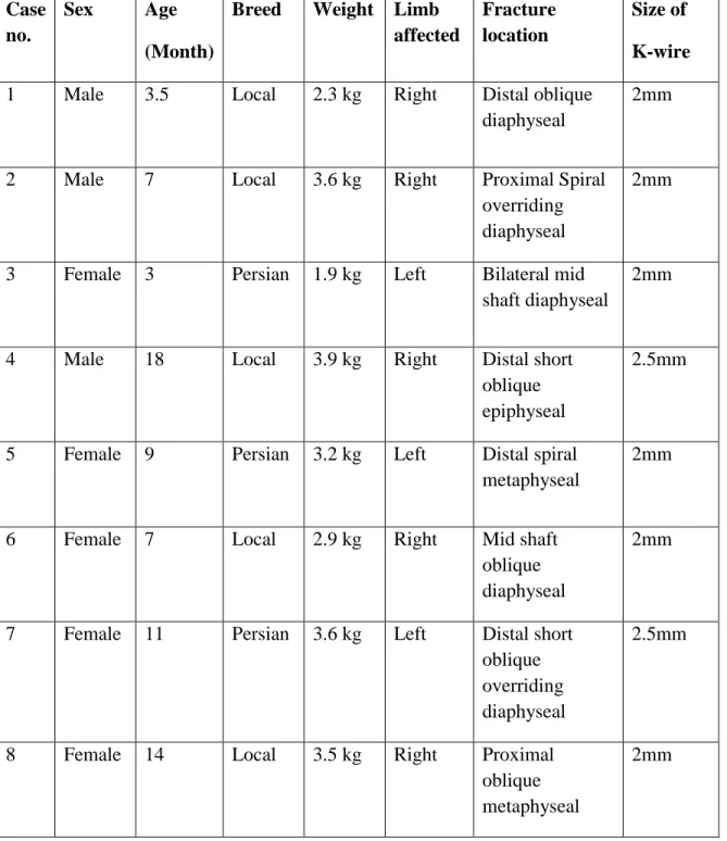
Thus, it was seen that the highest incidence was found at the age of less than 6 months in dogs and 6 months to 12 months in cats, while the lowest incidence was found at the age of more than 12 months in both dogs and cats. . The table showed that the incidence is higher in males than in females in the case of dogs and the incidence rate is higher in females than in males in the case of cats.
Evaluation of femur fracture management technique
- Overall description of femur fracture management in dogs Table 6: Overall description of femur fracture management in dogs
- Lameness grade evaluation
- Radiographic evaluation: Fracture healing was evaluated through radiographic examination on different interval on pre and post fracture management on 1 st , 15 th , 30 th
A lameness grade was assigned based on the severity of clinical signs on pre- and post-fracture management on 1st, 15th, 30th and 45th postoperative day to determine the response to treatment. All the cases, lameness grade was gradually improved on 45th post-operative day from grade 5 to grade 1, except in dog case number 3 (grade 3 on 45th day) and cat case number 3 (Death) (Table 8). Functional limb outcome: Functional limb outcome was evaluated on the 45th day of postfracture management and categorized as excellent, good, fair and poor.
All the cases noted functional limb outcome as excellent (Day 45) in postfracture management, except in dog case number 3, cat case number 3 where functional limb outcome was poor and good respectively. Radiographic evaluation: Fracture healing was evaluated by means of radiographic examination at different intervals on pre- and post-fracture management on 1st, 15th, 30th examination at different intervals on pre- and post-fracture management on 1st, 15th, 30th and 45th postoperative day. In all cases of dog and cat, secondary bone healing (periosteal callus formation) was noted (Day 30) (Table 9).
Intramedullary pinning technique of dog (Pre and Post-operative evaluation)
Intramedullary pinning technique of Cat (Pre and Post-operative evaluation)
CHAPTER-5 DISCUSSION
Incidence of long bone fracture in dogs and cats 1. Incidence of fracture according to species
- Incidence of fracture according to the breed
- Incidence of fracture according to the sex
- Incidence of fracture according to the age
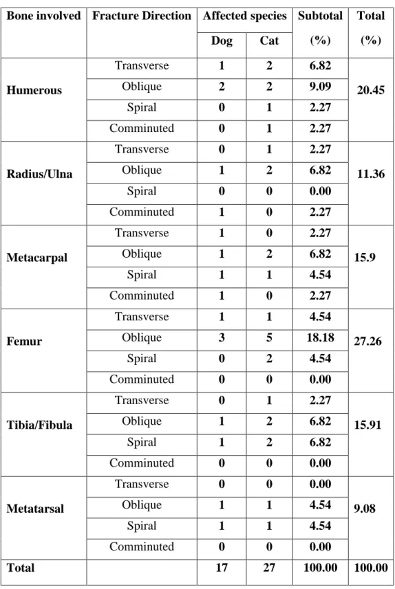
- Incidence of fracture according to the types and location of fracture
- Incidence of fracture according to the risk factors
The frequency of fracture distribution by breed may vary depending on the availability of breeds in a particular study area. Males predominated in dogs, while females predominated in cats. Regarding the gender distribution among the 44 cases of long bone fractures, 52.27% (n=23) were male and 47.73% (n=21) were female. experienced a fracture in this study. The higher incidence of fractures in males may be due to their greater activity and more aggressive nature compared to female dogs.
Female cats were more susceptible to breakage due to their tendency to mate in heat than female dogs. So it became clear between the report analysis that higher incidence was found below 6 months in dog and 6 months to 12 months in cat, whereas lower incidence was found in more than 12 months of age in both dog and cat. Alcantara and Stead (1975) analyzed 82% of fractures occurred between 2 and 10 months of age in dogs and cats.
The positive similarity between the present study and the Yardimci study that oblique fractures were more significant than other types of fractures. Minimum incidence of oblique fractures (6%) in cats reported in Langley-Hobb et al. 1996), which was contrary to the present study. In the current study, both dogs and cats, the femur was the most affected site (12/44 cases), and the frequency of femur fractures was significantly higher than other long bone fractures.
Braden et al., 1995 documented that among 1000 femur fracture cases, 77% were dogs and 23% were cats, which was in complete disagreement with the present study. The risk factors related to fracture observed in the present study that 47.73% (n=21) cases were caused by fall from height in cat. Fall from height has been seen either in the estrus season when seeking tendency to mate with another cat or want to catch the bird.
All cases of long bone fractures in the study showed maximum incidence of fractures in case of dog (20.45%, n=9) were responsible for car accidents due to either careless driving or fast running to fight with other dogs carelessly. 2006), Whitehair and Vasseur (1992) and Simpson and Lewis (2003) reported that the dog usually occurs due to motor vehicle accidents, which were in complete agreement with this study. The maximum of the cases was mainly due to the fall from height, especially for cats and car accidents, mainly for dogs, which similarity was found in two studies.
Evaluation of Femur fracture management 1. Intramedullary pinning technique
- Bone healing of IMP technique
- Complications of IMP technique
We encountered only two minor complications in the cat: one migration of the pin and one minimal infection at the suture site. Promoting bone healing by inserting an intramedullary pin, even if it comes into contact with bone fragments, bone marrow-derived pluripotent cells (Fossum, 2007). Unlike plate placement, intramedullary pinning insertion does not damage the periosteal blood, and operation time is halved (Daglar et al., 2007).
A positive correlation was found in the improvement of bone healing between age and time to achieve union. Because the immature animals have numerous arteries that perforate newly formed appositional bone running longitudinally over the periosteal surface observed by Johnson et al. This may be due to the extra forces on the broken leg in the post-operative period.
Major complications of fracture repair include delayed union, osteomyelitis, malunion, nonunion, premature physeal closure, and fracture-related sarcoma. In the present study, pin migration and minimal suture site infection were noted in the case of feline IMP. 35 Pin migration and nonunion were common complications in intramedullary pinning that were the outcome of several author's studies.
Pin migration may be due to uncontrolled movements during postoperative days and improper selection of the intramedullary pin diameter. In all cases the outcome was satisfactory, followed by weight-bearing status after the 45th postoperative day, except for the death of the cat in one case (case no. 3). From this study we have drawn a significant conclusion regarding the use of intramedullary pinning technique in femoral fractures in dogs and cats.
Intramedullary pinning technique can be successfully applied in dogs and cats under field conditions in Bangladesh. Open reduction internal fixation (ORIF) using intramedullary pins is safe and economical when the basic principles of fracture repair are used.
CHAPTER-6 CONCLUSIONS
CHAPTER-7
LIMITATIONS AND RECOMMENDATIONS
CHAPTER-8 REFERENCES
Use of IMEX SK circular external fixation hybrid constructs for fracture stabilization in dogs and cats. Treatment of long bone fractures with acrylic external fixation in dogs and cats: Retrospective study in 30 cases. Fragment reconstruction and bone plate fixation versus bridge plate fixation for the treatment of highly comminuted femoral fractures in dogs: 35 cases.
Incidence and classification of bone fracture in dogs and cats: a retrospective study at Veterinary Teaching Hospital, Khon Kaen University, Thailand. Management of femoral fractures in dogs with unilateral semicircular external skeletal fixator intramedullary pin tie-in configurations.
ANNEX
Biography