THE EFFECTS OF CARTILAGE THICKNESS ON CARTILAGE BEHAVIOUR IN INDENTATION TEST
KEVIN KONG TUCK HSIEN B041110071
BMCS
Email: [email protected]
Draft Final Report Projek Sarjana Muda II
Supervisor: DR MOHD JUZAILA ABD LATIF
Faculty of Mechanical Engineering Universiti Teknikal Malaysia Melaka
I
SUPERVISOR DECLARATION
“I hereby declare that I have read this thesis and in my opinion this report is
sufficient in terms of scope and quality for the award of the degree of Bachelor of
Mechanical Engineering (Structure & Materials)”
Signature : ………
Supervisor : DR MOHD JUZAILA BIN ABDUL LATIF
II
THE EFFECTS OF CARTILAGE THICKNESS ON CARTILAGE BEHAVIOUR IN INDENTATION TEST
KEVIN KONG TUCK HSIEN
This report was submitted in fulfillment of the requirement for the award of Bachelor of Degree of Mechanical Engineering with Honours (Structure &
Materials)
Faculty of Mechanical Engineering Universiti Teknikal Malaysia Melaka
III
DECLARATION
“I hereby declare that the work in this report was my own except for summaries and
quotations which have been duly acknowledged.”
Signature : ………
Author : KEVIN KONG TUCK HSIEN
IV
ACKNOWLEDGEMENT
In preparing this report, there were many people had involved helping me to
complete the report, so, I would like to express my grateful and thankful to them.
I am very appreciate the help from my final year project supervisor, Dr. Mohd
Juzaila Bin Abdul Latif who has gave a lot of useful idea, suggestions and knowledge
to me. He has spent his precious time in guiding me with his expertise and giving his
moral support.
My sincere appreciation also extends to all my friends and others who have
provided assistance at various occasions. Their views and tips were useful indeed.
Unfortunately, it was not possible to list all of them in this limited space. Once again
V
ABSTRACT
Osteoarthritis is the most common form of arthritis. Nearly one in two people
may develop symptomatic knee osteoarthritis by age 85 years in United State of
America. It causes pain, swelling, and reduced motion in human joints. It was
characterised by joint pain and malfunction of the joint, and then in further stage will
be the joint contractures, muscles atrophy and limb deformity spine. The factors that
cause osteoarthritis included overweight, joint injury, genetic defect in joint, etc. In
previous studies, articular cartilage is usually study only based on certain thickness
and flat cartilage surface to characterize the biomechanical properties. Therefore, this
project will study on the effect of the cartilage thickness in indentation experiment
and computational method in order to determine the biomechanical behaviours of
cartilage. The actual cartilage surface in human synovial joint will be in either
convex or concave shape or came in different size, this could affect the characterised
properties. Hence, this project is will be using indentation testing and computational
method of ABAQUS software in determining the biomechanical behaviours such as
the pore pressure and contact pressure of the cartilage caused by different thickness
of cartilage. As the thickness of the cartilage changes, the pore pressure and contact
pressure will be affected. It is suggested that further study of the biomechanics of
human cartilage and its applications will gain from the incorporation of existing
VI
ABSTRAK
Osteoartritis adalah bentuk yang paling biasa dalam arthritis. Hampir satu dalam dua
orang mungkin menghidapi osteoartritis lutut gejala mengikut umur 85 tahun di
United State of America. Ia menyebabkan rasa sakit, bengkak, dan mengurangkan
gerakan dalam sendi manusia. Ia dicirikan oleh sakit sendi dan kerosakan sendi, dan
kemudian di peringkat lanjut akan bersama contractures, otot atrofi dan anggota
badan kecacatan tulang belakang. Faktor-faktor yang menyebabkan osteoartritis
termasuk, kecederaan berat badan berlebihan sendi, kecacatan genetik pada sendi,
dan lain-lain. Dalam kajian sebelum ini, rawan artikular biasanya kajian yang hanya
berdasarkan ketebalan tertentu dan permukaan rawan rata untuk mencirikan
sifat-sifat biomekanik. Oleh itu, projek ini akan mengkaji tentang kesan ketebalan tulang
rawan dalam eksperimen lekukan dan kaedah pengiraan untuk menentukan tingkah
laku biomekanik rawan. Permukaan rawan sebenar pada sendi sinovia manusia akan
berada dalam keadaan yang sama ada cembung atau cekung atau datang dalam saiz
yang berbeza, ini boleh memberi kesan kepada ciri-ciri yang mempunyai ciri-ciri.
Oleh itu, projek ini akan menggunakan ujian lekukan dan kaedah pengiraan perisian
ABAQUS dalam menentukan tingkah laku biomekanik seperti tekanan liang dan
hubungan tekanan rawan yang disebabkan oleh ketebalan yang berbeza rawan.
Sebagai ketebalan perubahan tulang rawan, tekanan liang dan tekanan kenalan akan
terjejas. Adalah dicadangkan bahawa kajian selanjutnya biomekanik rawan manusia
dan aplikasinya akan mendapat manfaat daripada penubuhan teknik kejuruteraan
VII
TABLE OF CONTENT
CHAPTER CONTENT PAGE
SUPERVISOR DECLARATION I
DECLARATION III
ACKNOWLEDGEMENT IV
ABSTRACT V
ABSTRAK VI
TABLE OF CONTENT VII
LIST OF FIGURES VIII
LIST OF TABLES IX
LIST OF SYMBOLS X
LIST OF ABBREVIATION XI
CHAPTER 1 INTRODUCTION 1
1.0 Introduction 1
1.1 Problem Statement 2
1.2 Objective 2
1.3 Scope 2
CHAPTER 2 LITERATURE REVIEW 3
2.1 Osteoarthritis 3
2.1.1 Causes 7
2.1.2 Symptoms 8
2.1.3 Diagnosis 8
2.1.4 Treatment 9
2.2 Anatomy of Synovial Joint 10
2.3 Articular Cartilage 11
VIII
2.3.2 Functions of Articular Cartilage 14
CHAPTER CONTENT PAGE
2.3.3 Biomechanical Behaviour 15
2.4 Characterisation of Articular Cartilage
Biomechanical Properties 17
2.4.1 Thickness 17
2.4.1.1 Magnetic Resonance Imaging
and Computed Tomography 18
2.4.1.2 Indentation Testing 19
2.4.2 Mechanical Properties 21
2.4.2.1 Finite Deformation Biphasic Theory 22
2.4.2 2 Indentation Test of Previous Study 22
CHAPTER 3 METHODOLOGY 24
3.1 Introduction 24
3.2 Specimen Preparation 25
3.2.1 Phosphate Buffered Saline 25
3.2.2 Cartilage Specimen 25
3.3 Indentation Test Apparatus 26
3.4 Calibration Procedure 28
3.5 Indentation Test 30
3.6 Thickness Measurement of the Cartilage 31
3.7 Radius Measurement of the Cartilage 32
3.8 Computational Method 32
3.8.1 Implementation of Contact Dependent Flow 33
3.8.2 Development of Finite Element Model: Previous
Study 33
3.8.3 Development of Finite Element Model: Current
Study 34
CHAPTER 4 RESULT AND DISCUSSIONS 37
4.1 Introduction 37
4.2 Cartilage Thickness and Radius 38
IX
4.4 The Effects of Cartilage Thickness on Cartilage Behaviour
CHAPTER CONTENT PAGE
in Indentation Test 40
4.4.1 Contact Pressure 40
4.4.2 Pore Pressure 41
4.5 Discussion 43
CHAPTER 5 CONCLUSION 45
5.0 Conclusion 45
5.1 Recommendation 46
X
LIST OF FIGURES
FIGURE TITLE PAGE
2.1 Comparison between a healthy knee and knee with
osteoarthritis
5
2.2 Structure of synovial joint 11
2.3 Articular cartilage 12
2.4 Schematic diagram of the four zones of articular cartilage 14
2.5 The thickness of cartilage for triathletes and physically inactive
volunteers
19
2.6 Commonly used mechanical testing configurations a.
Unconfined compression, b. Confined compression c.
Compression test using and indenter
21
2.7 Indentation testing apparatus 23
3.1 a. Smaller pieces of cow b. Test specimen 26
3.2 Indentation method test rig 27
3.3 LVDT connected to analogue-to-digital converter 27
3.4 The schematic diagram of the indentation test rig 27
3.5 Indenter 28
3.6 Calibration setup 29
3.7 Calibration graph 29
3.8 Indentation test on the cartilage specimen 29
3.9 Indentation test result to determine cartilage thickness 31
XI
3.12 FE model of previous study 34
3.13 Axisymmetric FE model of cartilage specimen. a. flow
boundary condition of cartilage and b. convex curvature of
cartilage
35
4.1 Comparison of stress-relaxation 39
4.2 Comparison of creep-deformation 39
4.3 Contact pressure at cartilage surface for different thickness of
cartilage at different time a. 2 s and b. 1000 s
41
4.4 Pore pressure at Cartilage Surface for different thickness of
cartilage at different time a. 2 s and b. 1000 s
XII
LIST OF TABLES
TABLE TITLE PAGE
2.1 Crude prevalence of knee pain or a diagnosis of knee
osteoarthritis (OA)
6
2.2 Thickness of normal articular cartilage in human synovial
joints
20
3.1 Formulation of the PBS tablets used in this study 25
3.2 Material properties for FE model for previous study 34
3.3 Material properties for FE model for current study 35
XIII
LIST OF SYMBOLS
E – Young’s Modulus
k – Permeability
v – Poisson’s ratio
r – Indenter radius
s – Second (time)
s – Second (time)
V- Voltage
XIV
LIST OF ABBREVIATION
LVDT – Linear variable differential transformer
MRI – Magnetic resonance imaging
OA – Osteoarthritis
1
CHAPTER 1
INTRODCUTION
1.0 INTRODUCTION
Osteoarthritis is the most common form of arthritis. Nearly one in two people
may develop symptomatic knee osteoarthritis by age 85 years in United State of
America. It causes pain, swelling, and reduced motion in human joints. It can occur
in any joint in human body particularly hands, knees, hips, spine or knee. The main
cause of the osteoarthritis is the degeneration of articular cartilage. Articular cartilage
is a white dense connective tissue which covers the bone ends within a diarthrodial
joint in human body. The principal function of cartilage is to provide a smooth,
lubricated surface for articulation and distribute the load, minimize peak stresses on
subchondral bone, and provide a friction-reducing, weight-bearing surface.
Alterations associated with injuries, osteoarthritis, and other degenerative processes
vary normal structure-function and affected the biomechanical properties of articular
2
1.1 PROBLEM STATEMENT
In previous studies, articular cartilage is usually study only based on a single
thickness flat surface of cartilage specimen to the biomechanical behaviour.
Therefore, this project will study on the different thickness of cartilage in indentation
test in order to determine the biomechanical behaviours of cartilage.
1.2 OBJECTIVE
The aim of this study is to investigate the geometrical effects of cartilage
thickness on determining the biomechanical behaviours of articular cartilage.
1.3 SCOPE
The scope of the project includes:
To determine the thickness of cartilage specimen.
To study the effects of cartilage thickness on biomechanical behaviours of
3
CHAPTER 2
LITERATURE REVIEW
2.1 OSTEOARTHRITIS
Osteoarthritis (OA), is a syndrome of joint pain and dysfunction caused by
the degeneration of joint, and it influence human the most compare to any other joint
disease. It is a chronic condition due to the defect of cartilage. OA is one of the
leading cause of disability and the main causes to this disability burden due to the
hips or the knees. The commonness of OA is in the region of 10 to 20% of the adult
population (Fransen, 2011).
There are more than one billion US dollars are spent every year on the surgery for
joint disease in the United States, the majority of these are and mainly is caused by
OA. Similarly the health and economic impact in other countries are as dramatic. The
most common form of OA in Malaysia is knee OA. OA is strongly often caused by
the aging of human body and the Asian are aging rapidly. Furthermore, OA are also
caused by the heavy physical occupational activity, a required activity for many
people living in rural area in the countries that are still developing (Fransen, 2011).
The commonness of OA increases as the age increases and generally affects women
more frequently compared to men. In fact, OA is the precipitating diagnosis for more
than 90% of the increasing number of total hip or knee joint replacement operations
4
Degeneration of articular cartilage caused by osteoarthritis is shown in Figure 2.1. It
is one of the most common causes of pain and disability in the middle-aged group
and senior citizen. OA affects all people no matter which ethnic groups, it also
affects both gender no matter men and women, however OA occurs more frequently
in women, (Buckwalter, 2000) and it is the most common cause of crucial disability
in most populations of senior citizen. Lawrence et al have estimated that more than
20 million Americans have OA; the World Health Organization (WHO) estimates
that one out of ten of the world’s people over the age of 60 years suffer from OA,
and that eight out of ten people with OA have limitation of movement and one
quarter people will have difficulty in perform major daily activities (World Health
Report Archives, 1995–2000). Moreover, every country in Asia are aging rapidly. It
has been estimated that the percentage of people aged 65 years and over in Asia will
increase more than double in the next two decades, where it increases from 6.8% in
2008 to 16.2% in 2040. In most of the developed world, demographic change was a
slow process following by the steady socioeconomic growth over several era. In
many Asian countries, these changes will be compressed into two or three era.
(Kinsella, 2009). It was found that the commonness of either knee pain or knee OA is
high, particularly given that the group are young which usually 15 years or above,
5
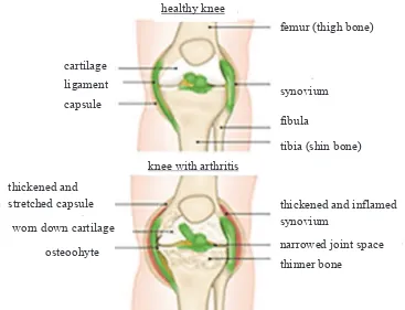
Figure 2.1: Comparison between a healthy knee and knee with osteoarthritis. Adapted from Rebecca Canvin, Bupa Health Information Team (2012)
healthy knee
femur (thigh bone)
synovium
fibula
tibia (shin bone) cartilage
ligament
capsule
thickened and stretched capsule
worn down cartilage
osteoohyte
thickened and inflamed synovium
narrowed joint space
6
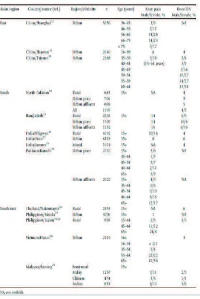
Table 2.1: Crude prevalence of knee pain or a diagnosis of knee osteoarthritis (OA). Retrieved from COPCORD Studies
7
2.1.1 Causes
Common risk factors for OA included aging, obesity, old joint injury, overuse
of joint, and the cause by genetics. However, the degeneration of articular cartilage is
not only caused by the result of aging and wear. It is also because of high force due
to collision and torsional loads which will increase the percentage of the
degeneration of normal joints. By the way, the person who have an unusual joint
anatomy, unstable joint, disturbances of joint or muscle irritation, or insufficient
muscle strength or endurance probably have a greater risk of degenerative joint
disease (Buckwalter and Mankin, 1998). In human, intensive physical exercises will
also cause osteoarthritis development (L'Hermette, 2006). Apart from aging, there is
some proof mostly from North American or European groups stated that obesity or
heavy occupational physical activity are the risk factors for symptomatic knee and
hip OA (Jensen, 2008). Furthermore, obesity, which was the another major risk , may
be less common, although it is increasing and likely to have a major effect on OA
commonness in the future. In the early stage of OA, the water content of cartilage
will increased and this compositional crisis has been shown to have deep
biomechanical effects. The increase of water content greatly reduces the intrinsic
Young modulus and increase the permeability of the collagen-proteoglycans solid
matrix of cartilage. This shows that the changes of these material properties will have
high effects on the method cartilage supports the loads which apply onto the surface
and the mode in which the interstitial fluid flows. Theoretical prediction made based
on the functional responses of cartilage by considering the increased hydration show
that cartilage may no longer function as the near-frictionless and wear resistant
bearing material of the joint. Thus a vicious cycle is set up which will caused further
breakdown of the tissue, possibly by increases in both the friction and wear rates of
8
2.1.2 Symptoms
The people that experience OA will first have a sense of unease when using
the joint. It has been described as stiffness or gelling. There will also have the feeling
of unstable and unsafe on walking which will cause pain. The degree and duration of
pain change but the pain of OA will be even worse while the joint are being use. This
pain may be only happen for a short period during waking hours or it may be occur
throughout. OA are mostly occurs in the hands, knees and hips. Hand OA is
commonly affected at the base of the thumb and the joints at the end of the fingers.
As time passes, defected joint will become red, swollen and tender, especially when
OA first arise. Slowly, for more than few years, joint will formed firm knob
swellings at the side which named as Heberden's nodes. For hip OA, the pain is often
occurs in the front of the groin, and sometimes around the thigh, buttock or even
down to the knee. Hip OA will have effect on walking. In knee OA, the pain is often
become more damage by frequently usage (Arthritis Foundation of Malaysia, 2010).
2.1.3 Diagnosis
Synovial joint are often being checked by using plain radiographs and the
diagnose of the joint with clinical syndrome of OA requires the presence of
long-range joint pain. The patients whose have OA will have difficulty of movement,
crepitus with motion and joint discharge. The most severely affected patients willl
develop joint deformation and subluxations. Patients with OA usually seek for
medical attention due to the pain of joint and the pain they experienced are often be
described as a deep aching poorly localized discomfort which has affecting for years.
The pain may vary with the changing of weather, the pain will be harder when
storms occurs or there is a drop in temperature, and the pain will also vary according
to the activity taken. Activity that will cause pain are often begins simultaneously or
after a short period of time when the joint is being use and may persist for hours after
end of activity. Some people will find out the symptoms of degenerative joint disease
following a small joint injury or strenuous physical activity, although the study of
9
OA the pain will be constant and may cause patients awake from sleep. As joint
degeneration progresses go on, patients may notice that they have loss motion slowly
and feel crepitus, or grating, catching and grinding sensations in the joint with
motion. Joint enlargement due to osteophyte formation, joint subluxation and
deformity occur later in the course of the disease.
The first signs of osteoarthritis include a decrease in the freedom of active joint
movement. Physicians frequently diagnose osteoarthritis based on the patient’s
history and physical findings. Imaging studies other than plain radiographs, including
bone scans, CT scans and MRI, and arthroscopic examination of joint surfaces, may
be helpful in evaluation of early stages of degenerative joint disease, but they rarely
are necessary for establishing the diagnosis (Buckwalter, 2000).
2.1.4 Treatment
There is no cure for osteoarthritis, but there are medications to help relieve
pain. The doctor may recommend physical therapy (PT) or occupational therapy (OT)
to help improve strength and function. Surgery may need to be conducted when a
person was in severe pain when he moves. Doctors usually use painkillers to relief
pain. The ideal painkiller such paracetamol is one of the painkillers that provides
pain relief with minimal side effects. Beside this, pain can also be relieved by using
other agents such as non-steroidal anti-inflammatory drugs (NSAIDs). However,
these agents may cause bad effects as it may cause stomach irritation or ulcer. One of
the common adverse effects of NSAIDs is it may cause stomach irritation or stomach
ulcer. It is still preferable that pain relieving medication in OA be used "as and when
required" rather than constantly. It is a smart move to lessen the mechanical burden
on the joint when a person had OA. Weight reduction is the key to lessen the burden
of OA if the patient is overweight. Muscle strengthening (quadriceps strengthening)
is vital to stop the progression of knee OA. The patient is always advised to get