Vol. 20, No. 4, November 2011 Expression of pab gene of M. tuberculosis 247
Cloning and expression of
pab gene of
M. tuberculosis isolated from
pulmonary TB patient in E.coli DH5
α
Tri Yudani M. Raras,1 Diana Lyrawati2
1 Department of Biochemistry-Molecular Biology, Universitas Brawijaya, Malang, Indonesia 2 Department of Pharmacy, Faculty of Medicine, Universitas Brawijaya, Malang, Indonesia
Abstrak
Latar belakang: Antigen38 Mycobacterium tuberculosis merupakan agen serodiagnostik yang potensial karena mengandung dua epitop spesiik untuk sel B. Mahalnya agen diagnostik menyebabkan lambatnya realisasi diagnosis TB secara cepat di negara berkembang. Kami memproduksi antigen 38 rekombinan yang berasal dari galur lokal yang kemungkinan dapat digunakan untuk memproduksi alat serodiagnostik TB yang ekonomis.
Metode: Gen pab diisolasi dari pasien TB paru di Malang, diklon ke plasmid pGEM-Teasy menjadi pMB38. Klon E.coli DH5α yang membawa pMB38 diseleksi di medium yang ditambah dengan X-Gal. Ekspresi pab dilakukan menggunakan pMBhis yang berasal dari pPRoExHTc dibawah kontrol promoter Trc dengan inang E.coli DH5α
Hasil: Pencocokan sekuen gen pab dari klon E.coli DH5α berwarna putih dengan gen pab dari M. tuberculosisH37Rv memperlihatkan homologi sebesar 98%. Protein rekombinan yang sudah dihilangkan signal peptidanya ditemukan di sitoplasma.
Kesimpulan: Gen pab dari pasien TB dapat diekspresikan secara intraseluler dengan sistem heterolog. (Med J Indones 2011; 20:247-55)
Abstract
Background: Mycobacterium tuberculosis antigen38 is a potent serodiagnostic agent containing two M. tuberculosis
-speciic B-cell epitopes. The high price of imported diagnostic agents hinders realization of fast clinical TB diagnosis in developing countries. Therefore, we produced recombinant antigen38 (recAg38M) from M. tuberculosis local strain, which
might be used to produce economical tuberculosis serodiagnostic kit.
Methods: Pab gene that was isolated from pulmonary TB patient in Malang was cloned into a plasmid vector
(pGEM-Teasy) to construct pMB38. The E.coli DH5α clone carrying pMb38 was selected on X-gal medium. The expression of pab
was mediated using pPRoExHTc under the control of Trc promoter and E.coli DH5α as host.
Results: Alignment of the pab sequence from the white E.coli DH5α clones with that of M. tuberculosis H37Rv showed
98% homology. The recombinant protein in which the signal peptide has been deleted to prevent the protein being secreted into medium was found in the cytoplasm.
Conclusion: pab gene of M. tuberculosis isolated from a TB patient could be expressed in heterologous system in E. coliDH5α. (Med J Indones 2011; 20:247-54)
Key words: Mycobacterium tuberculosis, Pab gene expression, recombinant antigen38
Correspondence email to: [email protected]
Mycobacterium tuberculosis (M. tuberculosis) Antigen38, a phosphate-binding protein, has a function that is similar to periplasmic phosphate-binding protein of Escherichia coli (E.coli) i.e. as an initial receptor for active transport.1 Among other antigens that were used
as tuberculosis serodiganostic agents, Antigen38, that
contains two M. tuberculosis-speciic B-cell epitopes has been shown as the most potent immunogen due to its high speciicity and sensitivity. 2The high price of
imported diagnostic agent hinders realization of fast clinical TB diagnosis in developing countries. One of the urgent goals of mycobacterial research in developing countries is to provide serodiagnostic agents. For such purpose, antigen need to be produced in a large scale. However, mass production of mycobacterial antigen
using conventional system encountered several problems such as high cost laboratory facility, dificulty in the protein identiication and reduction of immunogenicity
after puriication.3 An alternative solution to these
problems would be the production of recombinant
antigen in E.coli.
Therefore, in this study we aim to express the recombinant Antigen38 from M. tuberculosis local strain (recAg38M), in E.coli DH5a, which might be used to produce economical tuberculosis serodiagnostic kit.
METHODS
The antigen38 coding pab gene was ampliied from M. tuberculosis that was isolated from the sputum of pulmonary TB patient. The pab gene containing
was isolated from a patient who was suffering from pulmonary tuberculosis and hospitalized in Batu public hospital, Malang. Written consent was obtained from the patient. The study was approved by the Ethics Committee of Saiful Anwar Public Hospital. Plasmid pGEM-Teasy was purchased from Promega (USA). Plasmid pPRoExHTc was kindly given by Dr Rintis Noviyanti from Eijkman Institute.
Isolation and ampliication of pab gene from chromosomal DNA of M. tuberculosis
Bacterial DNA was isolated according to van der Zanden. 4
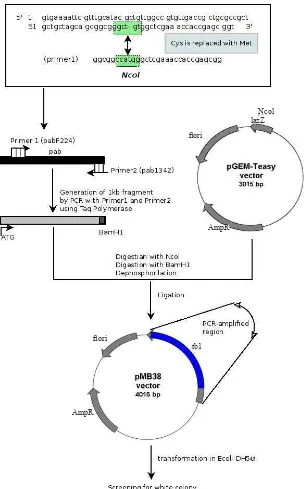
The pab gene was ampliied using PCR with modiication, i.e. without putative signal sequence (amino acid position 1-23) and Cys1 was replaced with Met residue, and was ampliied directly from the chromosomal DNA of M tuberculosis using two oligonucleotides, as forward (sense) and reverse (antisense) primers (igure 1).5
Primer 1: pabF224 (5’-GGCGGCCATGGGCTC GAA ACCACCGAGCGG-3’) carried an NcoI cleavage site and attached to a position 63 to 93 starting from initiation codon (ATG).
Primer 2: pab1342 3’ (5’-CCAGCAGGATCC GCA AAGCAGCCCGATGGC-3’) was located downstream from stop codon. It carries restriction site BamH1 and correspond to the sequence at the position +1.002 to +974 counted from initiation codon ATG.
Construction of plasmid pMB38
The pab gene was ampliied using PCR without the putative signal sequence. The ampliied pab gene was cloned into pGEM-Teasy vector (Promega, USA) according to
a standard method.6 The bacterial host for cloning and
expression was E.coli DH5α (F-recAI endAI gyrA96 thi-1 hsdr17(rk-mk-) sup44 relAIl(Φ80lacZDM15) ∆(lacZYA-arg F) U169). To maintain compatibility of the alignment with Antigen38, the deduced amino acid of the recombinant protein was renumbered so that Cystein that was replaced by Met became the irst residue. The new construct was then named pMB38. (Figure 1).
Screening of E.coli DH5α/pMB38
Plasmid pMB38 was then used to transform E.coli DH5α using heat shock method, 6 and the E.coli was spread on
LB (Luria broth) solid medium supplemented with IPTG and X-gal. 7 The white clones were selected and recultured
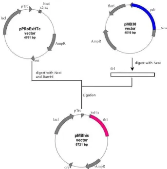
fragments. The 1130 bps fragment that corresponded to pab gene was then puriied using Qiagen kit (USA) and ligated with pPRoExHTc vector that previously was linearised using the same restriction enzyme, and both DNA segments were then ligated to produce pMBhis (Figure 2). The ligation product was used to transform E.coli DH5α. Selection of E.coli DH5α/pMBhis was conducted on ampicillin 100mg/mL containing LB. The correct clone was conirmed via PCR.
Expression ofrecAg38M in E.coli DH5a/pMBhis
E.coli DH5α/pMBhis was cultivated at 37°C in Ampicillin (100mg/ml) containing LB medium to an OD600 of 0.6. The expression of pab was
induced by addition of 0.6 mM isopropyl-B-D-thiogalactopyranoside (IPTG). After 3 hours cells were harvested by centrifugation at 6000xg for 15 min. The pellet was resuspended in 2.5 volume of 50 mM potassium phosphate buffer (pH 6.9) that contained 1 mM protease inhibitor, i.e. phenyl methyl sulphonyl luoride (PMSF), and the cells were broken using a sonicator at 40% for 20 seconds, 3 times. The cell extract was then centrifuged for 15 minutes at 8.000x g. The supernatant was removed and further centrifuged (Bench Top Ultracentrifuge) at 30.000xg for 30 minutes to separate the S30 extract. The S30 extract was taken and underwent another centrifugation at 100.000xg for 90 minutes, and the supernatant was called S100, while the pellet was called P100. All cell fractions were subjected to 12.5% SDS-PAGE.
Puriication of recAg38M using batch system
Vol. 20, No. 4, November 2011 Expression of pab gene of M. tuberculosis 249
Figure 1. Construction of plasmid pMB38 (derivate of pGEM-Teasy). Plasmid pGEM-Teasy was digested with NcoI and ligated with PCR ampliied product that was digested previously with NcoI and BamHI. The 3’ overhang of insert (product of BamHI digestion) should ligate with 5’ overhang of the plasmid (product of NcoI digestion) because there were only two different nucleotides within the cleavage site.
lazZNcol
lori
AmpR
tb1 lori
AmpR
Growth of E.coli DH5α/pMB38
To examine whether the expression of pMB38 inluence the growth of the host, growth tests were performed.
RESULTS
Construction of pMB38 and pMBhis
Primers were designed to modify pab gene by omitting the putative signal peptide. As a result, a
Raras and Lyrawati
250 Med J Indones
10
Figure 2. Construction of plasmid pMBhis (derivate of pPRoEXHTc). Plasmid
pMB38 was digested with NcoI producing insert (tb1) while pPRoExHTc was
double digested with NcoI and BamHI. The fragment tb1 was then ligated into
pPRoExHTc. To ensure that the insert (tb1) bound to the plasmid in the correct
direction, restriction test was perfomed.
Figure 2. Construction of plasmid pMBhis (derivate of pPRoEXHTc). Plasmid pMB38 was digested with NcoI producing insert (tb1) while pPRoExHTc was double digested with NcoI and BamHI. The fragment tb1 was then ligated into pPRoExHTc. To ensure that the insert (tb1) bound to the plasmid in the correct direction, restriction test was perfomed.
lori
AmpR
AmpR AmpR
lacl
lacl pTrc
pTrc
ori
tb1 tb1
Ncol 6xHis
Ncol
6xHis
pab
Figure 3. Amplified
pab
fragment from chromosomal DNA of
Lanes 1= DNA marker, 2-4= amplified
gene correspond to 970 bp.
1353 bp 1078 bp 872 bp
603 bp
310 bp
Figure 3. Ampliied pab fragment from chromosomal DNA of M. tuberculosis. Lanes: 1= DNA marker, 2-4= ampliied pab gene correspond to 970 bp.
Vol. 20, No. 4, November 2011 Expression of pab gene of M. tuberculosis 251
Forward sequence:
> gb|M30046.1|MSGPABA M.tuberculosis protein antigen b (Pab) gene, complete cds
Length=1993
Score = 1072 bits (580), Expect = 0.0 Identities = 586/592 (98%), Gaps = 0/592 (0%) Strand=Plus/Plus
Query 2 TGTCGCGNNTACCCCCGCGTCGTCGCCGGTGACGTTGNNGGANACCGGTAGCACGCTGCT 61 ||||||| |||||||||||||||||||||||||||| ||| ||||||||||||||||| Sbjct 274 TGTCGCGACTACCCCCGCGTCGTCGCCGGTGACGTTGGCGGAGACCGGTAGCACGCTGCT 333
Query 62 CTACCCGCTGTTCAACCTGTGGGGTCCGGCCTTTCACGAGAGGTATCCGAACGTCACGAT 121 |||||||||||||||||||||||||||||||||||||||||||||||||||||||||||| Sbjct 334 CTACCCGCTGTTCAACCTGTGGGGTCCGGCCTTTCACGAGAGGTATCCGAACGTCACGAT 393
Query 122 CACCGCTCAGGGCACCGGTTCTGGTGCCGGGATCGCGCAGGCCGCCGCCGGGACGGTCAA 181 |||||||||||||||||||||||||||||||||||||||||||||||||||||||||||| Sbjct 394 CACCGCTCAGGGCACCGGTTCTGGTGCCGGGATCGCGCAGGCCGCCGCCGGGACGGTCAA 453
Query 182 CATTGGGGCCTCCGACGCCTATCTGTCGGAAGGTGATATGGCCGCGCACAAGGGGCTGAT 241 |||||||||||||||||||||||||||||||||||||||||||||||||||||||||||| Sbjct 454 CATTGGGGCCTCCGACGCCTATCTGTCGGAAGGTGATATGGCCGCGCACAAGGGGCTGAT 513
Query 242 GAACATCGCGCTAGCCATCTCCGCTCAGCAGGTCAACTACAACCTGCCCGGAGTGAGCGA 301 |||||||||||||||||||||||||||||||||||||||||||||||||||||||||||| Sbjct 514 GAACATCGCGCTAGCCATCTCCGCTCAGCAGGTCAACTACAACCTGCCCGGAGTGAGCGA 573
Query 302 GCACCTCAAGCTGAACGGAAAAGTCCTGGCGGCCATGTACCAGGGCACCATCAAAACCTG 361 |||||||||||||||||||||||||||||||||||||||||||||||||||||||||||| Sbjct 574 GCACCTCAAGCTGAACGGAAAAGTCCTGGCGGCCATGTACCAGGGCACCATCAAAACCTG 633
Query 362 GGACGACCCGCAGATCGCTGCGCTCAACCCCGGCGTGAACCTGCCCGGCACCGCGGTAGT 421 |||||||||||||||||||||||||||||||||||||||||||||||||||||||||||| Sbjct 634 GGACGACCCGCAGATCGCTGCGCTCAACCCCGGCGTGAACCTGCCCGGCACCGCGGTAGT 693
Query 422 TCCGCTGCACCGCTCCGACGGGTCCGGTGACACCTTCTTGTTCACCCAGTACCTGTCCAA 481 |||||||||||||||||||||||||||||||||||||||||||||||||||||||||||| Sbjct 694 TCCGCTGCACCGCTCCGACGGGTCCGGTGACACCTTCTTGTTCACCCAGTACCTGTCCAA 753
Query 482 GCAAGATCCCGAGGGCTGGGGCAAGTCGCCCGGCTTCGGCACCACCGTCGACTTCCCGGC 541 |||||||||||||||||||||||||||||||||||||||||||||||||||||||||||| Sbjct 754 GCAAGATCCCGAGGGCTGGGGCAAGTCGCCCGGCTTCGGCACCACCGTCGACTTCCCGGC 813
Query 542 GGTGCCGGGTGCGCTGGGTGAGAACGGCAACGGCGGCNTGGTGACCGGTTGC 593 ||||||||||||||||||||||||||||||||||||| |||||||||||||| Sbjct 814 GGTGCCGGGTGCGCTGGGTGAGAACGGCAACGGCGGCATGGTGACCGGTTGC 865
Reverse sequence:
> gb|M30046.1|MSGPABA M.tuberculosis protein antigen b (Pab) gene, complete cds
Length=1993
Score = 1077 bits (583), Expect = 0.0 Identities = 591/597 (98%), Gaps = 1/597 (0%) Strand=Plus/Minus
Query 7 GGTC-ACGAGGCTAGCTGGAAATCGTCGCGATCAACGCGTCANACAACTTCACCACCGCG 65 |||| ||||||||||||||||||||||||||||||||||||| ||||||||||||||||| Sbjct 1287 GGTCAACGAGGCTAGCTGGAAATCGTCGCGATCAACGCGTCAGACAACTTCACCACCGCG 1228
Query 66 GGCGGCAGCGGCTGGAAATGAACCTGGTCGAGGAACGAGGCCTTGTTGCCGTCGGTGATC 125 |||||||||||||||||||||||||||||||||||||||||||||||||||||||||||| Sbjct 1227 GGCGGCAGCGGCTGGAAATGAACCTGGTCGAGGAACGAGGCCTTGTTGCCGTCGGTGATC 1168
Query 126 GCCCAGTGCANAAATGCCTGCAAGGTCTGCGCGGTGGCGGCGTCCTTTTGCCGGTTGTTG 185 |||||||||| ||||||||||||||||||||||||||||||||||||||||||||||||| Sbjct 1167 GCCCAGTGCAGAAATGCCTGCAAGGTCTGCGCGGTGGCGGCGTCCTTTTGCCGGTTGTTG 1108
Query 186 ACGATGGCGTACTCGTAGTTGATGATCGGGTAGCCGTCCGGGGCGGGCCCGTCGATCATC 245 |||||||||||||||||||||||||||||||||||||||||||||||||||||||||||| Sbjct 1107 ACGATGGCGTACTCGTAGTTGATGATCGGGTAGCCGTCCGGGGCGGGCCCGTCGATCATC 1048
Query 246 GAAATCGCCTGGTTCGCCGGGGTTTTCGATGCGAAGCCAGCCGCCGCGGCCTGAATGCTT 305 |||||||||||||||||||||||||||||||||||||||||||||||||||||||||||| Sbjct 1047 GAAATCGCCTGGTTCGCCGGGGTTTTCGATGCGAAGCCAGCCGCCGCGGCCTGAATGCTT 988
Query 306 TGCGCGTCGGGCAACAAGAAATTGCCAGAGCTATTGCCTAGTTGGGCCTCGCCGAGTCCC 365 |||||||||||||||||||||||||||||||||||||||||||||||||||||||||||| Sbjct 987 TGCGCGTCGGGCAACAAGAAATTGCCAGAGCTATTGCCTAGTTGGGCCTCGCCGAGTCCC 928
Query 366 CGTTGACTGGCCTGNTCNAGGAAGCTGATGCCGATATAGGCCACGCAGCCCGGTGTCTCG 425 |||||||||||||| || |||||||||||||||||||||||||||||||||||||||||| Sbjct 927 CGTTGACTGGCCTGGTCGAGGAAGCTGATGCCGATATAGGCCACGCAGCCCGGTGTCTCG 868
Query 426 GCGCAACCGGTCACCATGCCGCCGTTGCCGTTCTCACCCAGCGCACCCGGCACCGCCGGG 485 |||||||||||||||||||||||||||||||||||||||||||||||||||||||||||| Sbjct 867 GCGCAACCGGTCACCATGCCGCCGTTGCCGTTCTCACCCAGCGCACCCGGCACCGCCGGG 808
Query 486 AAGTCGACGGTGGTGCCGAAGCCGGGCGACTTGCCCCAGCCCTCGGGATCTTGCTTGGAC 545 |||||||||||||||||||||||||||||||||||||||||||||||||||||||||||| Sbjct 807 AAGTCGACGGTGGTGCCGAAGCCGGGCGACTTGCCCCAGCCCTCGGGATCTTGCTTGGAC 748
Query 546 AGGTACTGGGTGAACAAGAAGGTGTCACCGGACCCGTCGGANCGGTGCAGCGGAACT 602 ||||||||||||||||||||||||||||||||||||||||| ||||||||||||||| Sbjct 747 AGGTACTGGGTGAACAAGAAGGTGTCACCGGACCCGTCGGAGCGGTGCAGCGGAACT 691
Figure 6. SDS-PAGE of extracts from cells that expressed the Ag38-His fusion protein
Lanes: 1= molecular mass standard, 2= cell extract of E.coliDH5a/pMBhis before induction, 3, 6= 1and 2 hours after induction with 200mM IPTG, 4, 7= 1 and 2hours after induction with 400mM IPTG, 5, 8= 1and 2hours after induc-tion with 600mM IPTG.
Figure 7. SDS-PAGE of extract of cells producing the Ag38M fusion protein
Lanes: 1= cell extract of E.coliDH5α/pMBhis without IPTG in-duction, 2= after IPTG inin-duction, 3= the 8,000 x g supernatant, 4= the 8,000 x g precipitate, 5= Molecular mass standard (from top to bottom): galactosidase(118 kD), bovine serum albumin(90 kD), ovalbumin (50 kD), carbonic anhydrase (34 kD), b-lacto-globulin (26 kD), lysozyme (19 kD), 6= the 30,000 x g superna-tant (S30), 7= the 30,000 x g precipitate (P30), 8= the 100,000 x g supernatant (S100), 9= the 100,000 x g precipitate (P100) Figure 5. PCR-results of the ampliication of pab gene on several clones
of E.coliDH5a/pMBhis
Lanes: 1= Negative control, 2-8= Colonies 1-7, 9= tb1. Lane 3,4 and ,6 were positive showing the presence of frag-ment tb1.
1 2 3 4 5 6 7 8 9
For the expression of tb1, plasmid pMBhis was used and E.coli DH5α served as the host. Transformation of E.coli/
pMBhis resulted in several white clones only. Seven white colonies were picked and conirmation with PCR showed that four colonies carried the tb1 gene (Figure 5).
Expression of pab gene of M. tuberculosis in E.coli
In this study pab gene could be successfully expressed in E.coli DH5α. For this purpose E.coli DH5α/pMBhis was grown on LB liquid medium suplemented with Ampicillin (100 mg/mL). Expression was conducted by inducing the transcription process with addition of various concentration of IPTG (isopropyl-D-thiogalactopyranose). We used several concentration of IPTG (100 mM – 600 mM), and the cells were harvested 3 hours after induction with IPTG. The results of SDS-PAGE of the various expression with and without induction can be seen in Figure 6. Furthermore, SDS-PAGE of the supernatants and pellets after various centrifugation can be seen in Figure 7. The expression of pab was conirmed using western Blot against recAg38 from M tuberculosis H37Rv (data not shown).
Figure 9. Growth curve of wild type E.coli DH5α and E.coli DH5α/pMBhis
16 Figure 9. Growth curve of wild type DH5α DH5α/pMBhis
0 0.2 0.4 0.6 0.8 1 1.2
0 2 4 6 8 10
hours-E. coliDH5α
E. coliDH5α/pMBhis
Optical
Density
(OD)
1 2 3 4 5 6 7 8 9
1 2 3 4 5 6 7 8 9
Figure 8. Puriication of protein rec-Ag38M
S30 was mixed with matrix Ni-NTA, shaked gently for 1 hour and apllied onto ilter.
Lanes : 1= Protein marker, 2= Flow through (FL), 3= Wash I (W1), 4= wash II (W2),5= Elution I (E1), 6= Elution II (EII), 7= Elution III (EIII), 8=S30, 9= P30.
E.coli DH5α E.coli DH5α/pMBhis
185
kDa-Vol. 20, No. 4, November 2011 Expression of pab gene of M. tuberculosis 253
During the course of the expression experiment, the plasmid containing host showed a different behavior. The low rate of transformation of pMBhis into E.coli DH5α suggested that a large amount of the recAg38M within the cell somehow disturb the homeostasis of host cell, so that the growth of E.coli DH5α/pMBhis in the liquid medium was slightly slower than E.coli DH5α alone with a generation time of 2 hours compared to 1 hour (igure 9). Previous study has noted that the expression of recombinant protein retarded the growth of the host, such as in the case of expression of cymH gene of Klebsiella oxytoca in E.coli DH5α. 11
During the course of expression experiment, the pMBhis containing host has shown a different behaviour from the wild type. Comparison of growth curve of wild type E.coli DH5α and E.coli DH5α/pMBhis can be seen in Figure 9. Subcellular localization showed that recAg38M was partly located in the cytoplasm as the band corresponding to 37 kDa that was found in supernatant (S100) after centrifugation at 100.000xg (igure 7 lane 8) was faint, whereas most of protein was found in the precipitate (P100) (igure 7 lane 9). The question whether recAg38 is only temporarily attached to periplasmic membrane or becomes a trans-membrane protein is still open. Attempts to release the protein from the membrane using Triton X-100 failed (data not shown). This inding was in contrasts with the recAg38 from M. tuberculosis H37Rv, which was found 90% in the cytoplasm. 2
One crusial problem in producing recombinant protein is during puriication process. To obtain pure protein, several puriication steps are needed, so that at the end of the process the yield is low, and the proteins loss part of their immunogenicity. Hence there is a need to conduct a short and eficient puriication process so that most of the protein could be puriied with a high immunogenicity. This might be mediated through protein fusion with Histidin tag at the N-terminus that was puriied through Ni-NTA matrix. During the course of puriication, the Ni-NTA matrix was used to capture the Histidine tag to increase the protein resolution accordingly. The puriication of recAg38M using Ni-NTA afinity batch system showed that there were still contaminations of several proteins. Therefore, it is sugessted to repeat the puriication procedure with optimizations such as changing the buffer that could increase the purity of recombinant protein.
In conclusion, pab gene of M. tuberculosis isolated from TB patient could be expressed in heterologous system
in E.coli DH5α. Although the pab gene in our study genetically showed high homology to pab gene from M. tuberculosis H37Rv, further study was required to purify the protein, and to identify its biological function.
Puriication of recAg38M
Loss of protein during puriication was minimized by inclusion of a histidine tag at N-terminus, allowing puriication using a Ni-matrix to capture histidine tag. The band of the protein recAg38M did not appear in the lowthrough (Figure 8 lane 2) as well as during wash steps (Figure 8 lanes 3 and 4 ), in contrast, the bands that coressponded to recAg38M were found during elution steps (Figure 8 lanes 5-7). However, there were still many other protein that were copuriied so further washing may be warranted.
Growth of E.coli DH5α/pMB38
It was found that E.coli DH5α/pMB38 exhibited growth with slightly longer generation time (tD> 2 hours) compared to the host (tD= 1.5 hours) (Figure 9).
DISCUSSION
The present study aimed to express pab gene from a local strain of M. tuberculosis in heterologous system (E.coli) to achieve high antigen yields at potentially low cost. The construct was designed by omitting the putative signal peptide, so that the recombinant protein remained in the cytoplasm. Alignment of the ampliied PCR product of pab gene isolated from malang showed 98% homology with sequence of pab gene from M. tuberculosisH37Rv. Hence, it was expected that the recombinant protein recAg38M from Malang could have a similar immunological properties to recAg38 synthesized from M. tuberculosisH37Rv when they are in the future used as serodiagnostic agent. However, we did not conduct immunological experiment yet.5
The pab gene from Malang was successfully expressed
in E.coli DH5α, as the band did not present without IPTG induction. Therefore, the band that appeared after induction with IPTG must be the recAg38M protein. These was also conirmed using anti Ag38 (data was not shown). Nevertheless the amount of protein recAg38M was less than protein recAg38 originated from M. tuberculosis H37Rv, where Ag38 may account for 10% of the total protein.
The recombinant protein was dominated by the insoluble form, and only 25% of the protein was found in a soluble form. The failure to obtain recAg38M in a soluble form was the consequence of the fact that the expression of pab always lead to the formation of inclusion bodies that contain the insoluble form of the recombinant protein.2,5 It might be worthwhile
from Western University for valuable comments. This study was inanced by STRANAS grant from Indonesian government.
REFERENCES
Chang Z, Choudary A, Lathigra R, Quiocho FA. The 1.
immunodominant 38-kDa lipoprotein antigen of Myco-bacterium tuberculosis is a Phosphate-binding protein. J
Biol Chem. 1994;269;1956-68.
Wilkilson RJ, Haslov K, Rappuoli R, Giovannoni F, 2.
Narayanan PR, Desai CR, et al. Evaluation of the recombinant 38-kilodalton antigen of Mycobacterium tuberculosis as a potential immunodiagnostic reagent. J
Clin Microbiol. 1997;35:553-7.
Cheng VCC, Yew WW, Yuen KY. Molecular diagnostics in 3.
tuberculosis. Eur J Clin Microbiol Infect Dis. 2005; 24:711-20.
laboratory manual. New York: Cold Spring Harbour; 2001. Ausubel FM, Brent R, Kingston RE, Moore DD, Seidmann 7.
JG, Sminth JA, et al. Current protocol in molecular biology. New York : Wiley and Sons; 1997.
Young DB, Garbe TR. Heat shock protein and antigen of 8.
Mycobacterium tuberculosis. J Infec Immun. 2001;59:
3086-93.
Yasukawa C, Kanei-Ishii T, Maekawa J, Fujimoto T, 9.
Yamamoto, Ishii S. Increase of solubility of foreign protein
in E. coli by overproduction of bacterial thioredoxin. J Biol
Chem. 1995;270:25328-31.
Gubernator B, Seidler A, Rögner M, Szczepaniak A. 10.
Overexpression and reconstitution of a Rieske iron-sulfur protein from the higher plant. Protein Expr Purif. 2003;29:8-14. Mardining Raras TY. Study on the cyclodextrine metabolism 11.
in Klebsiella oxytoca M5a1. A molecular Approach.