Vol. 17 No. 2, p 53-57 EISSN: 2086-4094
Effect of
Mamordica charantia
L. Powder on Antioxidant
Superoxide Dismutase in Liver and Kidney of Diabetic Rats
TUTIK WRESDIYATI∗∗∗∗∗, TEGUH SURANTA SINULINGGA, YOLI ZULFANEDI
Department of Anatomy, Physiology, and Pharmacology, Faculty of Veterinary Medicine, Bogor Agricultural University, Darmaga Campus, Bogor 16680, Indonesia
Received October 22, 2009/Accepted May 7, 2010
The status of antioxidant superoxide dismutase (SOD) was reported decreased in the liver tissues of diabetic experimental Macaca fascicularis. This study observed effect of Mamordica charantia on the status of SOD in the liver and kidney of diabetic experimental rats. The SOD was localized using immunohistochemical technique. Male Wistar rats of negative control and diabetes mellitus (DM) group treated with 5 and 10% of M. charantia powder for 28 days. The DM condition was achieved by alloxan (110 mg/kg BW) induction. Charantia powder increased the status of antioxidant SOD in the liver and kidney of diabetic experimental rats. Aplication of M. charantia powder 10% gave better results than that of 5%. The results suggested that M. charantia powder can increase the status of antioxidant in the oxidative stress condition, such as diabetes mellitus.
Key words: Superoxide dismutase (SOD), Mamordica charantia, diabetes mellitus, liver, kidney, immunohistochemistry
___________________________________________________________________________
http://journal.ipb.ac.id/index.php/hayati DOI: 10.4308/hjb.17.2.53
_________________
∗ ∗∗
∗∗Corresponding author. Phone: +62-251-8626064,
Fax: +62-251-8629464, E-mail: [email protected] INTRODUCTION
Diabetes mellitus (DM) is a carbohydrate metabolic disorder that was signed with high blood glucose level, more than 140 mg/dl. World Health Organization (WHO) survey showed that Indonesia has high number of DM patients, on fourth rank in the world after India, China, and America. In 2010 the number of DM patients in the world will be 239 million and it will be 306 million in 2020 (Mandrup 1998). DM condition decreased the status of antioxidant enzyme and increased tissues alteration. It can lead to atherosclerosis and cataract (Szaleczky et al. 1999; Ferrari & Torres 2003). Wagenknecht et al. (1998) and Gerrity et al. (2001) also reported that DM patient has higher risk 2-6 times to get atherosclerosis than normal condition.
Wresdiyati et al. (2003) reported that DM conditions decreased the level of intracellular antioxidant copper,zinc-superoxide dismutase (Cu,Zn-SOD) in liver tissues of diabetic experimental Macaques (Macaca fascicularis). These alterations may account for the diabetic condition inducing production of reactive oxygen species-free radical. Increased levels of the reactive oxygen species, free radical, create a situation known as oxidative stress. This highly reactive oxygen can readily react with various biological macromolecules such as DNA, proteins, lipids, and caused protein destruction. The lesions in turn lead to various diseases and degenerative processes such as aging and carcinogenesis in human and animals (Halliwell & Gutteridge 1995).
Antioxidants play an important role in protection of cells against oxidative stress and maintain a balance
between the various toxic oxygen species (Touati 1992). The protection can be done by several ways such as prevention, stopping or decreasing of oxidations, as well as catalyzing free radicals by intracellular antioxidant enzymes (Mates et al. 1999).
The intracellular antioxidant enzymes comprise catalase, glutathione peroxidase, and three isoforms of SOD (Valko et al. 2007); Cu,Zn-SOD, Mn-SOD, and Fe-SOD. The SOD provides a primary defense against superoxide anion radical generated intracellularly. It was reported that SOD was immunohistochemically and immunocytochemically localized in the human and rat tissues (Dobashi et al. 1989; Wresdiyati & Makita 1997). SOD was also reported plays important role in physiological processes. Some cases of failed pregnancy in human was caused by the decreasing level of SOD (Sugino et al. 2000). Profile of SOD was also reported in pathological condition such as stress, diabetes mellitus, and hypercholesterolemia (Wresdiyati et al. 2002; Wresdiyati 2003; Wresdiyati et al. 2003; Wresdiyati et al. 2006a,b), in neoplastic tissues (Keller et al. 1991), and neuron of hypocampus in Alzheimer and Down’s syndrome patient (Furuta et al. 1995).
MATERIALS AND METHODS
Mamordica charantia Powder Production and Analysis. The present study used 18 days old M. charantia L. fruit. It was obtained from Balitro Cimanggu, Deptan Bogor. The M. charantia were blanched with alcohol at 50 oC for 1 minute, then the seeds were removed.
The remain materials were then sliced and dried using cabinet dryer at 60 oC for 16 hours, followed by grinding
to obtain M. charantia powder. Proximate analysis was done to the powder (AOAC 1995).
Treatment of Animals and Tissues Preparation. A total of 20 male Wistar rats (250 ± 5 g BW) were used for this study. The rats were obtained from BPOM Jakarta. The animals were adapted to the situation and conditions of the animal laboratory for 2 weeks, and then blood glucose was analysis before treatment. The rats were then randomly divided into four groups; (i) negative control group (A), (ii) positive control group/DM (B), (iii) and (iv) DM groups that treated by 5% (C) and 10% (D) of M. charantia powder (C). The treatments was done for 28 days. DM condition (>150 mg/dl), except group A, was achieved by alloxan induction in dose of 110 mg/kg BW. Drinking water was provided ad libitum.
Blood glucose analysis was done once a week using glucometer. Tissues sampling was carried out at the end of each treatment. Following cervical dislocation liver and kidney tissues were collected from each animal in all groups. And the tissues were then processed by paraffin standard method. Specimens were cut into 4 mm-thick sections and subjected to immunohistochemical technique for detection of Cu,Zn-SOD.
Immunohistochemistry. SOD was localized immunohistochemically as describe previously (Wresdiyati et al. 2003). The tissue sections were washed for 15 min with 3 changes of PBS between each step. After deparaffinization and rehydration, the tissue sections were exposed to 3% H2O2 for 10 min to inactivate endogenous peroxidase activity and then to 10% normal goat serum to block nonspecific binding. Following rinsing with PBS, the tissue sections were incubated in primary antibody of copper zinc-superoxide dismutase (Cu,Zn-SOD) at 4 oC.
The tissues were then incubated with enhanced labelled polymer peroxidase (Dako K1491). The reaction product of antigen-antibody was visualized using diamino benzidine (DAB). The tissue sections were then dehydrated with series of alcohol, and cleared with xylol. Finally, the sections were mounted with entelan. As control of staining, tissue sections were incubated with PBS
instead of Cu,Zn-SOD antibody. The tissue sections of control staining showed negative reaction with minimal background staining.
Observation and Data Analysis. The immunoreaction products of the Cu,Zn-SOD were observed by using a light microscope. The qualitatively observation of Cu,Zn-SOD content in the tissues was based on the brown colour intensity and the distribution of the positive reaction product of Cu,Zn-SOD in the liver and kidney tissues. While the quantitatively observation of Cu,Zn-SOD content was done by counted the number of hepatocytes, as well as renal tubule cells of kidneys that give different level of brown colour intensity.
RESULTS
Mamordica charantia Powder Analysis. M. charantia powder contain a large amount of soluble (25.38%) and insoluble (39.73%) dietary fibers, and carbohydrate 86.2% of dry weight (Table 1).
Blood Glucose Levels. M. charantia powder showed decreased blood glucose level of diabetic rats (Table 2). The 10% of M. charantia powder treatment decreased blood glucose to the normal level at 7 days after treatment, while the dose of 5% M. charantia powder treatment needs 14 days treatment to reach normal blood glucose level.
Immunohistochemistry of Antioxidant Cu,Zn-SOD. Cu,Zn-SOD was immunohistoche-mically localized in the nuclei and cytoplasm of the hepatocytes, as well as renal tubule cells (Figures 1 & 2). The positive reaction product of the enzyme in the liver and kidney tissues was showed brown colour in the tissues. The distribution and frequency of positive reaction product on the tissues of control group were compared qualitatively and quantitatively with those of the treatment groups. The observation of Cu,Zn-SOD in the tissues was based on the brown colour intensity in the cells and the distribution of the reaction product.
Table 1. Data of proximate analysis of Mamordica charantia
powder
Table 2. Blood glucose levels of diabetic rats treated with Mamordica charantia powder for 28 days
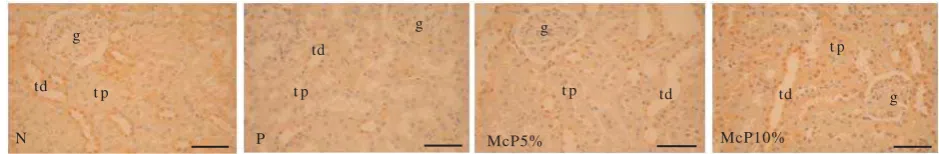
Qualitatively observation of Cu,Zn-SOD in the tissues of positive control (DM) group, showed the enzyme content decreased compare to the negative control group (Figure 1 & 2). It was shown by the intensity of brown colour reaction product in the positive control group lower than that of negative control group. The content of Cu,Zn-SOD in the DM groups treated with M. charantia powder increased compare to the positive control group. The treatment of 5% M. charantia powder gave slightly increasing on the content of Cu,Zn-SOD, while 10% M. charantia powder treatment showed remarkable increased of Cu,Zn-SOD content in the liver and kidney tissues of rats compared to that of positive control group (Figure 1 & 2). It showed by increased the brown colour intensity of positive reaction product to the intracellular antioxidant Cu,Zn-SOD.
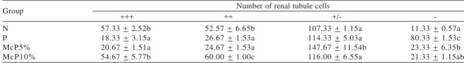
Quanitatively observation of Cu,Zn-SOD in the liver and kidney tissues also showed that the enzyme content in the DM is the lowest compared to other groups. It was shown by the number of hepatocytes and renal tubule cells that showed strong positive (+++) reaction to Cu,Zn-SOD in the DM group was significantly (P < 0.05) the smallest, while that of showed negative (-) reaction was significantly (P < 0.05) the biggest number (Table 3 & 4). The content of Cu,Zn-SOD in the liver and kidney of DM groups treated with 5 and 10% of M. charantia powder increased compare to the DM. It was shown by the number of hepatocytes that showed strong positive (+++) reaction to Cu,Zn-SOD increased significantly (P < 0.05) compared to that of positive control group. However, the number is still smaller compared to that of negative control group. It was also shown by the number of renal tubule cells that showed negative (-) reaction to Cu,Zn-SOD decreased significantly (P < 0.05) compared to that of positive control group. Both in liver and kidney tissues, the content of antioxidant Cu,Zn-SOD increased more remarkably in the DM group that treated with 10% of M. charantia powder than that of 5%.
DISCUSSION
The content of Cu,Zn-SOD in the liver and kidney tissues of DM groups showed the lowest level compared to other groups (Tables 3 & 4; Figures 1 &2). It was reported that lipid beta oxidation, which take place in cellular peroxisomes, increased at fasting, and diabetes conditions (Nilson et al. 1987; Hawkins et al. 1987). Under fasting stress, the number of peroxisomes increased 2.8 fold in the renal tubule cell of Japanese monkeys (Wresdiyati & Makita 1995). These conditions also increased cytochrome P-450 for fatty acid oxidation (Orellana et al. 1992). All those oxidations will create more free radicals as a side effects, so the conditions needs more antioxidant to neutralize free radicals. Subsequently the status of antioxidant especially Cu,Zn-SOD decreased in the liver tissues of diabetic Macaca fascicularis (Wresdiyati et al. 2003), in the pancreas tissues of diabetic rats (Wresdiyati et al. 2008), and in the liver and kidney tissues of diabetic rats in the present study.
Mamordica charantia powder treatment showed increased the antioxidant status in the liver and kidney tissues of diabetic rats (Figure 1 & 2; Table 3 & 4). The M.charantia powder may contain flavonoid and
N P McP5% McP10%
Figure 1. Photomicrographs of immunohistochemical localization of Cu,Zn-SOD in the rat liver tissues. N: negative control group, P: positive control group (DM), McP5%: DM+5% of Mamordica charantia powder, McP10%: DM+10% of Mamordica charantia powder. Scale = 50 µm.
g
td t p
N
g
td
t p
P
g
td t p
McP5%
g td
t p
McP10%
Figure 2. Photomicrographs of immunohistochemical localization of Cu,Zn-SOD in the rat kidney tissues. N: negative control group, P: positive control group (DM), McP5%: DM+5% of Mamordica charantia powder, McP10%: DM+10% of Mamordica charantia powder, g: glomerulus, tp: tubuli proximalis, td: tubuli distalis. Scale = 50 µm.
Table 3. The number of hepatocyte at different degree of antioxidant Cu,Zn-SOD content in liver tissues of rats (per view of 20 magnification)
Number of hepatocytes +++ ++ +/-Group
N P McP5% McP10%
54.0 + 3.54d 1.2 + 1.09a 14.8 + 1.96b 34.7 + 4.24c
41.7 + 2.91c 7.0 + 2.57a 32.1 + 4.04b 40.7 + 3.02c
polyphenol that scavenged free radicals (Halliwell & Gutteridge 1995), then cellular antioxidant Cu,Zn-SOD can be saved and subsequently the status in the tissues increased compared to that of DM group without treatment of M. charantia powder.
The other pathway is M. charantia powder treatment decreased blood glucose level. This effect was caused by a large amount of dietary fiber content in M. charantia powder (Table 1). It was reported that dietary fiber could maintain degenerative diseases such as diabetes, heart disease, and cancer (Thompson 1988). In this diabetes case, dietary fibers directly bind glucose in intestine and inhibit absorption of glucose. Beside of dietary fiber content of M. charantia powder, which able to decreased blood glucose level, it may also contain bioactive compound-charantin that has a hypoglychemic effect (Taylor 2002). Wresdiyati et al. (2008) also reported that M. charantia powder treatment could inhibit the rate of pancreatic β-cells damage in rats. Comprehensively, M. charantia powder treatment could maintain blood glucose level by insulin secretion of β-cells and it’s dietary fiber content. Subsequently, all oxidations reactions that create energy can be decreased, such as fatty acid oxidation. Then free radical production, as a side effect from these oxidations, decreased. This condition can also save the utility of intracellular antioxidant especially Cu,Zn-SOD to neutralize the free radicals, and subsequently the status in the tissues increased as shown in the liver and kidney of DM group that were treated with 5 and 10% of M. charantia powder. The dose of 10% M. charantia powder gave better results because the dose contains more dietary fibers, flavonoid, and polyphenol.
The study concluded that M. charantia powder decreased blood glucose level and increased the antioxidant status, especially Cu,Zn-SOD in the liver and kidney tissues of diabetic experimental rats. These results suggested that Mamordica charantia powder can be used as an alternative substitution material in order to produce some functional foods for maintain antioxidant status of diabetes mellitus patients.
REFERENCES
[AOAC] Association of Official Analytical Chemist. 1995. Method of Analysis. Washington: Association of Official Analytical Chemistry.
Dobashi K, Asayama K, Kato K, Kobayashi M, Kawaoi A. 1989. Immuhistochemical localization and quantitative analysis of superoxide dismutase in rat tissue. Acta Histochem Cytochem
22:351-365.
Table 4. The number of renal tubule cells at different degree of antioxidant Cu,Zn-SOD content in kidney tissues of rats (per view of 20 magnification) N: negative control group, P: positive control group (DM), McP5%: DM+Mamordica charantia powder 5%, McP10%: DM+Mamordica charantia powder 10%. Strong positive (+++), moderate positive (++), weak positive (+/-), and negative (-) reaction to the Cu,Zn-SOD.
Ferrari CKB, Torres EAES. 2003. Biochemical pharmacology of functional foods and prevention of chronic diseases of aging.
Biomed Pharm 57:251-260.
Furuta A, Price DL, Pardo CA, Troncoso JC, Xu ZS, Taniguchi N, Martin LJ. 1995. Localization of superoxide dismutase in Alzheimer’s disease and Down’s syndrome neocortex and hippocampus. Am J Pathol 146:357-367.
Gerrity RG, natarajan R, Nadler JL, Kimsey T. 2001. Diabetes induced accelerated atherosclerosis in swine. Diabetes 50:1654-1665.
Halliwell B, Guttridge JMC. 1995. Free radicals in biology and medicine. 3rd Ed. Oxford: Clarendon Pr. p 301.
Hawkins J, Jones WE, Bonner FW, Gibson GG. 1987. The effect of peroxisomes proliferators on microsomal, peroxisomal, and mitochondrial enzyme activities in the liver and and kidney. Drug Metab Rev 18:441-451.
Keller GA, Warner TG, Steimer KS, Hallewell RA. 1991. Cu,Zn superoxide dismutase is a peroxisomal enzyme in human fibroblasts and hepatoma cells. Proc Natl Acad Sci USA
88:7381-7385.
Mandrup-Poulsen T. 1998. Diabetes. British Med J 316:1221-1225.
Mates JM, Gomez CP, Castro IN. 1999. Antioxidant enzymes and human diseases. Clin Biochem 32:595-603.
Nilsson A, Pridz K, Rotveit T, Christiansen EN. 1987. Studies on the interrelated of microsomal omega-oxidation and peroxisomal beta-oxidation in rat liver with partially hydrogenated fish oil diet. Biochem Biophys Acta 920:114-119.
Orellana M, Fuentes O, Rosenbluth H, Lara M, Valdes E. 1992. Modulatios of rats liver peroxisomal and microsomal fatty acids oxidation by starvation. FEBS 310:193-196.
Sugino N, Nakata M, Kashida S, Karube A, Takiguchi S, Kato H. 2000. Decreased superoxide dismutase expression and increased concentrations of lipid peroxide and prostaglandin F2α in the decidua of failed pregnancy. Mol Human Reprod
6:642-647.
Szaleczky E, Prechl J, Feher J, Somogyi A. 1999. Alterations in enzymatic antioxidants defence in diabetes mellitus [a Rational Approach]. Postgrad Med J 75:13-17.
Taylor L. 2002. Bitter melon. In: Herbal Secret of The Rainforest. Austin: Sage Pr.
Thompson LU. 1988. Antinutrients and Blood Glucose. Food Tech 42:123-129.
Touati D. 1992. Regulation and protective role of the microbial superoxide dismutases. In: Scandalios (ed). Molecular Biology of Free Radical Scavenging Systems. Cold Spring Harbor Laboratory Pr. p 231-261.
Valko M, Leibfritz D, Moncol J, Cronin MTD, Mazur M, Telser J. 2007. Free radicals and antioxidants in normal physiological functions and human disease. The Int J of Biochem Cell Biol 39:44-84.
Wagenknecht LE, D’Agostino Jr RB, Haefner SM, Savage PJ, Rowers M. 1998. Impaired glucose tolerance, type 2 diabetes and carotid wall thickness. Diab Care 21:1812-1818. Wresdiyati T. 2003. Immunohistochemical study of oxygen-free
radical scavenger superoxide dismutase (Cu,Zn-SOD) in the liver of rats under stress condition. Biota 8:107-112. Wresdiyati T, Astawan M, Hastanti LY. 2006a. Profil
imunohistokimia antioksidan superoksida dismutase (SOD) pada jaringan hati tikus di bawah kondisi hiperkolesterolemia.
Wresdiyati T, Astawan M, Kesenja R, Lestari PA. 2008. Pengaruh pemberian tepung buah pare (Mamordica charantia L.) pada sel β dan SOD pankreas tikus diabetes mellitus. J Bahan Alam Ind 6:193-200.
Wresdiyati T, Astawan M, Nurwati VD. 2006b. Kondisi hiperkolesterolemia turunkan level antioksidan superoksida dismutase (SOD) pada jaringan ginjal tikus: suatu kajian imunohistokimia. J Sain Vet 24:168-176.
Wresdiyati T, Lelana RPA, Adnyane IKM, Noor K. 2003. Immunohistochemical study of superoxide dismutase (SOD) in the liver of diabetic experiment Macaca fascicularis. Hayati 10:61-65.
Wresdiyati T, Makita T. 1995. Remarkable increase of peroxisomes in the renal tubule cells of Japanese monkeys under fasting stress. Pathophysiology 2:117-182.
Wresdiyati T, Makita T. 1997. Immunocytochemical localization of Cu,Zn-SOD (Cooper, zinc-superoxide dismutase) in the renal tubules and glomerulus of rats kidney. Mol Biol Cell Suppl 8:342.