Hayati, September 2002, hlm. 85-88 Vol. 9, No. 3 ISSN 0854-8587
The Effect of Stress Condition on the Intracellular Antioxidant
Copper,Zinc-Superoxide Dismutase in the Rats Kidney:
an Immunohistochemical Study
TUTIK WRESDIYATI1*, KOICHI MAMBA2, I KETUT MUDITE ADNYANE1, UMI SITI AISYAH1
1Department of Anatomy, Faculty of Veterinary Medicine, Bogor Agricultural University, IPB Darmaga, Bogor 16001 2Department of Anatomy, Faculty of Agriculture, Yamaguchi University, Yoshida 1677-1 Yamaguchi City, Japan
Diterima 29 Januari 2002/Disetujui 28 Maret 2002
The intracellular antioxidant copper, zinc-superoxide dismutase (Cu,Zn-SOD) was immunohistochemically studied in the kidney of rats under fasting stress condition. A total of 45 male Wistar rats were used for this experiment. They were divided into three groups; control group, 3 days fasted and 5 days fasted stress groups. The Cu,Zn-SOD was observed qualitatively in the kidney tissues especially in the cytoplasm of renal tubule cells, as well as quantitatively in the nucleus. The results showed that Cu,Zn-SOD level decreased in the fasting stress group when compared to that of control group. The effect was more dramatic in the 5 days than the 3 days fasted group. The decrease of the enzyme in the kidney account for the involvement of the enzyme in the antioxidant defense system in the tissues of rats under fasting stress condition, in order to defense tissue damage from the free radical. These results suggested that fasting stress condition may increase oxygen-free radical and therefore it altered the kidney tissue and decreased their content of Cu,Zn-SOD.
___________________________________________________________________________
_________________
* Penulis untuk korespondensi, Tel. +62-251-626064, Fax. +62-251-505339, E-mail: [email protected]
INTRODUCTION
Increased levels of the active oxygen species, free radical, create a situation known as oxidative stress, which lead to a variety of biochemical and physiological lesions often resulting in metabolic impairment and cell death. These highly reactive oxygens can readily react with various biological macromolecules such as DNA, proteins, lipids, and caused protein destruction. The lesions in turn lead to various diseases and degenerative processes such as aging and carcinogenesis in human and animals (Ames & Shigenaga 1992).
Antioxidants, free-radical scavenger, play an important role in protection of cells against oxidatives stress and maintain a balance between the various toxic oxygen species (Touati 1992). The protection can be done by several ways such as prevention, stopping or decreasing of oxidations (Schuler 1990), as well as catalyzing free radicals by intracellular antioxidant enzymes (Mates et al. 1999).
The intracellular antioxidant enzymes comprise catalase, glutathione peroxidase, and three isoforms of superoxide dismutase (SOD); copper, zinc (Cu,Zn)-SOD, manganese (Mn)-SOD, and iron (Fe)-SOD. The SOD provides a primary defence against superoxide anion radical generated intracellularly. Wresdiyati and Makita (1998) reported Cu,Zn-SOD was immunocytochemically localized in the renal tubule of rats kidney.
Previous research (Wresdiyati & Makita 1995) reported that fasting stress condition altered renal peroxisomes both morphologically and increased on their number. Peroxisomes play an important role in certain oxidations such as lipid β-oxidation, D-amino acid oxidation, D-aspartate oxidation, L-α-hydroxyacid oxidation, urate oxidation, etc (Zaar 1992).
The increasing number of peroxisomes under fasting stress may induce and produce active oxygen-free radical.
This study was designed to examine the effect of stress condition on the Cu,Zn-SOD in the rats kidney. The intracellular antioxidant-Cu,Zn-SOD was observed immunohistochemically in the kidney tisssues. The observation was done qualitatively in the cytoplasm of renal tubule cells, glomerulus and medulla area. The observation was also done quantitatively in the nucleus of renal tubule cells, which was expected to give different degree of reaction product to Cu,Zn-SOD.
MATERIALS AND METHODS
Treatment of Animals. A total of 45 male Wistar rats (230-250 g) were used for this experiment. They were divided into three groups; control, 3-days fasting, and 5-days fasting stress group. All animals were placed in appropriate cages and stabilized with the housing condition for one week. The control group was fed with a rat commercial diet and drinking water ad libitum, while the fasting groups were given drinking water only for 3 and 5 days.
Tissue Preparation. After the treatment, all animals were decapitated, and pieces of tissues from kidney were fixed with 4% paraformaldehyde in phosphate buffer saline (PBS). Tissues were dehydrated through a graded ethanol series, and embedded in paraffin. Specimens were cut into 4 µm-thick sections and subjected to histopathological and immunohistochemical studies (Dobashi et al. 1989).
Immunohistochemical Staining. SOD was localized immunohistochemically as describe previously (Dobashi et al. 1989) with a modification. The tissue sections were washed for 15 min with 3 changes of PBS between each step. After deparaffinization and rehydration, the tissue sections were exposed to 3% H2O2 for 10 min to inactivate endogenous peroxidase activity and then to 10% normal goat serum to block nonspecific binding. Following rinsing with PBS, the tissue sections were incubated in primary antibody of copper,zinc-superoxide dismutase (Cu,Zn-SOD) at 40C. The tissues were then incubated with enhanced labelled polymer peroxidase (Dako K1491). The reaction product of antigen-antibody was visualized using diamino benzidine (DAB). The tissue sections were then counterstained with haematoxylin, dehydrated with series of alcohol, and cleared with xylol. Finally, the sections were mounted with entelan. As control of staining, tissue sections were incubated with PBS instead of Cu,Zn-SOD antibody. The tissue sections of control staining showed negative reaction with minimal background staining. Observation and Data Analysis. Haematoxylin and eosin stained tissue sections were observed under a light microscope. The cell condition and some histopathological signs of the tissues from the treatment groups were compared to that of the control group. Observation to the kidney tissues was done to the condition of renal tubule cells, glomerulus, and the interstitial cells area. The polymorphonuclear cells in the interstitial renal tubule cells of the kidney tissues were counted per view of 400 magnification. There are five views observa-tion per each sample. The number of polymorphonuclear cells in the tissues of control group was statistically (t-test) com-pared to that of fasted groups.
The immunoreaction products of the SOD were also ob-served by using a light microscope. The distribution and fre-quency of positive reaction product on the tissues of control group were compared qualitatively and quantitatively to that of the treatment groups. The qualitatively observation of Cu,Zn-SOD reaction product was done to the cytoplasm of renal tubule cells, as well as in the glomerulus and medulla area. The quantitatively observation of the enzyme was done to the nucleus of renal tubule cells. The reaction product of Cu,Zn-SOD in the nucleus was graded based on the colour intensity of reaction product, from brown (positive) to blue (negative) colour. There are four grades of reaction product; (i) strong positive (+++), strong brown colour, showed high concentration of the enzyme, (ii) moderate positive (++), light brown colour, (iii) weak positive (+/-), mixed light brown and blue colour, and (iv) negative reaction product, blue colour. The renal tubule cells in processing to death showed negative reaction product (blue colour) of Cu,Zn-SOD in their nucleus. The renal tubule cells in different degree of reaction product to Cu,Zn-SOD in the control and fasted groups were counted per view of 400 magnification. There are five views observation per each sample. The number of renal tubule cells in different degree in the control group was compared statis-tically (Anova) to that of fasted groups.
RESULTS
Histopathological Evidence. The haematoxylin and eosin (HE)-stained kidney tissues of control group showed general feature of kidney. The glomerulus was arranged in the cortex with distal renal tubule, loop Henle and proximal renal tu-bule. While medulla area of the kidney was filled mostly by ductuli renal and a few number of part of distal renal tubule. The tissues of both the 3 days and 5 days fasted stress groups showed some alterations. The nuclear of several renal tubule cells showed pycnosis. In the glomerulus and interstitial re-nal tubule cells of fasted groups showed infiltration of red blood cells and polymorphonuclear cells. The number of poly-morphonuclear cells was significantly increased in the fasted groups from the control group (Table 1). It was more remark-able in the 5 days fasted group than that of the 3 days fasted group. These data showed that an inflammatory process (Ringler 1996) occurred in the kidney tissues of rats under fasting stress.
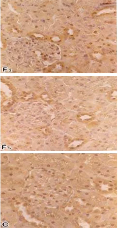
Cu,Zn-SOD Observation. The immunohistochemical lo-calization of Cu,Zn-SOD in the kidney yielded positive reac-tion product in the glomerulus, cytoplasm and nuclear of re-nal tubule, and a lesser amount in the medulla area. The reac-tion product of the enzyme in the kidney tissues of the fasted groups qualitatively showed significantly different from con-trol group (Table 2). The enzyme activity gradually decreased in the fasted groups than control group. The decreasing of Cu,Zn-SOD occurred in almost every part of the kidney, it was more clearly shown in the cytoplasm and nuclear of re-nal tubule cells (Figure 1). The enzyme gradually decreased related to the length of fasting stress in the glomerulus, renal tubule cells and medulla area (Table 2). In the fasted groups, especially in the 5 days fasted group, the decrease of SOD was more dramatic in the cytoplasm of proximal tubule cells than that of distal tubule cells.
One of new findings here is that the renal tubule cells in the process to death are negative to the Cu,Zn-SOD content. The negative reaction product was more clearly demonstrated in their nuclear than cytoplasm.
Table 1. The number of polymorphonuclear (inflammatory) cells, in the interstitial renal tubule cells of the male Wistar rats
Group Number of poplymorphonuclear cells
* Significantly different to the control group, P<0.001 (t-test)
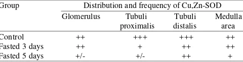
Table 2. The distribution and frequency of Cu,Zn-SOD in the kidney tissues of the male Wistar rats
Group Distribution and frequency of Cu,Zn-SOD +++: strong positive, ++: moderate positive, +: positive, +/-: weak positive
The decreasing of Cu,Zn-SOD content in the kidney tis-sues of rats under fasting stress condition was also quantita-tively shown significantly by decreasing amount of strong positive-cells, and the increase amount of negative-cells in the renal tubule cells. These results might be related to the length of fasting stress (Table 3).
The percent number of cells, in every degree of reaction product of Cu,Zn-SOD, to the total renal tubule cells was also showed the decrease of the enzyme more in the fasted groups than that of the control group (Figure 2).
DISCUSSION
The histopathological data in the kidney tissues under fast-ing stress condition showed some alterations and inflamma-tory process (Ringler 1996), such as the increasing of poly-morphonuclear cells (Table 1). While the content of Cu,Zn-SOD significantly decreased in the tissues of fasted groups than that of control group (Tables 2 and 3, Figures 1 and 2). The effect is more dramatic in the 5 days fasted group than that of the 3 days fasted group.
The fasting stress was reported altered kidney peroxisomes of the Japanese monkeys. The alterations are both morpho-logically and increase the number of peroxisomes (Wresdiyati & Makita 1995). It was evidence that some oxidations of the peroxisomes may also increase caused by the increasing of the organelle under fasting stress condition.
Peroxisomes play an important role in certain oxidations. Langseth (1995) reported that oxidations and reductions in peroxisomes and mitochondria resulting oxygen-free radical. So, fasting stress condition, which increased the number of peroxisomes, may increase peroxisomes oxidations. Further-more, it may induce and produce large amount of oxygen-free radical. The condition with highly reactive oxygen spe-cies, oxidative stress, can readily react with various biologi-cal macromolecules of cells including protein destruction. The lesions in turn lead to degenerative process (Halliwell & Gutteridge 1990). The alteration in the renal tubule cells and inflammatory process that occurred under fasting stress con-dition in this study showed the degenerative, cell death, proc-ess (Forrest et al. 1994) that caused by the highly oxygen-free radical resulting from the peroxisomes oxidations.
Table 3. The number of renal tubule cells in different degree of reaction product to Cu,Zn-SOD in the male Wistar rats under stress condition, per view of 400 magnification
Group Number of renal tubule cells in different degree of reaction product to Cu,Zn-SOD +++ ++ +/ -Control
Fasted 3 days Fasted 5 days
148.00b 101.67a 91.00a
104.330a 109.667a 135.330b
34.00a 40.00a 34.00a
45.67a 147.67b 201.33c Different superscript letters are significantly different (P< 0.05). +++: strong positive, ++: moderate positive, +/-: weak positive, -: negative
0 10 20 30 40 50
Control F-3 F-5
Group
% Number
+++ ++ +/-
-Figure 2. The percent number of renal tubule cells in different degree of reaction product to Cu,Zn-SOD (+++: strong positive, ++: mod-erate positive, +/-: weak positive, and -: negative) in the male Wistar rats under stress condition, per view of 400 magnifica-tion. F3: 3 days fasted group, F5: 5 days fasted group. Figure 1. The micrograph of Cu,Zn-SOD localization in the kidney of male
Wistar rats. F3: 3 days fasted group, F5: 5 days fasted group, and C: control group. The enzyme in the fasted groups showed quali-tatively decreased in the cytoplasm of renal tubule cells and glomerulus than that of control group. The decreasing was more remarkable in the proximal tubule cells than distal tubule cells. The Cu,Zn-SOD in the fasted groups was also decreased quantita-tively in the nucleus of renal tubule cells than that of control group.
It was reported in fasted animals, the blood glucose level decreased and released of fatty acids from adipose tissue into the blood stream is stimulated. The free fatty acids are incor-porated into organs, oxidized in the cells of these organs and utilized as energy sources instead of glucose (Ishii et al. 1980). Gaal (1993) also reported that fasting stress produce in im-mediate decrease in blood glucose accompanied by an increase of free fatty acid, total lipid, total cholesterol and urea in plasma.
The peroxisomal oxidizing system plays an important role in the oxidation of fatty acid originating from the adipose tissue. The activity of peroxisomal oxidation was showed in-crease more rapidly and markedly than that of mitochondrial oxidation during fasting stress condition (Ishii et al. 1980). The fasting stress was also reported increased the cytochrome P-450 that oxidize fatty acid, as well as peroxisomal β -oxida-tion, in the rat liver and kidney (Orellana etal. 1992).
The peroxisomal and cytochrome P-450 oxidations result-ing reactive oxygen species, superoxide anions (O2-) by cyto-chrome P-450 oxidation, and hydrogen peroxide (H2O2) by peroxisomal β-oxidation (Mates et al. 1999). The cytochrome P-450 oxidation was reported occurred in the endoplasmic reticulum (Orellana et al. 1992) and peroxisomes (Dhaunsi et al. 1992). So, the increase number of kidney peroxisomes under fasting stress (Wresdiyati & Makita 1995) may increase the number of reactive oxygen species, oxygen-free radical. Therefore, in order to defense the tissue damage from the oxidant, large amount of Cu,Zn–SOD were needed to cataly-ses the dismutation of the highly reactive superoxide anions to O2 and to the less reactive species H2O2. Subsequently, the Cu,Zn–SOD content in the kidney tissues decreased in fasted groups than that of control group. The gradual decreased of the enzyme related to the length of fasting stress. It suggested that reactive oxygen species more markedly increase in the longer time of fasting stress.
The decrease of the intracellular antioxidant Cu,Zn-SOD in the fasting groups was more remarkable showed in the proximal tubule cells than that of distal tubule cells. The proxi-mal tubule cells contain more of peroxisome organelle than that of distal tubule cells. So, the oxygen-free radical may also show more remarkable increase, and more Cu,Zn-SOD were needed to catalyze the free radical.
These results showed that fasting stress condition may increase reactive oxygen species, therefore it decreased the content of intracellular antioxidant Cu,Zn–SOD in the kidney tissues of rats. It might account for the involvement of intracellular antioxidant Cu,Zn-SOD, in the antioxidant
defense system of male Wistar rat kidney tissues under fast-ing stress condition, in order to protect tissue damage from the oxygen-free radical.
ACKNOWLEDGMENT
This study was supported in part by University Research for Graduate Education (URGE) Project Doktor Baru Batch IV No. 007/DB-IV/URGE/1999 (Ministry of Education) for Tutik Wresdiyati.
REFERENCES
Ames BN, Shigenaga MK. 1992. DNA damage by endogenous oxidants and mitogenesis as causes of aging and cancer. In: Scandalios (ed).
Molecular Biology of Free Radical Scavenging Systems. New York: Cold Spring Harbor Laboratory. p 1-22.
Dhaunsi GS, Gulati S, Avtar KS, John KO, Asayama K, Inderjit S. 1992. Demonstration of Cu,Zn-superoxide dismutase in the rat liver peroxisomes. J Biol Chem 267:6870-6873
Dobashi K, Asayama K, Kato K, Kobayashi M, Kawaoi A. 1989. Immu-histochemical localization and quantitative analysis of superoxide dismutase in rat tissue. Acta Histochem Cytochem 22:351-365. Forrest VJ, Kang YH, Mc Clain DE, Robinson DH, Ramakrishnan N. 1994.
Oxidative stress apoptosis prevented by trolox. Free Rad Biol Med
16:675-683.
Gaal T. 1993. Effect of fasting on blood lipid peroxidation parameters of sheep. Res Vet Sci 55:104-107.
Halliwell B, Gutteridge JMC. 1990. Role of free radicals and catalytic logam ions in human disease: an overview. Meth Enzymol 186:1-83. Ishii H, Horie S, Suga T. 1980. Physiological role of peroxisomal β
-oxida-tion in the liver of fasted rats. J Biochem 87:1855-1858.
Langseth L. 1995. Oxidants, Antioxidants and Disease Prevention. London: ILSI.
Mates JM, Gomez CP, Castro IN. 1999. Antioxidant enzymes and human diseases. Clin Biochem 32:595-603.
Orellana M, Fuentes O, Rosenbluth H, Lara M, Valdes E. 1992. Modulatios of rats liver peroxisomal and microsomal fatty acids oxidation by star-vation. FEBS 310:193-196.
Ringler DJ. 1996. Inflammation and repair. In: Jones TC (ed). Veterinary Pathology. Baltimore: Williams & Wilkins. p 113-157.
Schuler P. 1990. Natural antioxidants exploited commercially. In: Hudson BJF (ed). Food Antioxidants. London: Elsevier Applied Science. p 99-170.
Touati D. 1992. Regulation and protective role of the microbial superoxide dismutases. In: Scandalios (ed). Molecular Biology of Free Radical Scavenging Systems. New York: Cold Spring Harbor Laboratory. p 231-261.
Wresdiyati T, Makita T. 1995. Remarkable increase of peroxisomes in the renal tubule cells of Japanese monkeys under fasting stress. Pathophysi-ology 2:177-182.
Wresdiyati T, Makita T. 1998. Immunocytochemical localization of Cu,Zn-SOD (Cooper, zinc-superoxide dismutase) in the renal tubules and glom-erulus of rat kidney. Mol Biol of the Cell 8:342.
Zaar K. 1992. Structure and function of peroxisomes in the mammalian kidney. Eur J Cell Biol 59:233-254.