Genotype distribution of methylenetetrahydrofolate reductase A1298C and
C677T gene in Indonesian infertile men
Dwi A. Suryandari,1 Yurnadi,1 Budi Wiweko,2 Luluk Yunaini1
1 Department of Medical Biology, Faculty of Medicine ,Universitas Indonesia, Jakarta, Indonesia
2 Department of Obstetric and Gynecology, Faculty of Medicine, Universitas Indonesia and Cipto Mangunkusumo Hospital,
Jakarta, Indonesia
Abstrak
Latar Belakang: Methylenetetrahydrofolate reductase (MTHFR) merupakan enzim penting untuk membentuk folat dan metabolisme methionin, sehingga enzim ini sangat dibutuhkan untuk sintesis DNA dan metilasi. Varian dari MTHFR C677T dan A1298C dapat menurunkan folat dalam plasma dan meningkatkan suseptibilitas terhadap spermatogenic arrest. Penelitian ini bertujuan untuk menganalisis polimorisme gen MTHFR SNP A1298C dan C677T dan hubungannya dengan infertilitas pria oligozoospermia dan azoospermia di Indonesia.
Metode: Penelitian ini merupakan penelitian cross sectional dengan mengambil darah 3 mL pada pria oligozoospermia dan azoospermia sejumlah 150 orang. Gen MTHFR dianalisis menggunakan teknik polymerase chain reaction (PCR) dengan primer spesiik. Penelitian dilakukan dengan teknik PCR-RFLP menggunakan enzim restriksi MboII dan HinfI. Analisis PCR-RFLP gen MTHFR digunakan untuk mendeterminasi alotip gen MTHFR SNP A1298C dan SNP C677T pada kelompok pria oligozoospermia dan azoospermia dalam populasi Indonesia.
Hasil: Hasil penelitian menunjukkan bahwa distribusi alotip gen MTHFR SNP A1298C tidak berbeda bermakna (p>0,05) antara kelompok oligozoospermia dan azoospermia. Selanjutnya, distribusi alotip gen MTHFR SNP A677T antara kelompok oligozoospermia dan azoospermia juga tidak berbeda bermakna (p > 0.05).
Kesimpulan: Polimorisme gen MTHFR pada SNP A1298C dan C677T tidak berhubungan dengan infertilitas pria
oligozoospermia dan azoospermia di Indonesia. (Med J Indones 2012;21:23-7)
Abstract
Background: Methylenetetrahydrofolate reductase (MTHFR) is an important enzyme of folate and methionin metabolism, making it crucial for DNA synthesis and methylation. Variants of MTHFR C677T and A1298C gene result in reduced plasma folate levels and increase the susceptibility to spermatogenic arrest. This research aims to analyses MTHFR C677T and A1298C gene polymorphism in Indonesian infertile men with azoospermia and oligozoospermia.
Methods: This cross sectional study takes 3 mL of blood from 150 infertile men with oligozoospermia and azoospermia.
MTHFR gene is analyzed using polymerase chain reaction technique (PCR) with speciic primers. PCR-RFLP analysis of
the MTHFR gene using restriction enzymes MboII and HinfI determines allotypes, both of SNP A1298C and C677T in oligozoospermia and azoospermia in Indonesian population.
Results: The results show that the distribution of allotypes of MTHFR gene SNP A1298C and A677T is not signiicantly
different (p>0.05) between patient groups with oligozoospermia and azoospermia.
Conclusion: MTHFR gene polymorphisms, both of SNP A1298C and C677T are not associated with male infertility in Indonesian men including patients with severe oligozoospermia and azoospermia. (Med J Indones 2012;21:23-7) Keywords: DNA methylation, MTHFR, spermatogenic arrest
Correspondence email to: [email protected]
consists of 2.2 kbp and encodes a 70 kDa enzyme involved in folate metabolism as a cofactor for the methylation of homocysteine to methionin.
DNA methylation is a process of adding a methyl group to cytosine bases that will alter the structure of DNA without changing the composition of the nitrogenous bases.2,3
The MTHFR gene is polymorphic in 20 points and
two of them exert a very signiicant effect on enzyme activity, i.e. at positions C677T and A1298C. MTHFR gene polymorphism in position 677 causes a change of alanine to valine and in position 1298 of glutamate Spermatogenic arrest or interruption of the process
of spermatogenesis in the seminiferous tubules is a phenomenon, which causes azoospermia or severe oligozoospermia (sperm count < 2 million/ejaculate). Spermatogenic arrest can be caused by external factors such as diet, high temperatures, chemicals, cigarette smoke, alcohol, and psychological stress and by internal factors such as loss of one of the genes, which control the process of spermatogenesis, e.g., SRY, AZF, or MTHFR genes.1
to alanine. These changes cause a decrease in enzyme
levels in the blood, which will inluence the disregulation
of folic acid metabolism that resulted from errors in the methylation of genomic DNA.4-7
Many studies on MTHFR gene polymorphism indicated its association with colon cancer, leukemia, schizophrenia, migraine, Down syndrome and other neurological disorders.8-12 Studies on the association of infertility with MTHFR gene variations have also been conducted in African, American, Hispanic, European, Romanian and Indian populations. They showed that a change to valine reduces homocysteine levels with implications on spermatogenesis in populations in Romania and India, but not in other populations.13,14
In the population of the United States most individuals are found with homozygous dominant genotype: frequency in Mediterranean / Hispanic > Caucasian
> African / Afro-American populations. Homozygous
recessive genotype was found in the Romanian population at 35.7% among infertile men. The MTHFR gene mutation causes the inactivation of the germ cells and will eventually lead to spermatogenic arrest. Increase in oxidative status in reproductive tissues was also shown as a result of DNA damage in spermatozoa.10
It is known that the polymorphism is inluenced by
race. Therefore, in the Indonesian population we should know whether the frequency and distribution of MTHFR gene polymorphism and variation of this gene affects spermatogenesis in infertile men. This data will be considered in the management and handling of infertile men and in providing a solution for infertile men who want a child through assisted fertilization program.
METHODS
Subjects
This study was conducted from February until October 2011 and research subjects were male patients diagnosed with infertility. All subjects gave their informed consent before participating in this research. The diagnosis of infertility, which included azoospermia and severe oligozoospermia was determined by an andrologist. Participants were infertile subjects who followed the IVF program at the Yasmin Clinic, Cipto Mangunkusumo Hospital, Jakarta, Indonesia.
Study design
The design of this study was a cross-sectional study
with laboratory approach. The sample used in this
study was 3 mL of peripheral blood. According to the minimum formula of Sastroasmoro and Ismael,15 the total amount of samples is 138 for oligozoospermia and azoospermia. This study used DNA preparations that we obtained from previous studies entitled correlation microdeletion of AZFc genes with sperm quality of azoospermic and severely oligozoospermic men who followed the ICSI program (Ethical Approval No: 62/ PT02.FK/ETIK/2011).
Blood samples were obtained from Yasmin Clinic, Cipto Mangunkusumo Hospital (RSCM) and analyzed for MTHFR gene using PCR technique followed by RFLP technique conducted at the Department of Medical Biology, Faculty of Medicine Universitas Indonesia (FMUI).
Genomic DNA isolation
The DNA isolation method of this study is commonly used in the Department of Medical Biology FMUI.16 An amount of 3 mL of peripheral blood containing EDTA was transferred into a falcon tube (15 mL) containing 9 mL of red blood cell (RBC) lyses solution (1.45 M NH4Cl, 5 mM anhydrous EDTA, and 0.1 M KHCO3), then inverted 2-3 times and incubated at room temperature for 10 minutes. During the process of
incubation, the tube was inverted 3-4 times as much. The
sample was then centrifuged in an Expender Centrifuge 5702R at a speed of 1,500 rpm for 10 minutes at room temperature. The supernatant was removed by leaving the pellet in the form of mononuclear leucocytes.
The above steps were repeated to obtain a white pellet, which did not contain red blood cells. Then, to the pellet was added 2 mL of Nuclei Lyses Solution (NLS)
(10 mM Tris-HCl pH 8, 0.25 mM EDTA, and 0.5%
SDS), then pipetted until it became a homogeneous solution. Further samples were incubated in the water
bath (NESLAB, RTE-111) at 37°C for 30-60 minutes
until the solution was completely homogeneous, marked through the absence of small granules. Then were added 1.3 mL of protein precipitation solution (ammonium acetate 5 M) and vortexed with an Auto
Vortex (Stuart Scientiic SA 6) for 15-20 seconds. The
lysate was centrifuged at 3000 RPM for 15 minutes at 4oC. Centrifugation produced a light-brown precipitate of protein, the supernatant containing DNA. Then, the
supernatant was poured into a new falcon tube illed
with 2.3 mL of cold isopropanol. The tube was inverted
for 20-30 times until it looked like a collection of DNA strands. The samples were incubated at -20oC overnight
(16-18 hours).
precipitate appeared white. The supernatant was removed and then added 1.3 mL of cold 70% ethanol, the tube was inverted to wash the DNA. DNA solution was centrifuged at 3,000 RPM for 5 minutes at 4oC. The supernatant was discarded and the DNA dried in the open air by reversing the tube for approximately
2 hours. DNA was rehydrated with 200-300 mL of TE solution (Tris-HCl EDTA) and then incubated in a
water bath at 37oC for 2 hours. The solution was then transferred into a tube with 1.5 mL of sterile expender.
Then, DNA solution was stored at -20oC until further examination.
MTHFR DNA ampliication and PCR-RFLP
technique
Ampliication MTHFR gene was performed
by using forward primer 5’- CTTGGGGAG
CTGAGCACTACT-3’ and reverse primer 5’-
C A C T T T G T G G A C C A T T C C G G T T - 3 ’
for SNP A1298C and forward primer
5’-TGAAGGAGAAGGTGTTGCGGG-3’ and reverse
primer 5’-AG GACGGTGCGGTGAGAGTG-3’ for
SNP C677T. PCR method was performed using
PCR GotaqTM Core System kit consisting of 25 mM MgCl 2, 5x Green Gotaq, dNTP mix and Gotaq
Polymerase. The volume of each reagent was 50 mL
consisting of 100-500 ng genomic DNA, 20 ng of each oligonucleotide primer, 1x Green Gotaq, 2.5
mM MgCl2, 200 mM dNTP mix (mixture of dATP,
DTTP, dCTP and dGTP) and 1.25 U Tag DNA
polymerase. The DNA sample was amplified through
30-35 cycles with initial denaturation at 94°C for 5
minutes, then entered into the cycle consisting of denaturation, annealing and elongation. At the end of the cycle performed lengthening the time extension
72°C for 7 minutes. After the DNA amplification
process was completed, the amplicon was stored at
4°C. The results of electrophoresis was band size
340 bp (C677T) and band size 133 bp (A1298T).
Amount of 10 µl amplicon DNA (C677T) results by PCR digested using 1 unit digestion enzyme (HinfI) and the amount of amplicon DNA (A1298T) results by PCR also digested using 1 unit MboII. The DNA incubated at a temperature of 37oC for 3 hours. The results of digested DNA were separated by electrophoresis on 3% agarose gel (Promega) containing 1 µL ethidium bromide (0.5 mg/mL) in 1X TAE buffer solution
(0.04 M Tris-acetate, 0.002 M EDTA pH 8, 0). DNA
digested in 5 µL mixed with 2 µL tracking dye (0.25% bromophenol blue, 0.25% xylene cyanole, 25% sucrose), then inserted into the electrophoresis wells. Bands of DNA fragments were then separated by electrophoresis at 80 volts for 60 minutes. As markers a 100 bp DNA
ladder was used. Results of electrophoresis bands of DNA fragments were observed by UV illuminator and photographed with a Polaroid camera.
Statistical analysis
This research used nonparametric statistical analysis, to evaluate distribution and relations between two populations of genotype and allele of MTHFR gene
Chi-square test was used.17
RESULTS
DNA ampliication to detect MTHFR gene
polymorphism
Interpretation of digestion results are shown by different patterns. RFLP analysis for the A1298C on a 163 bp PCR fragment with MboII: Digestion of the 163 bp
fragment of 1298 AA genotype gives ive fragments of
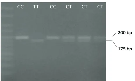
56, 31, 30, 28 and 18 bp, whereas the 1298CC genotype results in four fragments of 84, 31, 30, and 18 bp. The 18 bp fragment ran off the gel (Figure 1). RFLP analysis for the C677T on a 307 bp PCR fragment with HinfI digestion of the 307 bp fragment of 677 CT genotype gives two fragments of 200 and 175 bp, genotype CC gives one fragment of 200, whereas the 677 TT genotype results in four fragments of 175 bp (Figure 2).
Figure 1. RFLP analysis for the SNP A1298C
Figure 2. RFLP analysis for the SNP C677T
84 bp
56 bp
Distribution of MTHFR SNP A1298C in oligozoospermic and azoospermic patients
The distribution of genotypes and allele frequencies of SNPs A1298C MTHFR gene on oligozoospermia and azoospermia is shown in Table 1 and Figure 3. We can see the genotype distribution and allele frequency of MTHFR gene in the groups with oligozoospemia and azoospermia. Table 1 indicates that genotype has distribution of MTHFR gene in oligozoospermia and azoospermia shows a spreading pattern that is not
lattened. In the oligozoospermia group, AC genotype
has highest proportion (63.45%), AA genotype has lower proportion (35.58%), and CC genotype lowest proportion (0,97%). In the azoospermia group, AC genotype has highest proportion (86.96%), CC genotype has lower proportion (13.04%), and CC genotype has lowest proportion (0.00%).
Table 1. Comparison of genotype distribution and allele frequency of MTHFR gene SNP A1298C between oligozoospermia and azoospermia groups.
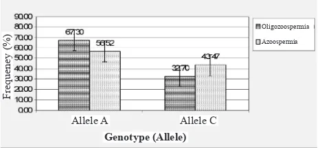
Furthermore, for the allele frequency, in the oligozoospermia group, A allele has high frequency (67.30%), whereas C allele has low frequency (32.70%). In the azoospermia group, A allele has high frequency (56.52%), whereas C allele has low frequency (43.47%). As a whole in the population, this indicates that A allele has high frequency (64.00%), whereas
C allele has low frequency (36.00%). By chi-square
(χ) test, allele frequency of the MTHFR gene in the
oligozoospermia group was not signiicantly different
from the azoospermia group (p>0.05). This condition indicates that allele frequency of MTHFR gene SNP A1228C is not disposition and MTHFR gene does not contribute to the pathogenesis of male infertility.
Figure 3. Allele frequency of SNP A1228C MTHFR gene on oligozoo -spermia and azoo-spermia groups (chi square, p> 0.05).
Distribution of MTHFR SNP C677T in oligozoospermic and azoospermic patients
The distribution of genotypes and allele frequencies of SNPs C677T MTHFR gene on oligozoospermia and azoospermia are shown in Table 2 and Figure 4. We can see the genotype distribution and allele frequency of MTHFR gene in oligozoospemia and azoospermia groups. Table 2 indicates that genotype distribution of MTHFR gene in oligozoospermia and azoospermia
shows a spreading pattern that is not lattened. In the
oligozoospermia group, CC genotype has highest proportion (68,26%), CT genotype has lower proportion (30,77%), and TT genotype has lowest proportion (0,97%).
Table 2. Comparison of genotype distribution and allele frequency of MTHFR gene SNP C667T between oligozoospermia and azoo -spermia groups.
In the azoospermia group, CC genotype has highest proportion (58.69%), CT genotype has lower proportion (39.15%), and TT genotype has lowest proportion (2.16%). Furthermore, for the allele frequency, in the oligozoospermia group, C allele has high frequency (83.65%), whereas T allele has low frequency (16.35%). In the azoospermia group, C allele has high frequency (78.26%), whereas T allele has low frequency (21.74%). As a whole, in the population indicates that C allele has high frequency (82.00%),
whereas T allele has low frequency (18.00%). By
chi-square (χ) test, allele frequency MTHFR gene in the
oligozoospermia group was not signiicantly different
from the azoospermia group (p>0.05). This condition indicates that allele frequency of MTHFR gene is not disposition and MTHFR gene does not contribute to the pathogenesis of male infertility.
Figure 4. Allele frequency of SNP C677T MTHFR gene in oligozoo -spermia and azoo-spermia groups (chi square, p> 0.05)
Frequeney (%)
Oligozoospermia
Azoospermia
Allele A Allele C
Genotype (Allele)
Frequeney (%)
Allele C Allele T
Oligozoospermia
Azoospermia
Genotype (Allele)
Group Number AA AC CC Allele Frequencies
Oligozoospermia 104 37 66 1 67.30 32,70 (35.58%) (63.45%) (0.97%)
46 6 40 0 56.52 43,47
(13.04%) (86.96%) (0.00%)
150 43 106 1 64,00 36,00
A(%) C(%)
Azoospermia
Total
83.65 16.35
78.26 21.74
82.00 18.00
Group Number CC CT TT Allele Frequencies
C(%) T(%)
Oligozoospermia
Azoospermia
Total
104
46
150
71 32 1
(68.26%)
(58.69%)
(30.77%) (0.97%)
(39.15%)
27 18 1
(2.16%)
DISCUSSION
Table 1 and Figure 3 indicate that the distribution of genotype MTHFR gene A1298C in oligoozoospermia
and azoospermia shows a spreading pattern. By
chi-square test, allele frequency of MTHFR gene A1298C between oligozoospermia and azoospermia groups
was not signiicantly different (p>0.05). Table 2 and
Figure 4 indicate that the distribution of genotype MTHFR gene C677T in oligoozoospermia and azoospermia shows a spreading pattern. By chi-square test, allele frequency of MTHFR gene C677T between oligozoospermia and azoospermia groups was not
signiicantly different (p>0.05).
Several studies have shown a signiicant statistical
correlation between MTHFR polymorphisms and male infertility,18 whereas other researchers did not
ind any signiicant association.19 These discrepancies could be explained by the fact that, in some patients with male infertility, the reduction of MTHFR activity
may be linked to other modiications, like, for instance,
abnormal methylation of its promoter region. Indeed,
epigenetic modiications at several genes, such as
methylation of the promoter region and the subsequent
down-regulation of gene expression, have been shown
to act similarly and in some cases in parallel to genetic mutations in a number of pathologies.20
In these studies both mutant genotypes at C677T or A1298C are very little with the frequency of less than 2% in the azoospermia or oligozoospermia groups. This suggests that the spermatogenic arrest in Indonesian men may not be caused by a mutation in the MTHFR gene. Therefore, further research should be conducted to determine the role of other genes as AZF, Boule and DAZL genes that play a role in controlling the mechanism of the process of spermatogenesis.
In conclusion, we found no association of metylenetetrahydrofolate reductase A1298C and C677T gene polymorphism and male infertility in Indonesian infertile men with azoospermia and oligozoospermia. Such studies need to be extended to get a better predictive association of the genotype and environment interaction in spermatogenic arrest.
Acknowledgment
We thank to Directorate of Research and Community
Service University of Indonesia (DRCS-UI) that have
supported this research through a competitive research grant (RUUI 2010).
REFERENCES
1. O’Brian KL, Varghese AC, Agarwal A. The genetic causes of male factor infertility: a review. Fertil Steril.
2010;93(1):1-12.
2. Foresta C, Moro E, Ferlin A. Y chromosome microdeletions and alterations of spermatogenesis. Endocr Rev.
2001;22(2):226-39.
3. Goyette P, Pai A, Milos P, Frosst P, Tran P, Chen Z, et al. Gene
structure of human and mouse methylenetetrahydrofolate
reductase (MTHFR). Mamm Genome. 1998;9:652-6.
4. Schwan B, Rozen R. Polymorphism in the methylenetetrahydrofolate reductase gene: clinical
consequences. Am J Pharmacogenomic.
2001;1(3):189-201.
5. Ma J, Strampfer MJ, Giovannucci E, Artigas C, Hunter DJ, Fuchs C, et al. Methylenetetrahydrofolate reductase polymorphism, dietary interactions and risk of colorectal
cancer. Cancer Res. 1997;57(6):1098-102.
6. Roffman Jl, Weiss AP, Deckersbah T, Freudenrich O, Henderson DC, Purcell S, et al. Effects of the methylenetetrahidrofolate reductase (MTHFR) C677T polymorphism on executive function in Schizophrenia. Schizophr Res. 2007;92(1-3):181-8.
7. Stankova J, Laurance AK, Rozen R. Methylenetetrahydrofolate reductase (MTHFR); a novel
target for cancer therapy. Curr Pharm Des.
2008;14(11):1143-50.
10. Kumar J, Das SK, Sharma P, Karthikeyan G, Ramakarishnan
L, Sengupta S. Homocysteine levels are associated with MTHFR A1298C polymorphism in Indian population. J
Hum Genet. 2005;50(12):655-63.
11. Florin M, Militaru M, Crisan T, Victor L, Anghel R. Methylenetetrahydrofolate reductase A1298C polymorphism and male infertility in a Romanian population
group. Maedica. 2009;4(4):294-9.
12. Khazamipour N, Noruzinia M, Fatehmanesh P, Keyhanee M, Pujol P. MTHFR promoter hypermethylation in testicular biopsies of patiens with non obstructive azoospermia : the role of epigenetics in male infertility. Hum Reprod.
2009;24:2361-4.
13. Dordevic V, Ljujic M, Nestorovic A, Ristanovic M,
Radojkovic D. Combined effect of GSTM1 gene deletion, GSTT1 gene deletion and MTHFR C677T mutation in male infertility. Arch Biol Sci Belgrade. 2010;63(3):525-30.
14. Singh K, Singh SK, Raman R. MTHFR A1298C polymorphism and idiophatic male infertility. J Postgrad
Med. 2010;56:267-9.
15. Sastroasmoro S, Ismael S. Dasar-dasar metodologi
penelitian klinis. Edisi ke-2. Jakarta: CV Sagung Seto; 2002. p. 270-1. Indonesian.
16. Maniatis T, Fritsch EF, Sambrook J. Molecular cloning: a laboratory manual. 2nd ed. New York: Cold Spring Harbor
Laboratory Press; 1989. p. 85, 159-61.
17. Medis R. Statistical hand book for non-statistician. London: McGraw-Hill Book Co.; 1975. p. 60-1.
18. Singh K, Singh SK, Sah R, Singh I, Raman R. Mutation C677T in the methylenetetrahydrofolate reductase gene is associated with male infertility in an Indian population. Int J Androl. 2005;28:115–19.
19. Paracchini V, Garte S, Taioli E. MTHFR 677C>T
polymorphism, GSTM1 deletion and male infertility: a
possible suggestion of a gene/gene interaction? Biomarkers. 2006;11:53–60.
20. Baylin SB, Herman JG. DNA hypermethylation in
tumorigenesis: epigenetics joins genetics. Trends Genet.