http://mji.ui.ac.id
Biocompatibility of various hydroxyapatite scaffolds evaluated by
proliferation of rat’s bone marrow mesenchymal stem cells: an in vitro
study
Achmad F. Kamal,1 Diah Iskandriati,2 Ismail H. Dilogo,1 Nurjati C. Siregar,3 Errol U. Hutagalung,1 R. Susworo,4 Achmad A. Yusuf,5 Adang Bachtiar6
1 Department of Orthopaedic and Traumatology, Faculty of Medicine, Universitas Indonesia, Cipto Mangunkusumo Hospital, Jakarta, Indonesia
2 Primate Research Center Bogor Agricultural University, Bogor, Indonesia
3 Department of Anatomical Pathology, Faculty of Medicine, Universitas Indonesia, Cipto Mangunkusumo Hospital, Jakarta, Indonesia 4 Department of Radiology, Faculty of Medicine, Universitas Indonesia, Cipto Mangunkusumo Hospital, Jakarta, Indonesia
5 Department of Histology, Faculty of Medicine, Universitas Indonesia, Jakarta, Indonesia 6 Faculty of Public Health, Universitas Indonesia, Jakarta, Indonesia
Abstrak
Latar belakang: Scaffold (biomaterial) adalah rangka yang digunakan dalam transplantasi sel punca mesenkimal. Sebelum digunakan perlu dilakukan uji biokompatibilitas secara in vitro melalui pengujian toksisitas langsung dan tak langsung MTT assay [3-(4,5-dimethylthiazol-2-yl)-2,5-diphenyltetrazolium bromide]. Penelitian ini menguji toksisitas beberapa scaffold hidroksiapatit yang banyak digunakan di Indonesia.
Metode:Scaffold yang diuji adalah hidroksiapatit-kalsium sulfat (HA-CaSO4) (scaffold I), pasta HA nano-partikular (scaffold II), granul HA sintetik (scaffold III), granul HA bovine (scaffold IV), dan morsellized bovine xenograft (scaffold V). Pada uji kontak langsung, granul HA dimasukkan ke dalam plate yang berisi sel punca mesenkimal, sedangkan pada uji MTT, ekstrak masing-masing scaffold HA yang dimasukkan ke dalam plate berisi sel punca mesenkimal, selanjutnya diinkubasi dan dievaluasi. Pada uji MTT digunakan fenol 20 mg/mL dan 100 mg/mL sebagai kontrol positif. Penilaian morfologi, gangguan perlekatan, dan pertumbuhan sel diamati setiap hari hingga hari ke-7.
Hasil: Perubahan morfologi dan jumlah sel belum terlihat pada pengamatan 24 jam (hari pertama) setelah kontak langsung dengan scaffold. Pada pengamatan hari ke-7 tampak gangguan perlekatan sel terhadap substrat plastik, perubahan morfologi sel, dan proses kematian sel terutama pada scaffold I, scaffold II, dan scaffold V. Pada uji MTT, hanya scaffoldI, fenol 20 mg/mL, dan fenol 100 mg/mL yang menunjukkan hambatan proliferasi sel lebih dari 50% pada 24 jam dan hari ke-7. Ekstrak scaffoldII, III, IV, dan V tidak memengaruhi viabilitas dan proliferasi sel punca mesenkimal (persentase inhibisi < 50%). Ekstrak scaffoldII, III, IV, dan V juga terbukti tidak sitotoksik serta menunjukkan biokompatibilitas yang baik, tidak ada perbedaan yang bermakna antar kelompok scaffold (p > 0,05).
Kesimpulan: Berbagai scaffold yang diuji memiliki kandungan bahan dasar yang sama, tetapi efek toksiknya berbeda. Scaffold IV (granul HA bovine) memberikan efek toksik yang paling rendah terhadap sel punca mesenkimal tikus pada uji toksisitas langsung dan uji MTT. (Med J Indones. 2013;22:202-8. doi: 10.13181/mji.v22i4.600)
Abstract
Background: Scaffold (biomaterial) biocompatibility test should be performed in vitro prior to in vivo stem cell application in animal or clinical trial. These test consists of direct and indirect toxicity test (MTT assay [3-(4,5-dimethylthiazol-2-yl)-2,5-diphenyltetrazolium bromide]). Those tests were used to identify cell morphological changes, cell-substrate adhesion impairment, and reduction in cell proliferation activity.
Methods: The tested scaffolds were hydroxyapatite-calcium sulphate (HA-CaSO4) (scaffold I), nano-particular HA paste (scaffold II), synthetic HA granule (scaffold III), bovine HA granule (scaffold IV), and morsellized bovine xenograft (scaffold V). Direct contact toxicity test and MTT assay [3-(4,5-dimethylthiazol-2-yl)-2,5-diphenyltetrazolium bromide] were performed on those groups. In direct contact toxicity test, we put granules of various scaffolds within plates and incubated together with mesenchymal stem cells (MSCs). In MTT assay we included phenol 20 mg/mL and 100 mg/mL group as positive control. Morphology, cell adhesion impairment, and cell growth were monitored daily until day-7. Cells counting in the direct contact toxicity test was conducted on day-7.
Results: There were no changes on 24 hours observation after direct contact. On day-7, an impairment of cell adhesion to plastic substrates, changes in cell morphology, and cell death were observed, especially in scaffold I, scaffold II, and scaffold V. In MTT assay, only scaffold I, phenol 20 mg/mL, and phenol 100 mg/mL showed more than 50% inhibition at 24-hour and 7-day-observation. Extracts from scaffold II, III, IV, and V did not affect the viability and proliferation of bone marrow MSCs (inhibition value < 50%). Scaffold II, III, IV and V were proven non-cytotoxic and have good biocompatibility in vitro, no statistical signiicant differences were observed among the scaffold groups (p > 0.05). Conclusion: We understand which scaffold was nontoxic or the least toxic to MSCs in vitro. Scaffold IV (bovine HA granule) showed the least toxic effect to rat’s bone marrow MSCs on direct contact test and MTT assay. (Med J Indones. 2013;22:202-8. doi: 10.13181/mji.v22i4.600)
Scaffolds used for tissue engineering should have good biocompatibility and no potential of serious immunological or foreign body reaction.1,2 In the last
years the intensity of research on biomaterials and their application in tissue engineering or regenerative medicine is rapidly increasing.3 Today, various synthetic
bone replacement materials, scaffolds, are available, such are hydroxyapatite-calcium sulphate (HA-CaSO4), nano-particular HA paste, synthetic HA granule, bovine HA granule, and morsellized bovine xenograft. Hydroxyapatite (HA), the main inorganic component of natural bone, has been widely investigated, because its material simulate the composition and mineralogical structure of natural bone.2,4,5 Based on literature, HA
which has high biocompatibility and bioainity will
be slowly integrated and replaced by host bone.6
Therefore in our clinical setting, we have applied HA alone or in combination with demineralized bone matrix and or bone marrow mesenchymal stem cells (MSCs) for bone defects due to bone tumor or trauma, fracture non-union and spinal fusion. Ideally, a scaffold should undergo in vitro biocompatibility test before it is implanted in animal, used in clinical trial or sold to market.7 Yet,we have no data about biocompatibility of
those scaffolds commonly used.
In vitro biocompatibility tests offer preliminary evaluations of newly developed materials, reducing the probability of untoward effects when tissue engineering, animal tests, or clinical trials are undertaken.3,8Those tests
may be performed by direct and indirect contact toxicity. In direct contact toxicity test, we put granules of various scaffolds within plates and incubated together with MSCs. It is useful to identify cell morphological changes, cell-substrate adhesion impairment, and reduction in cell proliferation activity. It may also determine correlation between toxic effect of scaffold and cell death.8-10
Indirect toxicity test was performed using MTT [3-(4,5-dimethylthiazol-2-yl)-2,5-diphenyltetrazolium bromide] assay. This assay measure cellular metabolic activity via NAD(P)H-dependent cellular
oxidoreductase enzymes and may, under deined conditions, relect the number of viable cells (cell
proliferation). Tetrazolium dye assays may also be used to measure cytotoxicity (loss of viable cells) or cytostatic activity (shift from proliferative to resting status) of potential medicinal agents and toxic materials.11 In MTT assay we included phenol red 20
mg/mL and 100 mg/mL as positive control.12
Our study was oriented towards the evaluation of cytotoxicity in the presence of various HA scaffolds
(direct contact test) and extraction luids of such HA
scaffold to MSCs in vitro.
METHODS
The scaffolds tested in this study were divided into hydroxyapatite-calcium sulphate (HA-CaSO4) (scaffold I), nano-particular HA paste (scaffold II), synthetic HA granule (scaffold III), bovine HA granule (scaffold IV), and morsellized bovine xenograft (scaffold V). We used phenol red 20 mg/mL and 100 mg/mL as positive control in MTT assay.
Harvest of bone marrow
Five male Sprague-Dawley (SD) rats aged 8-12 weeks with average weight of 269 ± 15 grams were prepared for harvesting of bone marrow MSCs. All procedures undertaken in this study have been approved by Institutional Animal Care and Use Committee (IACUC) PT Bimana Indomedical Bogor and ethical approval from Universitas Indonesia number 131/ PT02.FK/ETIK/2011. After bone marrow harvesting, we performed subculture of bone marrow MSCs and used subculture passage 1 for this in vitro study.
After euthanized, disinfection was done with 10% povidone iodine and 70% alcohol from mid-body to the entire region of the right and left lower extremities which had been shaved previously. Incision was made around the proximal femur in the border of body-extremity and the anterolateral approach to the femur was done. Skin was sharply separated from muscles, pulled toward the foot and cut at the ankle region. Disarticulation of the hip and ankle joint was done, the extremities of each rat were marked and put in Roswell Park Memorial Institute (RPMI 1640) (Gibco, USA) transport medium. Tibia and femur were separated aseptically at the knee joint in biosafety cabinet. Proximal tibial growth plate was cut together with the attached muscles, and the tibia was separated
from ibula. All muscles and connective tissues of
femur were detached from the bone and the femoral condyle was cut.
Isolation and culture of bone marrow MSCs
Bone marrow cells were taken using modiied Dobson
method by putting the bone in 25 mL polypropylene
conical lask. The lasks were centrifuged at 750 x g
http://mji.ui.ac.id culture plates with concentration of 107 cells per well,
and cells were incubated and evaluated.
Subculture procedure of bone marrow MSCs
Subculture of MSCs was performed on day 14 after
isolated and cultured MSCs reached 80% conluent.
Growth medium in culture disc was removed, then the attached monolayer cells to the plastic substrate were washed with 5 mL PBS (Invitrogen, USA) to remove fetal bovine serum (FBS). One mL 0.05% trypsin was added to the culture disc and incubated for 5 min at 37°C. Subsequently, one mL growth medium was added to the culture disc to make trypsin inactive. Cells and medium were moved into a 15 mL
polypropylene conical lask and centrifuged at 750 ×
g for 5 min. Formed pellets were resuspended in 10 mL DMEM growth medium. Cells were counted with hemocytometer and let grown back with concentration of 105 cells/mL in the 25 mL T lask.
Direct contact toxicity test: cell morphology
Granules of various HA scaffolds I-V with dimension of 0.1 g/mL were incubated together with MSCs with density of 10,000 cells (5,000 cells/mL in duplo). As control, MSCs were incubated without scaffold with the same initial density. Incubation was done at 37°C with 5% CO2 and 95% O2 concentration. Monitoring of cell morphology, MSC-substrate adhesion impairment, and reduction in cell proliferation activity was done daily up until day-7. At day-7, we evaluated reduction in cell proliferation activity by cell counting using trypan blue staining and Neubauer hemocytometer.
Indirect contact toxicity test: MTT assay
Granules of various HA scaffolds I-V (in triplo) with dimension of 0.1 g/mL were incubated in Dulbecco’s
modiied Eagle’s medium (DMEM) (Gibco, USA) for seven days. Each of HA scaffold containing media was
iltrated with 0.2 µm ilter and then placed in 96 well culture plate containing 5,000 MSCs per-well. About 50 µg
MTT reagent was added to each well and incubated for 4 hours at 37°C. Formazon crystals formed was dissolved by adding 0.1 NHCl in isopropanol. Absorbance value was then read using microplate reader (BioRad USA) at 595 nm wave length. Media without cell and phenol red (concentration of 20 and 100 mg/mL) was used as negative and positive control.
Statistical analysis
The MTT assay was performed two times at day-1 and day-7. Statistical analysis was performed using SPSS
software and one way analysis of variance (ANOVA) test. Differences between the means were considered
statistically signiicant when p < 0.05.
RESULTS
Direct contact toxicity test
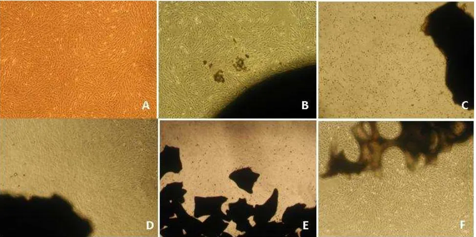
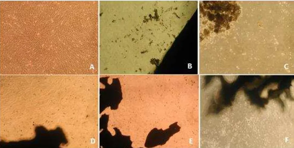
At day-1 (or 24 hours) after direct contact with scaffold, there was no cell morphological change and MSC-substrate adhesion impairment. Morphology and density of cells in each group relatively equal to control (Figure 2). At day-7, we found cell adhesion impairment to plastic substrate, cell morphological changes, cell death or reduction in cell proliferation in all scaffolds, it was very clear in scaffold I, II and V (Figure 3). Scaffold III showed inhibition of cell proliferation slightly higher than 50% (53.5%). Only scaffold IV (bovine HA granule) had less than 50% inhibition of cell proliferation (Figure 1).
Indirect contact test:MTT assay
MTT assay is an important method to evaluate the cytotoxicity of scaffold extract as scaffolds slowly released component in aqueous environment. Table 1 showed the results of MTT assay at day-1 (24 hours) and day-7,
relection of the number of viable cells (cell proliferation),
scaffolds I-V, culture media as negative control, phenol red 20 mg/mL and 100 mg/mL as positive control. MSCs could proliferated well in extract of scaffold II, III, IV, V and in negative control. In other words, extracts of scaffold II to V did not affect the viability and proliferation of MSCs with inhibition value < 50 (non-toxic). No statistically
signiicant differences were observed among all scaffolds (p > 0.05). But, among the non-toxic scaffolds, the least
inhibition of cell proliferation at 24 hours and day-7 was shown by scaffold IV.
Scaffold I, phenol 20 mg/mL, and phenol 100 mg/mL showed inhibition of cell proliferation more than 50% at
hour-24 and day-7. Statistically signiicant differences
were observed among the scaffold I, phenol 20 mg/mL, and phenol 100 mg/mL to control (p < 0.05). Although scaffold I showed inhibition value more than 50%, it was not as toxic as the positive control phenol.
DISCUSSION
In vitro cytotoxicity test usually used mouse ibroblast L929 and human osteoblast like cell (HOB). L929 is a cell line commonly used in cytotoxicity testing and HOB is a relevant cell line for application in bone regeneration.8 Our study which evaluated the
Figure 1.
Figure 2.
Toxicity effect of scaffolds I-V evaluated by proliferation of rat’s MSCs. At day-7, only scaffold IV showed proliferation inhibition < 50% (27.9%). Scaffold II showed 100% proliferation inhibition to MSCs
Morphology of MSCs in direct contact test of various scaffolds. At 24 hours after direct contact with scaffold, no cell morpho-logical change and MSC-substrate adhesion impairment or cell death observed. Morphology and density of cells relatively equal to control. A: control, B: scaffold I, C: scaffold II, D: scaffold III, E: scaffold IV, F: scaffold V
because MSCs had ibroblasts-like shape and this study
was a preliminary study to choose which one the non-toxic scaffold needed for further MSCs implantation.
The aim of this in vitro study with direct contact test and MTT assay was to evaluate cytotoxicity of various scaffolds to MSCs and also to provide information about cell viability.13 Every scaffold (biomaterial)
should be tested before it is implanted as scaffold, alone or in combination with demineralized bone matrix and or stem cells, in animal tests or clinical trials,8 because
it may reduce the probability of undesired effects such as; cell death, serious cell functional impairment,8,14
and disturbance of healing process (fusion/union),7
http://mji.ui.ac.id Figure 3.
Table 1.
Morphology of MSCs in indirect contact test of various scaffolds (MTT Assay). At day-7 adhesion impairment of cells to substrate, morphological changes, and cell death or reduction in cell proliferation in all scaffold was observed, especially at scaffold I, II, and V. A: control, B: scaffold I, C: scaffold II, D: scaffold III, E: scaffold IV, F: scaffold V
Result of MTT test at hour-24 and day-7
OD: optical density Scaffold
hour-24 Day-7
OD I OD II OD III Mean Inhibition % OD I OD II OD III Mean Inhibition %
I 0.024 0.037 0.064 0.042 52.3 0.026 0.019 0.037 0.027 69.3
II 0.050 0.048 0.055 0.051 42.0 0.034 0.044 0.077 0.052 40.9
III 0.056 0.070 0.081 0.069 21.6 0.089 0.090 0.104 0.094 -6.8
IV 0.062 0.062 0.093 0.072 19.3 0.089 0.102 0.106 0.099 -12.5
V 0.052 0.059 0.060 0.057 35.2 0.047 0.047 0.093 0.062 29.5
Phenol 20 mg/mL 0.002 0.001 0.003 0.002 97.7 0.002 0.003 0.001 0.002 97.7
Phenol 100 mg/mL 0.002 0.001 0.003 0.002 97.7 0.002 0.003 0.002 0.002 97.7
Control 0.102 0.097 0.065 0.088 0.0 0.102 0.097 0.065 0.088 0.0
toxicity and cell death, reduced cell proliferation, altered morphology, and impaired adhesion.8,9
It was already known that biocompatible HA scaffolds are promising materials for tissue engineering because they offer a tridimensional support and provide template for cell proliferation and tissue formation.15 In other words,
HA may intergrate with host bone in bone defect or comminuted fracture.7 Three of ive scaffolds tested are
produced by big company and have been sold in Indonesia, the other ones are still in research. Nevertheless, there
were no biocompatibility data of those scaffolds yet, it was the reason why we performed this study.
The tested scaffold is deined as toxic if it caused more than 50% cell death (inhibition value > 50 / IC50).An increase or decrease in cell number indicates the degree of toxicity of the biomaterial. IC50 is the concentration of the tested scaffold or biomaterial able to cause the death of 50% of the cells.11 The higher number of cell
In our study, at 24 hours after direct contact with scaffold, no change of cell morphology, decrease in cell number, or cell death was observed. It was supported by morphology and density of cells relatively equal to control group. Various inhibition of cell proliferation and change of cell morphology occured at day-7 evaluation. Scaffold I, II, and V inhibited cell proliferation very clearly. Scaffold III was slightly toxic but less than scaffold I,II, and V. From our study, only scaffold IV showed inhibition of cell proliferation less than 50%, therefore, it is no longer toxic to MSCs. Dias, et al13
stated that if there is no changes in morphology and activity of cells that were observed in cells culture in contact with scaffold/biomaterial, that scaffold is cytocompatible.
We found that toxic effect to the MSCs in direct contact test occured gradually. Dissolution of scaffold materials result in cell environment changes that impair cell viability. It may be assumed that there are other component in those scaffolds (eg. glycerol and calcium sulphate) which may cause cellular toxicity.8,10,14 In addition, dissolution of
HA-calcium sulphate will produce acidity in cell environtment.16 Ions released from calcium sulphate
material may produce hyperosmolarity that may cause cell proliferation. Therefore, scaffold must have appropriate environtment for cell proliferation, pH and physiological osmolarity in order to prevent toxic effect to cells.14
The indirect contact test using MTT assay utilized extract from each scaffold.8 As we mentioned that
scaffold contains additives, certain component of low molecular weight and initiator fragment that may affect cells viability.8,9 In present study, MTT assay
revealed that none of the extracts from the tested scaffolds affected the viability of the cells, except extract of scaffold I and phenol red which showed inhibition of cell proliferation more than 50%. As positive control, phenol red was responsible for the cytotoxicity. It was consistent with Zhu, et al12 who
said that there were no cellular toxicity, impairment of cell adhesion to the plastic substrate, and decrease in cell viability found in other four scaffolds. That result is also similar to Zheng17 about MTT assay
tri-calcium phosphate (TCP) which stated cell proliferation increased in both TCP and bone marrow MSCs groups. In addition, Zheng mentioned that the time of cell proliferation between the negative control
group and the experimental group has no signiicant
difference.17
Among extract of four non-toxic scaffolds, scaffold IV showed the least inhibition value to
cell proliferation, and inversely it improved cell proliferation better than on the control group.
That inding was also similar to Zheng’s study. We
thought that there were probability of weakness in cellular environment (quality of cells, culture media, and condition of incubation) that may affect the interpretation of scaffold toxicity if calculation is
based only in inal cell count. Therefore in this study,
inhibition percentage was determined by reference
to inal cell number in control group.
In direct contact test we found that 4 of 5 tested scaffolds were toxic, inversely in MTT assay 4 of 5 scaffolds were nontoxic or only one scaffold that showed more than 50% inhibition value. Many researchers mentioned that a scaffold might have higher toxic effect than scaffold extract which may explain different result of both tests. Therefore, from these tests we are able to determine which scaffold is nontoxic or the least toxic to MSCs in vitro.
In conclusion, although scaffolds contain similar main ingredient, they show different degree of toxicity. Scaffold IV (bovine HA granule) showed the least toxic effect to rat’s bone marrow MSCs on direct contact test and MTT assay. Finally, from those tests we were able to determine and choose certain scaffold before its application to in vivo study.
Acknowledgment
I would like to express my deep gratitude to Silmi Mariya for advice and suggestion in methodological aspect of this research; Kurniadi for typing and data
input. I hereby afirm that there is no conlict of interest
in this research.
REFERENCES
1. Lehmann G, Palmero P, Cacciotti I, Pecci R, Campagnolo L, Bedini R, et al. Design, production and biocompatibility of nanostructured porous HAp and Si-HAp ceramics as three-dimensional scaffolds for stem cell culture and differentiation. Ceramics-Silikaty. 2010;54(2):90-6. 2. Hollister SJ, Taboas JM, Schek RM, Lin CY, Chu TM.
Design and fabrication of bone tissue engineering scaffolds. In: Hollinger JO, Einhorn TA, Doll BA, Sfeir C, editors. Bone tissue engineering. Bocaraton: CRC Press; 2005.p.167-94.
3. Radu A, Eleonora C, Lucian A, Georgeta C, Virginia V, Cristiana T. In vitro biocompatibility testing of implantable biomaterials. Roum Biotechnol Lett. 2008;13(4):3863-72. 4. Nuss KM, von Rechenberg B. Biocompatibility issues with
modern implants in bone – a review for clinical orthopedics. Open Orthop J. 2008;2:66-78.
http://mji.ui.ac.id of carbonated apatite with an anorganic bovine xenograft
in particulate forms in a canine maxillary augmentation model. Clin Oral Implants Res. 2010;21(12):1334-44. 6. Bong S, Choon K, Kug S, Hyuk J, Hyun S, Sung S.
Osteoduction at porous hydroxyapatite with variuos pore
conigurations. Biomaterials. 2000;21:1291-8.
7. Rhodes N. Biocompatibility testing of tissue engineered products. Vox Sanguinis. 2004;87:161-3.
8. Idris BS, Danmark S, Wistrand F, Arvidson K, Albertsson AC, Bolstad AI, et al. Biocompatibility of polyester
scaffold with ibroblast and osteoblast-like cells for bone
tissue engineering. J Bioactive Compatible Polymers. 2010;25(6):567-83.
9. Silva GA, Marques AP, Gomes ME, Coutinho OP, Reis R, editors. Cytotoxicity screening of biodegradable polymeric systems. Texas: CRC Press; 2004.
10. Louisia S, Stromboni M, Meunier A, Sedel L, Petite H. Coral grafting supplemented with bone marrow. J Bone Joint Surg Br. 1999;81(4):719-24.
11. Fotakis G, Timbrell JA. In vitro cytotoxicity assays: comparison of LDH, neutral red, MTT and protein assay in hepatoma cell lines following exposure to cadmium chloride. Toxicol Lett. 2006;160(2):171-7.
12. Zhu Y, Zhang X, Zhu J, Zhao Q, Li Y, Li W, et al. Cytotoxicity of phenol red in toxicity assays for carbon nanoparticles. Int J Mol Sci. 2012;13(10):12336-48.
13. Dias RCM, Goes AM, Serakides R, Ayres E, Oreice RL.
Porous biodegradable polyurethane nanocomposites: preparation, characterization, and biocompatibility tests. Mat Res. 2010;13(2):211-8.
14. Kan I, Melamed E, Offen D. Integral therapeutic potential of bone marrow mesenchymal stem cells. Curr Drug Targets. 2005;6(1):31-41.
15. Mastrangelo F, Nargi E, Carone L, Dolci M, Caciagli F, Ciccarelli R, et al. Tridimensional response of human follicular dental stem cells onto synthetic hydroxyapatite scaffolds. J Health Sci. 2008;54(2):154-61.
16. Rauschmann MA, Wichelhaus TA, Stirnal V, Dingeldein E, Zichner L, Schnettler R, et al. Nanocrystalline hydroxyapatite and calcium sulphate as biodegradable composite carrier material for local delivery of antibiotics in bone infections. Biomaterial. 2005;26(15):2677-84. 17. Zheng W. Preparation and characterization of tri-calcium