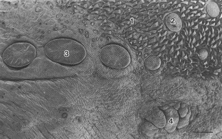
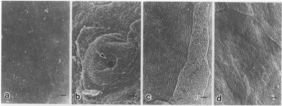
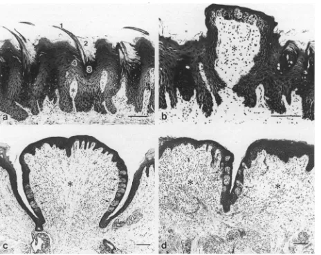
Morphology of the dorsal lingual papillae in the lesser mouse deer, Tragulus javanicus
Bebas
6
0
0
Teks penuh
Gambar

Dokumen terkait
The Extent of the Gravid M e rino Uterus In Relation to the Vertebral Column in the Dorsal Recumb ent Position and theW eights of the Gravid Uterus and Foetus in Relation to the
Relationship of the total cells number to follicle bulb and dermal papilla dimensions Derived variables Area density ratios Diameter ratios Papilla ratios DISCUSSION Bulb diameters
621 HUNT—PHYLOGENETICS OF POSEIDONAMICUS FIGURE15—Illustration of characters used in the phylogenetic analysis.1, 2,Characters 2 and 30, with arrows indicating posterior
Basale Figure 3/: dorsal margin with 1 spinous bristle at about 2h joint length, and 2 terminal bristles lateral spinous, medial longer, bare; lateral surface with 5 spinous bristles
Whitish opaque, a diffuse yellow dorsal band, the lateral region likewise yellowish shaded; no shields; joints 2 and 3 subdorsallyblotched in smoky bla,ck, the marksjoining
C: G-III, showing a strong iNOS positive cytoplasmic staining mainly on the surface epithelium, gastric pits, the isthmus blue arrows and in the basal areas black arrows.. D: G-IV,
The following pathological processes were identified in the gills of carp and zander: change in the shape of the lamellae of the secondary epithelium, hyperplasia of the gill
atm¼anterior transverse muscle, cf¼fiber-form muscle of the corona, dlm¼dorsolateral muscle, dlr¼dorsolateral retractor, dr¼dorsal retractor, lr¼lateral retractor, ma¼mastax muscles,
