226
Soesanto et aI. Med J Indonesteft
Atrial
Myxoma
with Mitral
Regurgitation
Amiliana
M. Soesanto,
Hamed Oemar, Omar Fauzi
Abstrak
Tumor primer jantung amat jarang terjadi.
Dari
semua tumor jantung, tumor milcsoma merupakan jenis yang paling banyak dijumpai. Diagnosis cukup sulit ditegakkan mengingat gejala dan tanda klinisdai
mil<soma ini tidak spesifik, bahkan dapat menyerupai penampakan klinis penyakit jantung lain yang lebih umum dijumpai. Pada kasus yang jarang, Hinis miksoma atrtum kiri dapat disertai keadaan insufisiensimital
seperti yang dilaporkan pada laporan ini. Kecuigaan terhadap adanya miksoma merupakan faktor penting agar diagnosis mil<soma tidak terluputl<nn. Saat ini pemeriksaan ekokardiografi mempunyai kontribusi yang besar karena kemampuan' nya memberikan keakuratan diagnosis miksoma atriumkii.
Dilaporkan seorang wanita 43 tahun dengan kcluhan sesak napas dan batuk-batuk yang berlangsung sejak setahun yang lalu.Dai
ekokardiografi dua dimensi tampak pembesaran atrium kiri dengan massa yang bergerakdanmenempel melalui tangkaiyang pendekke septuminteratrium. Setelah operasi ditemukantumoryang besarmernenuhi sebagian besar ruang atrium kiri. Pemerilcsaan histopatologi mmyimpulkan bahwa penarnpakannya sesuai dengan miksoma.Abstract
Primary tumors of the heart are yery rare; among these, myxonuts of the left
atium
(l,A) are the most common one. The clinical diagnosis isdfficult
since myxoma causes non-specific signs and symptoms, and its clinical manifestations often mimic other common cardiac diseases. Rarely, LA myxoma coincides with or even manifests asmital
regurgitation. Nevertheless, a high index of suspicion remains the most important element in diagnosing myxoma. The advantage in echocardiography modality has conffibuted an easy, simple, and accurate diagnosis of LA myxoma. A 43-year-old woman complained of breathlessness and cough lastingfor
one year. Echocardiography showed a large LA consisting of a mobile mass with short pedicle attached to the interatrial septum. The mass moved throughout a cardiac cycle. On operarton, alarge tutnor occupying most part of the lefi atrial cavity wasfound, The tumorwas completely removed and the pathological examination concluded the tumor as a myxoma.Keyw ords : Myxoma, M itral Re gur gitation, Echocardio graphy.
INTRODUCTION
Left atrial
myxomas
areuncommon
and presentwith
non-specific
clinical
cardiac symptoms and signs.
Therefore,
acorrect
diagnosis is essential to reduce themortality and
morbidity from serious
complications.l
In
most cases,this condition
has beenincorrectly
diag-nosed as
other common cardiac diseases.
However,
nowadays,with the
advancesof noninvasive technique
like
ultrasonography, an
accurate,cost
effeclive,
and safediagnostiè procedure
has beenprovided.2
The clinical
and hemodynamic features
of left
atrial
myxoma usually
mimic
those
of mitral stenosis,3
but
it
has beenreported
that afew
cases alsocoincide
with
mitral
regurgitation.4 This
paperillustrates
the featuresDepartment of Cardiology, Faculty of Medicine, University
of
Indonesia./National Cardiac Center Harapan Kita, J akarta, Indonesiaof left atrial
myxoma
with mitral
regurgitation
manifestation, which
was detectedby cardiac
Doppler
andechocardiography.
CASE REPORT
A
43-year-old woman
wasadmitted to
theEmergency
Unit
of
theNational
Cardiac Center'Harapan
Kita' on
January
31,
1997,with
the complaints
of
breathless-ness andcough.
For one
year
shehad been
suffering
from
exercise intolerance
which
developed gradually
andprogressively
until one
month
prior to
hospitaliza-tion.
There were also dyspnea oneffort, orthopnea,
andunproductive
coughwhich
was more intensein
upright
position.
Additional complaints
were
nausea,vomit-ing,
general
fatigue, anorexia, and intermittent low
gradefever.
She lost herweight about
13kg
within one
year.
History
of fainting, convulsion, or
neurological
disorders was denied.
Shealso denied any
history
of
Vol 6, No 4, October - December 1997
Physical examination
on
admission revealed
analert,
pale,
and
thin
women. Circumoral
cyanosis was
ob_ served.The
blood
pressurewas
ulse rate was 100beatsiminute,
regula
was30 /minute, regular.
There
was
waselevation
of jugular
venous
pressure.
The point
of
maximal impulse
was
locatedjust
2 cm lateral
to the
left midclavicular line
atthe
fourth
intercostal
space.A
heaving
on left
sternal border
atthe
4th
intercostal
space
was observed.
On
auscultation,
the
first
heart sound was accentuated at the apex,but neither gallop,
opening snap, nor tumor
plop
sound was audible.Theie
were
a
grade
316apical holosystolic munnur which
radiated
to the
left
mid-axillar
line and,
a
grade2/6
apical mid-diastolic murmur.
These
murmurs
varied
with
the
changeof position,
the
systolic
murmur
was more accentuateFine rales
was h Theliver was en
The
spleen
andwas no
pretibial
edemaor neurological abnormality.
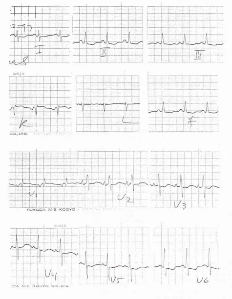
electrocardiogram
showed
sinus
rhythm,
100
beat/minute,
right
QRS axis
deviation with
left
atrial
The chestX-ray
revealedcar-Vo,left atrial
enlargement, and enouscongestion (Figure
2).The
two
dimensional
echoca
left
atrial (LA)
enlargement,
whi
argeecho mass
inside the atrial
c
wasLA Myxomawith
MR
227toto'.
The
mitral
and the tricuspid valves were not
repaired,
becauseit
wasconsidered
that there were nosignificant regurgitation
of
both valves after
tumor
removal.
The tumor weighed
73.8 grams and
measured about 8x
6 x 4 cm.It
had a rathersmooth
surface,yellowish
white
in
color,
with firmly
elastic
in
consistency
(Fig_ ure4).
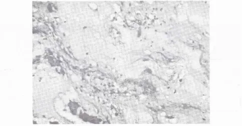
Microscopically it
was observed that thetumôr
cells
were round,polygonal
andclustered, which
were surrounded bymyxoid
andhyaline materials.
Scatteredfoci of calcification
werenoted. This histological
ap-pearance was consistentwith
the diagnosisof myxoma
(Figure
5).Three
days
after the
surgery, echocardiography
was revealed a residualmild
mitral regurgitation
and amild
tricuspid regurgitation,
with normal
left
ventricular
contractility.
The sizeof
theLA
cavity
was reducedto
almost
normal. Five
days after surgery, thepatient
was dischargedwithout
anycomplaint.
DISCUSSION
The incidence
of primary tumor of
the heart
is
about0.0017
Vo-\.028Vo;r
wjth
30Vo -50Vo"sf
those
are
The
clinical
manifestations
of left
atrial myxoma are
the resultsof obstructive
or/
andembolization factors.
They
also
causeanemia, fever,
erythrocyte
seglobulinemia,2,s-protein
have been repofted.a,l I Someof
these constitu_tional
symptoms
were observedin
ourpatient.
Emboli
can arisefrom
tumor fragmentation or
detach_ment
of
theentire tumor,
or from thrombi.ll
Obstruc_considered
asurgency;
surgical
removal
is
indicated
whenever
a diagnosisof
left atrial
myxoma is
made.tl
Soesanto et al. Med J Indones
[image:3.595.66.542.88.701.2]Ué
Vol 6, No 4, October - December 1997
Figure 2, The Chest X-ray revealed cardiomegaly, a prominent of left atrial
segment, and
signof venous
congesrton.LA Myxomawith MR
Figure 3' M mode echocardiogram demonstrated an echo dense mass just behind the anterior leaflet of mitral valve (right
),
onparasternal mass
prolapsed into
Long axis viewthe
oftwo-D atrioyentricular echocardiography a annulusdurinj
huge echo dense mass occupying almost most part oJ'LAwas observed. The [image:4.595.31.529.49.706.2] [image:4.595.196.524.72.426.2]230 Soesanto et al.
Figure 4. Gross specimen of the large tumor excised from the left atrium (
Ieft
), and during the tumor renloval(
risht
)Med J Indones
t.r, I
"r'z
f/
[image:5.595.63.547.442.694.2]"r/a
Vol 6, No 4, October - December 1997
symptom
to
be
sudden,
intermittent, and related
tobody position.8't0
In
our
case
almost
all
signs
andsymptoms mentioned
aboveoccurred.
Based on thosefindings
andthe
absenceof
history
of
rheumatic fever
the diagnosis
of
LA
myxoma
should have
beencon-sidered.
The
accentuatedfirst
heart
sound heardin
this patient
might
be
dueto
the late
onsetof mitral
valve closure
resulting
from
tumor
prolapsethrough
themitral
valve
orifice,
asreported
by
Greshlick et
a1.12Mobile
andpedunculated
left
atrial myxoma
may prolapseinto
themitral
valve
orifice,
resulting
in
obstruction
to
atrioventricular blood
flow
andmitral
regurgitation.
A
possibleexplanation
fbr
the occurrence ofmitral
regur-gitation is
the uncoaptation
stateof
themitral
leaflets
caused
by the large solid tumor
stretching the
mitral
annulus.4Accentuation
of
the
systolic murmur
in
anupright position,
asfound
in
our patient, might
be dueto the
downward
shift
of
thetumor which
caused moredilatation
to themitral
annulus and augmentation of theregurgitant
flow.4 This
was proved
during
the
opera-tion
that after tumor removal,
there wasno
significant
mitral regurgitation.
In
this
case,it
appearsthat
con-gestive heart
failure might
have been causednot only
by mitral
obstruction but
alsoby
mitral
regurgitation.
Presently
two dimensional
echocardiography
is
con-sidered to be the
most appropriate
screening anddiag-nostic imaging
modality
for
most cardiac
tumors,
particularly
-y*o-ur.ll
With this
technique,
mitral
stenosis can beexcluded,
andtumor prolapse through
theatrioventricular
valve may be demonstrated.Tumor
prolapse
in
ôur case can be accurately demonstratedby
echocardiography;
the tumor
was located
behind
theanterior
mitral
leaflet
and
it
moved
into
the
left
ventricle
during
diastole. l3LA Myxomawith
MR
231Acknowledgments
The
authorswish
tothank
Dr. M.
Yusak
for giving
uspermission
to illustrate his patient,
and
Dr.
I.
Made
Nassar
for
helping
us toillustrate
thehistopatological
features.
REFERENCE
l.
NasserWK,
Davis RH,Dill
JC, Tavel ME, Helman CH, FeigenbaumH,
FischC. Atrial
myxoma and pathologicfeatures in 9 cases. Am Heart J 1972;83:694-703. 2. Sutton MJ, Marcier LA, Giuliani ER, Lie JT. Atrial myxoma,
a review
of
clinical experiencein
40 patients. Mayo Clin Proc 1980;55:371-6.3. Bulkley BH, Hutchins
GM
:Atrial
myxomas, afifty
year review. Am Heart J 1979;97:639- 43.4. Oemar
H,
MasaokaT,
KanazawaI,
YuasaA,
Eno
S,Tsuchioka
Y,
et
al.
A
caseof
giantleft
atrial myxoma combined with mitral regurgitation. Hiroshima J Med Sci1982:31:165-73.
5. Colucci WS, Braunwald E. Primary tumors of the heart. In:
Braunwald
E,
ed.
Heart disease. Philadelphia:
Saunders,1992;
l45l-62.
6. Kabban SS, Cooley DA. Atrial myxoma, surgical considera-tion. J Thorac Cardiovasc Surg 1973 ;65:731-6.
7. Goodwin JF. Diagnosis of left atrial myxoma. Lancet1963;
l:464-7.
8. Greenwood WF. Profiles of atrial myxoma.
Am
J Cardiol 1968;21:367-75.9. Goodwin JF. The spectrum of cardiac tumors . Am J Cardiol 1968;21:307 -13.
10. Harvey W. Clinical aspects of cardiac tumors . Am J Cardiol 1968;21:328- 43.
I
l.
Kirklin JW, Barratt-Boyes BG. Cardiac tumors. In: KirklinJW, Barratt-Boyes BG, eds. Cardiac surgery. New York :
Churchill Livingstone, 1993; 1636-42.
12. Greshlick AH, Leech G, Mills PG, Leatham A. The loud first
heart sound in left atrial myxoma. Br Heart J 1984; 52:403.
13. Bass
NM,
Sharratt GP. Left atrial myxoma diagnosed by echocardiography, with observation on tumor movement. Br