278 Typhoid Fever and other Salmonellosis
Salmonella
species
in
a neonate
with
necrotizing enterocolitis
R. Chaudhryl, N. Sharmal, S. Bhushan2,
v'K'
Paul2,M'
Singh2, P' Panigrahi3Med J Indones
P-17
Abstrak
ggu, berat 1,249 kg, masuk ke unit perawatan intensif neonatus metabolik, serangan apnoe, tinia lunal</encer dan tes darah samar ai enterokolitis nekrotikans (BeIl stage l)' Tiga sampel tinja (hari 3 serotip Salmonella sP. yang berbeda dari ketiga sampel tinja
erg hari 30) bersama-sama dengan E' coli dan Staphylococcus dan kit identffikasi cepat (BioMérieux, Vitek, Inc)' Reaksi rantai
pada sampel tinja dan darah. SampeL tinja memberikan hasil PCR' Walaupun demikian, sampel darah ibu memberikan hasil gobatan suPofiif, pemberian antibiotika sefotaksim dan amikasin' mikroba multipeL dan infektif dari NEC serta penggunaan teknik ungkin akan memberikan panclangan baru lentartg patogenesis NEC' Abstract
we report, a male preterm baby of 31 weeks gestationweighing
L249
atal intensive care unit (NICU) ctt AIIMS, New Delhi. He hatl abelominal tlistension, melabolic acidosis,apnoic
that was positive for occult blootL' on the basis of above clinical features thebaby
haveNec
Il stage I)' Three samples of stool(ilay 9, 19, j0 ) antl one sample of btood (day
1
9' S' typhi day I 9 and S.senftenberg day 30) were isolated atd
egative Staphy-ction (PCR) fortive for S. tYPhi
ever mother's blood was found to be negative by PCR for S' typhi' taxime tmd amikacin ancl was discharged after a period of two
pid molecular techniques such as PCRfor identification of eti' to the pathogenesis of Necrotiling enterocolitis (NEC)'
INTRODUCTION
Necrotizing enterocolitis (NEC) is a disorder that oc-curs in newborns of low birth weight babies (< 1200
necrosisl,2. Association of NEC is well known with E.coli and with other enteric pathogens3' Salmonella infection in premature baby with NEC have been re-ported earlièr
with
single serotype namely Salmo-nella i.e. S. thompson4's'We are reporting here a unique case
of
necrotizing enterocolitisin
a newborne baby where more than one serotypeof
Salmonella sp' were isolated from stool along with E. coli, thus highlighting polymicro-bial etiology of NEC in premature baby.CASE REPORT
A male baby was born on 29 Dec 1996by emergency caesarean and was admitted to the nursery. He was a preterm baby
of
31+wks. gestation wetghing 11249kg. Indication
for
emergency LSCS was imminent eàlampsia, baby had apgar score atlmin
of 7/10 and at 5 min of 9/10. He was lethargic from day 1 and im-provedwith
neloxone.He
developed hyperbiliru-tinemia on day 5 (serum billirubin level12.9 mg/dl) for which phototherapy was given for 48 hrs.On day 8 he had abdominal distension, persistent metabolic acidosis and apnoea. His stool
for
occult Dept. of Microbiologyt , Pediatrics2, AII India lnstitute ofSuppl
I
-
1998blood was also positive. The baby was diagnosed as
having NEC and antibiotic therapy was started with Cefotaxime, Amikacin, Vancomycin and Metronida-zole. However, despite these he developed
perfora-tion on day 11, patient was managed conservatively.
A
left
side drain was complicated by feacal fistula.He was also having congenital cardiac anomaly in the
form of PDA on day 16, chest infection developed on day 48 and infective diarrhoea on day 60t1. The baby was started on injection of Cefotaxime and Amikacin for infective diarrhoea and he gradually improved.
MATERIALS
and METHODSQuantitative method
for
isolation of stoolflora
Stool samples were collected on day
9,
19 and 30.Aseptically collected stool sample weighing 0.1 gm
in
sterile vial and 1ml of
sterile normal saline was added with 2-4 sterile glass beads and then vortexed. Ten fold dilution (10-t to 10-8 ) were made and plated10
ml of
each dilution To 5Vo sheep blood agar andMacConkey agar
for
aerobic organism and Brain heart infusion agarfor
anaerobic organisms. Both aerobic and anaerobic organism were identified byusing conventional, biochemical test and rapid auto-mated tests such as AP120 E test, API staph/strep and Rapid ANA
II system
(bioMérieux Vitek, Inc).PCR
for
stool and bloodi)
Isolntion and detectionof
DNAfrom
stool and blood samplesThe DNA was isolated from 200
pl
of diluted stool sample and 200 pl of blood using QIA amp blood kit, according to manufacturer instructions. To the sam-pIe 25pl
of QIAGEN protease and 200 pl bufferAL
were added, vortexed and incubated in water bath at
70'C
for
10 min. 210 1tlof
isopropanol or ethanol was added to the sample and mixed again by vortex-ing.A
QIA amp spin column was plâcedin
a 2 ml collection tube. The above mixture was applied onQIA amp spin column and centrifuged at BÔOO rpm
for
1 min. The QIA amp spin column was placed ina clean 2 ml collection tube and washed with 500 pl
of
bufferAW
It
was then centrifugedfor
1 min at8000 rpm. The
QIA
amp column was placedin
a clean 2 ml collection tube, again 500 pl of buffer AWwas added and centrifuged for 2 minutes at 8000 rpm. Then the QIA amp spin column was placed in a clean 1.5 ml microfuge tube, DNA was eluted with 50 pl of buffer
AE
(Pre heatedto
70'C). And incubated atPoster
Session
279room temp.
for
1 min and centrifuged for 2 minutesat
8000 rpm. The eluent containedDNA
and was used for PCR.ii) PCR
for
S.typhiPCR was performed according to methods described before by us6. The reaction mixture for the first round of PCR contained 5 pl of DNA, 25 picomoles of
RKI
and RK2, primers, 200pl
of (each) all fourDeoxyri-bonucleoside Triphosphates, 0.75 units of Taq
polym-erase (Bangelore Genei, India
)
and standard PCRbuffer (100 mM Tris-HCl, 1.5 mM MgCl2, 50 mM KCl, 0.1. Gelatin, pH 8.3) in a final volume of 25 pl.
Amplification
in
an automated DNA thermal cycler (MJ. Research Inc. US) consisting 40 cycles at 94"Cfor 1 min (Denaturation ) 57'C for 1 min 15 sec.(
an-nealing) and72"C for 3 min (Polymerization ). After
the reaction, 7.5
pl
of amplified products of the first PCR was transferred to a IInd reaction mixture (17.5pl), containing 50 picomoles (each) of RK3 and RK4 primers
for
the nested PCR7. The nested PCR wasperformed for 35 cycles at94"C
for
I
min, 68'C for1 min and 15 sec and1Z"C for 3 min.
iii)
Detectionof
PCR produxtsThe DNA fragments of the flagellin gene of S.typhi
amplified by the PCR were identified by agarose gel electrophoresis, 8
pl
of the amplified products from the both roundsof
PCR were electrophoresed on alVo gel
for
60 min at constantI00
volts with TBE buffer (90 mM Tris borate, 2mM EDTA) molecularsized marker (lambda Hind
III
Banglore, Genei,In-dia)
wasrun
concurrently.The
gels stained with ethidium bromide were examined under U.V.illumi-nation for the presence of 486 or 342 bp bands.
RESULTS
l)
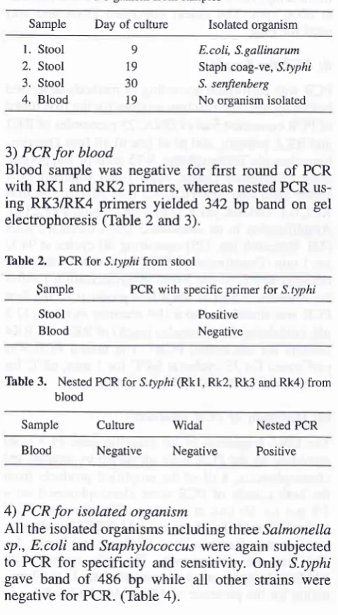
Isolation of organismfrom stoolThree samples of stool which were cultured on differ-ent dates yielded E.coli and S.gallinarLrm on day 9,
coagulase negative Staphylococcr"Ls and S.tltphi on
day 19 and only S.senftenberg on day 30. Isolated
Salmonella.rp. were further confirmed by
API
20E(Table 1).
2) PCR
for
stoolAll
the three stool samples were processed by PCRfor S.typhi.
A
specific band of 486 bp was detectedonly in the second sample (day 19) which was also
positive
for
S.typhi isolation, whilefirst
and third280
Typhoid Fever and other SalmonellosisThble
1.
Isolated organism from samplesMed J Indones
Blood culture was found to negative for S.typhi iso-lation as well as for nested PCR. Widal test for both
baby and mother was also negative
in
a titre<
1:40 for TO and TH antigens.6) To trace the source of S.typhi in this baby, samples
of all the medical and paramedical staffs were screened for S.typhi by Widal test culture and PCR. The stool sample collected from one
of
the paramedical staff working that time in the neonatal intensive unit was positive by PCR for S.typhi where as stool culture re-mained to be negative for S.typhi.DISCUSSION
Necrotizing enterocolitis is complicated by
bactere-mia
or
peritonitis3. Early reports implicating some bacteria were tempered when recovered organism were those that normally, colonize the intestinal tractsof premature infants. Such colonizing organisms in-cluding aerobes (coagulase negative staphylococcus,
facultative anaerobes (Enterobacteriacae and
entro-cocci) and strict anaerobes (Bacteroides, Clostridium
sp). An enrichment of certain colonizing bacteria in
cohorts of symptomatic infants has been reported8-lO in NEC, but considerable overleap with the flora con-tent of unaffected infants usually existll. Overgrowth
of a single predominant stool organism has been im-plicated in NEC developmentl2'13.
This is remarkable that NEC has been associated with most
well
known enteric pathogensla.In
a few in-stances in which enteropathogens such as Salmonella sp. and enterotoxigenic E.coli were identified in out-breaks among neonates, presenting symptoms were diarrhoeas'ls. Whereas this patients did not presentwith diarrhoea initially.
In
this caseof
NEC Salmonella sp. were isolatedthrice from stool samples. However the unique find-ing was that all Salmonella.rp. were of different
se-rotypes, identified
by
rapid systemof
biochemical (BioMérieux Vitek, Inc.). Isolated S.typhi from fecalsamples were further confirmed by PCR. In order to trace the source of S.typhi, mother's blood/stool proc-essed for culture and PCR were repofted to be nega-tive. Samples
(
stool and blood)
of
all the medical and para medical staffs were also subjected to cultureand PCR for S.typhi. Out
of
which oneof
the paramedical staff was found to be positive for S.typhi by PCR in both stooV blood whereas cultures remained negative. Her Widal test was also positive for TO and TH antibodies in hightitres (> 360).
Sample
Day ofculture
Isolated organisml.
Stool2. Stool 3. Stool 4. Blood
9 19
30
19
E.coli, S.gallinarum Staph coag-ve, S.typhi
S. senftenberg No organism isolated
3) PCR
for
bloodBlood sample was negative
for first
roundof
PCRwith RK1 and RK2 primers, whereas nested PCR us-ing RK3/RK4 primers yielded 342 bp band on gel electrophoresis (Table
2
and3).Table
2.
PCR for,S.Drpl,i from stool$ample PCR with specific primer for S.typhi
Stool Blood
Positive Negative
Table
3.
Nested PCR lor S.typhi (Rkl, Rk2, Rk3 and Rk4) from bloodSample Culture Widal Nested PCR
Blood Negative Negative Positive
4) PCR
for
isolated organismAll
the isolated organisms including three Salmonella sp., E.coli and Staphylococcus were again subjected to PCRfor
specificity and sensitivity. Only S.typhigave band
of
486bp while
all
other strains were negative for PCR. (Table 4).Table
4.
PCR for isolated organismIsolatated organism PCR with specific primer for S.typhi Rkl and Rk2
l.
E.coli2.
S.galinarum3.
Staph coag-ve4.
S.typhi5.
S.senftenbergNegative Negative Negative Positive Negative
5) Mother's blood
for
isolationof
S.typhi was [image:3.595.45.287.94.534.2] [image:3.595.49.281.561.697.2]Suppl
I
- 1998
The transient existence of S.gallinarum and S.typhi in stool
is amazing,
both of these serotypes were laterreplaced
by
third typeof
Salmonella sp. i.e.S.sen-ftenberg (Gp-E).
It
appears that multiple serotypesof
Salmonella.çp. were circulating
in
the environment thus competing with each other for their localisation in the gut of premature infants. One of these is ableto colonize in the intestinal flora depending on
avail-ability
of
suitable nidusfor
their existencein gut
mucosa. PolymicrobiaUmultifactorial aetiologyof
nectrotizing enterocolitis in premature baby is high-lighted in the etiopathogenesis of this disease. Use
of
rapid molecular techniques such as PCR for identih-cation of etiological agents is indicated in tracing the source
of
infection andfor
specific management ofnectrolizing enterocolitis.
In addition to stool culture, PCR for amplification
of
Salmonella sp. specific
DNA
sequences usingspe-cific
primers may emerge as a rapid diagnostic tool for identification of etiological agents in NEC. Thushelping
to
provide specific and appropriate rapid management of premature babies.REFERENCES
1. Lloyd JR. The etiology ofgastrointestinal perforation in the newboms. J Paediatr Surg 1996; 4:77.
Stevenson JK, Graham CB, Oliver TK, Goldenberg VE. Neo-natal Necrotizing Enterocolitis: A report of Twenty One cases
with Fourteen survivors. Am J of Surgery 1969; ll8:260. Kliegman RM. Neonatal necrotizing enterocolitis implica-tions for an infectious disease. Pediatr Clin North Am 1979; 26: 327.
Willoughby RE, Pickering LK. Necrotizing enterocolitis and
Poster
Session
281infection. Clin Perinatolo gy 1994; 2t: 307 -15.
5. Stein H, Beck J, Solomon A, et al. Gastroenteritis with ne-crotizing enterocolitis in premature babies. BMJ 1972; 2:
616.
6. Chaudhry R, Laxmi BV Nisar N, Ray K, Kumar D. Stand-ardisation of Polymerase chain reaction of Salmonelln typhi fever. J Clin Pathol 1997;50(5):437.
7. Chaudhry R, Lakshmi BY Nisar N. Kumar D, Dey AB. Ap-plication of nested polymerase chain reaction for Salmonella typhi in the diagnosis of typhoid fever. (submitted) 1997 .
8. BelI MJ, Feigin RD, Ternberg JL, et al. Evaluation of gastro-intestinal microflora in necrotizing enterocolitis. J Pediatr
1978;92: 589.
9, Hill HR, Hunt CE, Mastsen JM. Nosocomial colonisation with klebsiella, lype 26, in a neonatal intensive care unit
as-sociated with an outbreak of sepsis, meningitis and necrotiz-ing enterocolitis. J Pediatr 1974;85: 415.
10. Powell J, Bureau MF, Pare C, et aI. Necrotizing enterocolis-tis: Epidemic following an outbreak of Enterobacter cloacae type 3305573 in a neonatal intensive care unit. Am J Dis Child 1980; 134:1152.
11. Rotbart HA, Nelson WL, Glode MP, et al. Neonatal rotavirus - associated necrotizing enterocolitis : Case control study and prospective surveillance during an outbreak. J Pediatr 1988; 112: 8'1.
12. Bell MJ, Rudinsky M, Brotherton
T
et al. Gastrointestinal microecology in the criticallyill
neonate. J Pediatr Surg 1984; 19:745.13. Hoy C, Millar MR. Mackay P, et al. Quantitative changes in faecal microflora preceding necrotizing enterocolitis in pre-mature neonates. Arch Dis Child 1990; 65: 1057.
14. Rotbart HA, Levin MJ. How contagious is necrotizing en-terocolitis? Pediatr Infect Dis J 1983; 2: 406,
15. Cushing AH. Necrotizing enterocolitis with Escherichia coli heat-labile enterotoxin. Pediatrics 1983: 7 1 : 626.
2
3