EOSINOPHIL PROFILE of ELEMENTARY STUDENT, CAUSED by SOIL TRANSMITTED HELMINTHS INFECTION at SD NEGERI 026559 BINJAI,
SUMATERA UTARA
Oleh
Nurfida Khairina Arrasyid
UNIVERSITAS SUMATERA UTARA FAKULTAS KEDOKTERAN
EOSINOPHIL PROFILE of ELEMENTARY STUDENT, CAUSED by SOIL TRANSMITTED HELMINTHS INFECTION at SD NEGERI 026559 BINJAI,
SUMATERA UTARA
Nurfida K.A, Yoan C.P, Lambok S
Parasitology Dept., Medical Faculty, University of Sumatera Utara, Medan
Abstract
Background Soiled Transmitted Helminths ( STH ) infection, mainly caused by A. lumbricoides, T. trichiura and hookworm is still mostly found on children in Indonesia. Helminthic infection can effect malnutrition, anemia and growth and development disturbances on children.
On helminthic people not only immunoglobulin is increased but also the eosinophil. Eosinophil attaches to the helminth by Ig G/Ig A and release cathionic protein, MBP and neurotoxin. Mast cell and eosinophil will bind the surface of parasite by Fc -R, ,Fc -R, Fc∂-R and release toxin.
Helminthic infection initiates increasing level of eosinophil. Pardo et al, 2006, from his study on African imigrant who was suffered from helminthic infection found 27 % is followed by eosinophilia, 16 % among of them were ancylostomiasis and 17,2 % were schistosomiasis.
Purpose of this study is to know percentage of increasing eosinophil level on helminthic infection people in order to know the serve of eosinophil on immunity to helminthic infection.
Objective This study evaluates eosinophil level on to the occurrence of soil-transmitted helminthiasis in elementary students in Binjai City.
Methode Eosinophil level were examined by thin blood smear and stools by Kato’s-Katz.
Result On this study was found the incresing level of eosinofil > 5% were 72,7 % on askariasis students, 60 % on trichuriasis and 75 % on mixed infection of ascariasis and trichuriasis. There were 11 students infected by A. lumbricoides, 45 students by T. trichiura and 15 students by mixed infection of
A. lumbricoides and T. trichiura.
I. Introduction
Helminthiasis is still common found on children in Indonesia, especially Soil
Transmitted Helminths infection.. Ascaris lumbricoides is the most causes of STH
infection then follow by Trichuris trichiura and hookworm. Besides of growth and
development disturbunces, infection of the three species of STH can result another
bad efect.on children.3,7
Helminthic infection is usually chronic and deaths of host will harm the
parasite. Chronic infection of parasite will give persistent antigent stimulation which
will increase immunoglobulin level of the blood and form immune complex. On
helmintiasis people, besides of increasing level of immunoglobulin, the eosinophil
level will increase too.1,12
Immune response of helmintic infection is not effective and difficult to
understand. This difficulty may be cause of complex size and structure of the
helmitnhs. Size of the helminths is too large for phagosited but can arranged in layers
by Ig E and Ig A. Helminthic infection instructed immune system into Th2 response
and produce Ig E, Ig A and chemotactic Th2 cytokine into eosinophil and mast
cell.5,8,9
Eosinophil attaches to the helminths through Ig A/Ig E and release cathionic
protein, MBP and neurotoxin. Mast cell and eosinophil will bind surface of parasite
through Fc -R, Fc -R dan Fc -R and release their toxic content.1,12
Bejon, P, Mwamgi, T.W. et all reported that increasing of eosinophil level can be a
sensitive sign of parasite infection. Helminthic infection can trigger increasing of
eosinophil level was assured by Pardo et al, 2006, from their study of immigrant
African whom suffer from helminthiasis. Pardo stated 27% of that immigrant were
eosinophilia, 16% of them were ancylostomiasis and 17,2% were schistosomiasis.2,9
On their reports, Quinell, R.J. et all, 2004, stated 14 people with
ancylostomiasis have increasing level of eosinophil.11
Based on the states of the researchers above, it is need to make a study to see
percentage of increasing level of eosinophilia on helminthic infection people and to
II. References
Helminth is a multicelluler parasite so that it is difficult to know immune
response which work on it. Ovington, K.S. and Behm, C.A. (1997) stated that
helminthic infection will trigger Th2 released. One of its way is by activating
eosinophil as a response to kill the parasite.1,2,8
Eosinophil is part of white blood cells. 2-5% of blood cells without allergic
consists of eosinophil. Besides its fagositing, eosinophil can be stimulating to
degranulate mast cell, basophil and release mediator such as arisulfastese dan
histaminase. Mechanism of of eosinophil to kill parasite is through
antibody-dependent cellular citotoxicity (ADCC).8,9,10
Major basic Protein (MBP), Eosinophil Cationic Protein (ECP), Eosinophil
Derived Neurotoxin (EDN) dan Eosinophil Peroxidase will toxis and destroy target
cell if release. Helminths which have been arranged layer by Ig E will bind with
eosinophil and through degranulating process will release toxis protein.1,6
These facts show that eosinophil serves on immunity into helminthic infection.
III. Materials and Methods
The study was done on March 2008 on students of junior schools class 1 up to
class 6 SDN 026559 Binjai, Sumatera Utara by doing feces examine and leucosit
count on blood smear.
The study was started by taking feces sample. Every student was gave a 10 cc
pot as container of the feces which would bring by the students the day tomorrow.
On the same day with collected feces sample, blood smear of every students whom
follow this study was done.
First, top of the finger of the student was desinfected with alcoholated cotton.
After dry, jabed by using hemolet. The first blood drop was cleaned by dry cotton.
Then put one blood drop on the object glass, put the sliders object glass in front of
the blood drop, let the blood diseminated then push forward. Wait untill dry and then
fixation with methanol. After that decant with giemsa suspension, let for 15 minutes.
After 15 minutes, washed with aquadest. Then dry up.After dry, examine below the
microscope to count eosinophil amount.3,10,12
Examining of the feces was done by using Kato’s –Katz. Put 100 mg of a
excessive water sucked with tissue and then let it on the temperature room for 15
minutes. Then, examine below the microscope to see the eggs of helminths.4
IV.Result and Discussions
The samples were taken on March 2008 on students of class I- VI SD Negeri
025669, Binjai, Kabupaten Langkat Sumatera Utara. From 211 students of class I-VI,
only 141 students were checked, they were 8 students of class I, 31 students of class
II, 25 students of class III, 26 students of class IV, 29 students of class V, and 21
students of class VI. Another 71 students were out, cause did not fullfill the criteria.
On table 1, could be seen that only 11 from 141 students of SD Negeri
026659 Binjai whom had been checked, were infected by Ascaris lumbricoides.
Increasing of eosinophil level more than normal range was found on 8 infected
students. Eosinophil level 6-9% was found on 4 infected students (2,86%) and
eosinophil level >9% was found on 4 infected students (2,86%). Eosinophil level 9%
found on 3 students whom in their feces found egg of Ascaris lumbricoides < 2000/gr
feces and 1 student with 2.000 – 10.000 eggs/ gram feces.
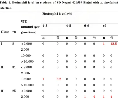
Table 1. Eosinophil level on students of SD Negeri 026559 Binjai with A. lumbricodes
infection.
Eosinophil level (%)
1-3
4-5
6-9
>9
Class
*n
Egg
amount (per
gram feces)
n % n % n % n %
I 8 < 2.000 0 0 0 0 0 0 1 12.5
2.000-10.000 0 0 0 0 0 0 0 0
> 10.000 0 0 0 0 0 0 0 0
II 31 < 2.000 0 0 0 0 0 0 0 0
2.000-10.000 1 3.2 0 0 0 0 0 0
> 10.000 0 0 0 0 0 0 0 0
III 25 < 2.000 0 0 0 0 0 0 0 0
Eosinophil level (%)
1-3
4-5
6-9
>9
Class
*n
Egg
amount (per
gram feces)
n % n % n % n %
10.000
> 10.000 0 0 0 0 0 0 0 0
IV 26 < 2.000 0 0 0 0 2 7.7 2 7.7
2.000-10.000 0 0 0 0 0 0 0 0
> 10.000 0 0 0 0 0 0 0 0
V 29 < 2.000 0 0 0 0 0 0 0 0
2.000-10.000 0 0 0 0 1 3.4 0 0
> 10.000 0 0 0 0 0 0 0 0
VI 21 < 2.000 2 9.5 0 0 0 0 1 4.8
2.000-10.000 0 0 0 0 0 0 0 0
> 10.000 0 0 0 0 0 0 0 0
Total 140 2 1.43 1 0.71 4 2.86 4 2.86
* n, sample amount
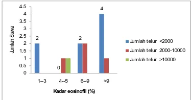
Table 1 shows that 11 students with ascariasis, 8 students (72,7%) have
eosinophil level > 5%. This result is according to the study of Ovington, K.S. dan
Behm, A.C. (1997), which was stated that eosinophil serves in immune response to
helminthic infection.
Increasing of eosinophil level on students with ascariasis in this study,
supports the statement of Mc. Donald A.S. et al (2002), in their verification of
2 0 2 4 0 0.5 1 1.5 2 2.5 3 3.5 4 4.5
1--3 4--5 6--9 >9
Ju m la h S is w a
Kadar eosinofil (%)
Jumlah telur <2000
Jumlah telur 2000-10000
Jumlah telur >10000
Picture1.Eosinophil level on students with ascariasis.
Increasing eosinophil level more than normal range ( not allergyc) on children
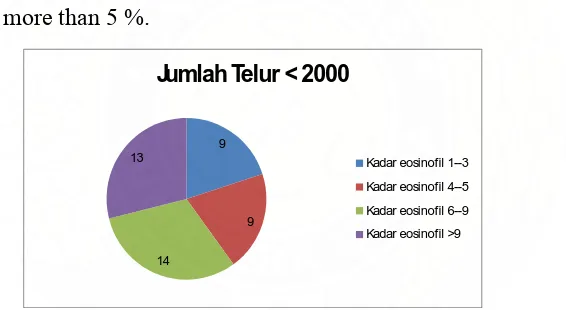
with T. trichiura infection can be seen on table 2. From 45 students with T. trichiura
infection, 14 students (10%) have eosinophil level 6-9% and 13 students (9,3 %)
eosinophil level more than 9%. The result is according to the study of Pardo et al
(2006) that there is increasing level of eosinophil on geohelminthic infection of
immigrant African in Gran Canaria.9
Table 2. Eosinophil level on students of SD Negeri 026559 Binjai with T. trichiura
infection.
Eosinophil level (%)
1-3 4-5
6-9 >9 Clas
s *n
Egg
amount
n % n % n % n %
I 8 < 2.000 1 12.5 0 0 0 0 0 0
2.000-5.000 0 0 0 0 0 0 0 0
> 5.000 0 0 0 0 0 0 0 0
II 31 < 2.000 0 0 2 6.5 4 12.9 6 19.4
2.000-5.000 0 0 0 0 0 0 0 0
> 5.000 0 0 0 0 0 0 0 0
III 25 < 2.000 2 8 1 4 3 12 2 8
2.000-5.000 0 0 0 0 0 0 0 0
> 5.000 0 0 0 0 0 0 0 0
IV 26 < 2.000 1 3.9 2 7.7 2 7.7 1 3.9
2.000-5.000 0 0 0 0 0 0 0 0
> 5.000 0 0 0 0 0 0 0 0
Eosinophil level (%)
1-3 4-5
6-9
>9
Clas
s *n
Egg
amount
n % n % n % n %
2.000-5.000 0 0 0 0 0 0 0 0
> 5.000 0 0 0 0 0 0 0 0
VI 21 < 2.000 3 14.3 2 9.5 3 14.3 0 0
2.000-5.000 0 0 0 0 0 0 0 0
> 5.000 0 0 0 0 0 0 0 0
Total 140 9 6.4 9 6.4 14 10 13 9.3
* n, sample amount
Table 2 shows, either more than 50 % students with T. trichiura infection or
more than 50% students with Ascaris lumbricoides infection has same eosinophil
level which is more than 5 %.
9
9
14 13
Jumlah Telur < 2000
Kadar eosinofil 1--3
Kadar eosinofil 4--5
Kadar eosinofil 6--9
Kadar eosinofil >9
Picture 2. Eosinophil level on students with T. trichiura infection.
Picture 2 shows, neither the students’ infected feces of T. trichiura nor the students’
infected feces of A. lumbricoides have egg amount which more than 2.000 /gram.
Table 3. Eosinophil level on students of SD Negeri 026559 Binjai
with mixed infection (A. lumbricoides and T. trichiura).
Eosinophil level(%)
1--3 4--5 6--9 >9
Class *n Egg amount
n % n % n % n %
* n, sample amount
2.000-10.000 0 0 0 0 0 0 0 0
> 10.000 0 0 0 0 0 0 1 13
II 31 < 2.000 0 0 1 3.2 0 0 0 0
2.000-10.000 0 0 0 0 3 9.7 0 0
> 10.000 1 3.2 0 0 0 0 1 3.2
III 25 < 2.000 0 0 0 0 0 0 0 0
2.000-10.000 0 0 0 0 2 8 1 4
> 10.000 0 0 0 0 0 0 0 0
IV 26 < 2.000 0 0 0 0 1 3.8 1 3.8
2.000-10.000 0 0 0 0 1 3.8 0 0
> 10.000 0 0 0 0 0 0 0 0
V 29 < 2.000 2 6.9 0 0 0 0 0 0
2.000-10.000 0 0 0 0 1 3.4 1 3.4
> 10.000 0 0 0 0 0 0 0 0
VI 21 < 2.000 0 0 0 0 1 4.8 1 4.8
2.000-10.000 0 0 1 4.8 0 0 0 0
> 10.000 0 0 0 0 0 0 0 0
Total 140 3 2.1 2 1.4 9 6.4 6 4.3
On table 3 can see, from 20 students (75%) with mixed infection of A. lumbricoides
and T. trichiura, 15 students have increasing level of eosinophil > 5%.
From all of the students’ feces which were examined, there was not found egg of
hookworm. This maybe cause the students had been usually used footgear so that they
can be avoided from hookworm infection.
Although cannot eliminate another causes which can increase eosinophil level, the
result of this study had been shown that there is increasing level of eosinophil on
students with helminthicinfection, especially students with ascariasis, trichuriasis and
mixed infection as more than 50%.
Conclusion
In SD Negeri 026659 Binjai, many students still have helminthic infection, especially
Increasing level of eosinophil > 5% is found as much as 72,7% on A. lumbricoides
infection, 62 % on T. trichiura infection and 75% on mixed infection students.
Acknowledgments
Researcher thanks to the headmaster and all the teachers of SD Negeri 026659, M.
Yusuf, SH, M. Hum, Dean of Medical Faculyi of Sumatera Utara University, Timbul