The effect of excessive disodium ethylene diamine tetraacetic acid (Na
2EDTA)
anticoagulant concentration on leukocytes profile in peripheral blood
examination
Usi Sukorini1, Tri Ratnaningsih1, Diannisa Ikarumi2
Abstrak
Akurasi pemeriksaan profil lekosit dipengaruhi beberapa faktor preanalitik diantaranya konsentrasi antiokoagulan. Antikoagulan yang paling sering dipakai pada pemeriksaan darah rutin adalah EDTA. Ketidaksesuaian perbandingan konsentrasi EDTA dengan bahan darah berefek terhadap hasil pemeriksaan darah tepi diantaranya parameter lekosit. Penelitian ini bertujuan untuk mengetahui apakah terdapat perbedaan antara hasil pemeriksaan profil lekosit pada bahan darah dengan berbagai konsentrasi antikoagulan Na2EDTA yang berbeda. Penelitian ini merupakan penelitian potong lintang. Bahan penelitian berupa 33 sampel darah vena mahasiswa Fakultas Kedokteran UGM Yogyakarta. Dua mL darah dibagi ke dalam 4 tabung Na2EDTA. Tabung pertama berisi Na2EDTA konsentrasi standar, 2 mg/dl, tabung yang lain secara berurutan berisi Na2EDTA dengan konsentrasi 4 mg/dl, 6 mg/dl, and 8 mg/dl. Sebelumnya dibuat sediaan hapus langsung dari setetes darah tanpa antikoagulan (sebagai kontrol). Darah dalam keempat tabung tersebut segera dilakukan pembuatan sediaan hapus dan diperiksa profil hematologi lekositnya menggunakan SYSMEX SE-9500 automatic analyzer. Terdapat perbedaan yang bermakna dari hitung lekosit, hitung jenis lekosit absolut dan prosentase monosit pada konsentrasi Na2EDTA yang berlebihan. Prosentase netrofil relatif meningkat dan terdapat perbedaan yang bermakna. Prosentase limfosit, eosinofil dan basofil tidak berbeda secara bermakna. Pemeriksaan morfologi lekosit menunjukkan perubahan yang bermakna berupa tepi sitoplasma yang irreguler, vakuolisasi, dan lobus nukleus yang irreguler akibat pengaruh konsentrasi Na2EDTA yang belebihan. Disimpulkan bahwa penggunaan konsentrasi Na2EDTA yang berlebihan pada preparasi spesimen darah menyebabkan perubahan profil lekosit sesuai peningkatan konsentrasinya. Konsentrasi standar tidak mempengaruhi hitung lekosit dan hitung jenis lekosit serta morfologinya, kecuali berpengaruh terhadap tepi sitoplasma yang irreguler dan lobus nukleus yang irreguler. (Med J Indones 2007; 16:168-75)
Abstract
Accuracy of leukocytes profile assessment is influenced by several pre analytical factors, among others, the anticoagulant concentration. EDTA is one of the most frequently used anticoagulant in peripheral blood examination. Several references stated that inappropriate concentration of EDTA anticoagulant in blood sample may affect the result of leukocytes profile in peripheral blood examination. The aim of this study was to evaluate whether there are differences among leukocytes profile in peripheral blood examination specimens, which were prepared with excessive Na2EDTA anticoagulant in different concentration. This study was conducted in Faculty of Medicine, Gadjah Mada University. Blood samples from 30 subjects were taken using vein puncture. Two millimeters blood was divided into 4 Na2EDTA-containing tubes. Before that, one drop of blood without Na2EDTA anticoagulant was used to make blood film right after vein puncture, as control. Each tubes contained different concentration of anticoagulant. The first tube contained Na2EDTA in standard concentration 2 mg/ml; the remaining tubes contained 4 mg/ml, 6 mg/ml, and 8 mg/ml respectively. These samples were immediately examined using SYSMEX SE-9500 automatic cell counter to measure the total and differential leukocytes count; and were stained with Wright staining for morphological examination under the microscope. These procedures were done before 20 minutes of vein puncture. There were significant decrement of total leukocytes count, absolute differential leukocytes count and monocyte percentage following excessive Na2EDTA administration. Neutrophil percentage was found to be relatively increased and the difference was significant. Lymphocyte, eosinophil and basophil percentages were not significantly different. Morphological examination showed significant increment in irregular cytoplasm margin, vacoulation and irregular nuclei lobes following excessive Na2EDTA administration. It is concluded that excessive concentration of Na2EDTA used in blood specimen preparation, will lead to changes in leukocytes profile as the concentration increased. Standard Na2EDTA anticoagulant concentration did not alter any leukocytes count and morphology, except for irregular cytoplasm margin and irregular nuclei lobes. (Med J Indones 2007; 16:168-75) Keywords: Na2EDTA, anticoagulant, leukocytes profile, leukocytes count, leukocytes morphology
1
Department of Clinical Pathology, Faculty of Medicine, Gadjah Mada University, Yogyakarta, Indonesia. 2
Peripheral blood examination is one of the important laboratory procedures in order to establish the diagnosis of various diseases, such as infection, tumor, degenerative diseases and others. This examination is usually used as first line procedure, because it is easy and relatively affordable for the patient.
One of the most important and routinely examined blood elements is the leukocyte. Leukocytes perform their role as host defender against various invaders. Abnormality in their quantitative and qualitative
profile may lead to the understanding of body’s
response mechanism to foreign body. The leukocytes profile includes examination of cell morphology; and cell count that consists of total leukocytes count and differential leukocytes count.
Many pre analytical factors can influence the result of leukocytes profile examination, one of it is the concentration of anticoagulant being applied. National Committee for Clinical Laboratory Standards (NCCLS) has recommended EDTA as anticoagulant for full blood cell counts and white blood cell differential analysis,principally because of its cell preservation properties.1
Most commonly used EDTA salt in hematology examination are disodium EDTA (NA2EDTA) with concentration of 1.4 – 2.0 mg/ml blood, dipotassium EDTA (K2EDTA) with concentration of 1.5 – 2.2 mg/ml blood, and tripotassium EDTA (K3EDTA) with concentration of 1.5 – 2.2 mg/ml blood.2
The aim of this study is to know whether there are differences among leukocytes profile in peripheral blood examination specimens, which were prepared with excessive Na2EDTA anticoagulant in different concentration.
METHODS
The design of this study was a qualitative non-experimental, performed by cross sectional method. It was conducted during September until November 2004 in the Faculty of Medicine, Gadjah Mada University.
There were 30 blood samples obtained from subjects with the following inclusion criteria; male, Malay race, age between 18 to 24 years old, healthy (proven by anamnesis and vital sign examinations), had been given informed consent prior to the study and agreed to join this research voluntarily as subjects, and the result of routine hematology examination using automatic
cell counter was within reference range. The exclusion criteria consist of history of allergy, recent disease, chronic disease and recent intake of medicine especially those that may influence hematology profile.
Variables being measured in this study include independent and dependent variables. Independent variables were Na2EDTA anticoagulant in different concentrations, which were standard concentration (2 mg/ml), 2x (4 mg/ml), 3x (6 mg/ml), and 4x (8 mg/ml) of standard concentration. For morphology examination, there was non-Na2EDTA specimen, as control in addition to the other 4 variables.
Dependent variables consist of leukocytes profile elements, that comprises total leukocytes count, differential leukocytes count and morphology variation of leukocytes. The differential leukocyte count was consists of absolute count and percentages of neutrophil, lymphocytes, monocytes, eosinophil and basophil. The evaluation of leukocytes morphology consists of irregular cytoplasm margin, vacuolation and irregular nuclei lobes.
Two ml of blood was drawn from the veins of antecubital fossa using aseptic technique of vein puncture for each subject. The blood samples were divided into 4 groups in 4 different tubes, 0.5 ml in each tube. These four tubes were prepared containing Na2EDTA in standard concentration, 2x, 3x and 4x of standard concentration, respectively. Cell counting using SYSMEX SE-9500 automatic cell counter and blood smear specimen preparation were done as soon as possible before 20 minutes from blood sampling to minimize bias with the effect of preparation time. For the morphology examination, control was made from the first drop of blood without anticoagulant. Principally, SYSMEX SE-9500 occupies flow-through system that identifies cell on the basis of cell size and intracellular density by using Direct Current (DC) and Radio Frequency (RF) method.3
Data analysis was performed by using ANOVA (Analysis of Variant) for normally distributed data and nonparametric tests (Kruskal-Wallis and Mann-Whitney) for not normally distributed data.
RESULTS
Total Leukocytes Count
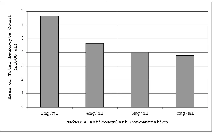
The graph 1 showed that the mean of total leukocytes count decreased significantly as the Na2EDTA con-centration increased beyond the standard concon-centration.
The percentage of total leukocytes count (x103 L) in 3 groups of excessive Na2EDTA anticoagulant con-centration is lower than that in the control group (standard Na2EDTA anticoagulant concentration) (p<0.0001).
The result means that excessive concentration of Na2EDTA will damage the leukocytes thus they cannot be detected any longer by the automatic cell counter, which result in the decrement of the total leukocytes count.
Differential Leukocytes Count
Absolute Differential Leukocytes Count
Figure 2 showed that the mean of absolute differential leukocytes count were decreasing significantly as the Na2EDTA anticoagulant concentration increased beyond the standard concentration (p < 0.05). This result is linear with the result of the total leukocytes count analysis, because as we know, differential leukocytes count were the building blocks of the total leukocyte count. Therefore, decrement in the amount of differential leukocytes will consequently decreasing the amount of total leukocytes.
The result signified that excessive concentration of Na2EDTA anticoagulant will damage the leukocytes in which Na2EDTA distorted cellular morphology, thus the cells cannot be detected by the automatic cell counter, which result in the decrement of absolute differential leukocytes count.5 Some authors said that pseudoleukopenia may due to the gatting out of large platelet-WBC masses.6,7
Figure 1. The relationship between mean of total leukocytes count (x103 L) with an increasing Na2EDTA anticoagulant concentration 0
1 2 3 4 5 6 7
2mg/ml 4mg/ml 6mg/ml 8mg/ml
Na2EDTA Anticoagulant Concentration
Mean of Total Leukocyte Count
Figure 2. The relationship between mean of absolute differential leukocytes count (x103 L) with an increasing Na2EDTA anticoagulant concentration
Percentages of Differential Leukocytes Count
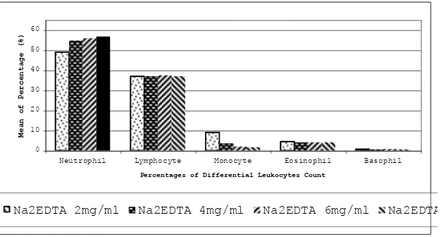
Figure 3 showed that the mean of differential leuko-cytes count percentages were not decreasing significantly as the Na2EDTA anticoagulant concentration increased beyond the standard concentration (p>0,05), except for monocyte percentage (p<0,05). Monocyte percentage was the only variable that showed linear result with its absolute count.
The mean of neutrophil count percentage in the excessive group is higher than that in the control group (p<0,05). Neutrophil percentage was increasing as the anticoagulant increased, which was not con-comitant with its absolute count result. Actually, the
absolute neutrophil count did not increase. Neutrophil percentage was relatively increased because the proportions of other cells absolute counts in total leukocyte population were decreasing more sharply than absolute neutrophil count. We assume that this decrement was due to cellular susceptibility, so probably they were not recognized by automatic cell counter as lymphocyte, eosinophil and basophil.
Percentages of lymphocyte, eosinophil and basophil were found to be varied and not significantly different among groups with standard and excessive Na2EDTA concentration. These results were also not linear with their absolute count results.
Figure 3. The relationship between mean of percentages of differential leukocytes count (%) with an increasing Na2EDTA anticoagulant concentration
0 10 20 30 40 50 60
Neutrophil Lymphocyte Monocyte Eosinophil Basophil Percentages of Differential Leukocytes Count
Mean of Percentage (%)
Morphology of Leukocytes
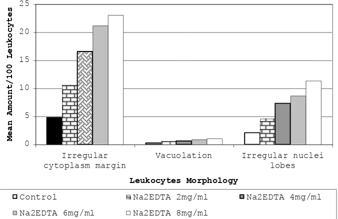
Figure 4 showed that the trend of mean of cells with irregular cytoplasm margin, vacuolation and irregular nuclei lobes per 100 leukocytes were increasing significantly (p<0,05) following the administration of standard and excessive Na2EDTA concentration, as compare to the control.
The descriptions of the leukocytes morphologic variables assessment were presented in Figure 5, 6 and 7. Since
we were not able to see the trend of morphologic changes following different excessive Na2EDTA administration, we preferred to choose some of these figures, only to represent the view of the evaluated variables.
Figure 5 showed a lymphocyte with irregular cytoplasm margin. Unlike the smooth and regular cytoplasm margin found in normal lymphocyte, this irregular cytoplasm margin of lymphocytes, protrudes, perhaps due to hypertonic environment.
Figure 4. The relationship between mean of leukocytes morphologic variables per 100 leukocytes with various Na2EDTA anticoagulant concentration
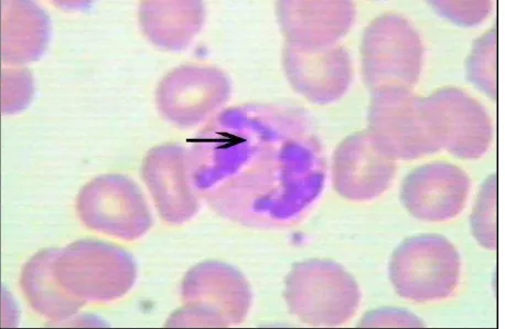
Figure 5. Photograph of Microscopic view at 100x magnifications. Irregular cytoplasm margin found in lymphocyte. Specimen Preparation with Na2EDTA 6mg/ml
0 5 10 15 20 25
Irregular cytoplasm margin
Vacuolation Irregular nuclei
lobes
Leukocytes Morphology
Mean Amount/100 Leukocytes
Control Na2EDTA 2mg/ml Na2EDTA 4mg/ml
Figure 6 and 7 represents anticoagulant-induced vacoulation and irregular lobulation found in neutrophil. In the microscopic examination, we found that lymphocyte experienced irregularity of cytoplasm margin more than other leukocyte elements. On the other hand, neutrophil was the most cells that underwent irregular nuclei lobes, while vacoulation mostly appeared in neutrophil and monocyte.
Figure 6. Photograph of Microscopic view at 100x magnifications. Vacoulation found in neutrophil. Specimen Preparation with Na2EDTA 4mg/ml. Specimen Preparation with Na2EDTA 6mg/ml
Figure 7. Photograph of Microscopic view at 100x magnifications. Irregular nuclei lobes found in neutrophil. Specimen Preparation with Na2EDTA 8mg/ml
DISCUSSION
Though unclear, cellular properties of each cell such as cytoskeleton and granularities, may have specific roles in determining the results of absolute counts and percentages following excessive Na2EDTA administration. The basic principle of SYSMEX SE-9500 automatic
cell counter in the detection of cells can also influence the result.
In clinical setting, the decrement of total leukocytes count is known as leukopenia. This quantitative alteration had been reported in some pathologic conditions such as decreased production and increased destruction of leukocyte following infection, post chemotherapy, glucocorticoid or ACTH administration and hyper-thyroidism.4
Erroneous percentages of differential leukocytes count may also result in misdiagnosis. In this study, we found that there were excessive anticoagulant-induced neutrophilia and monocytopenia. Pathologic neutrophilia may be due to generalized or localized infections, inflammatory response to tissue destruction, neoplastic growth, metabolic disorders, acute haemorrhage and corticosteroid or lithium administration, while mono-cytopenia can be found in overwhelming infections and following glucocorticoids administration.4,8
These findings indicate that excessive Na2EDTA administration may cause misdiagnosis, because of the misinterpretation of leukocytes profile results. Patient with leukocytosis such as in acute bacterial infection and inflammation, might be misinterpreted as having leukopenia because of excessive Na2EDTA administration to the blood specimen.
In patient with monocytosis such as in tuberculosis, dengue hemorrhagic fever in 6th and 7th days, syphilis and inflammatory responses,4 the result of peripheral blood examination may changed into monocytopenia if the specimen was prepared with excessive Na2EDTA, thus will lead to misinterpretation of the
patient’s condition.
Like other human cells, leukocytes are eucaryotes cells that possessed cellular components, including plasma membrane, cytoplasm, cytoskeleton, organelles and nucleus. One of the cell components that is important to be focused on, in relation to the irregularity of cytoplasm margin and nuclei lobes, is the structure of phospholipid bilayer plasma membrane in the outer part of the cell and the nucleus membrane.9
erythrocytes membrane. In other words, EDTA which is a chelating agent that binds to calciumin preventing blood coagulation, is able to influence the stability of erythrocytes membrane, and perhaps also leukocytes membrane.10
Our knowledge about leukocytes membrane, its properties and the possible relationship with Na2EDTA was not so extensive compare with those of erythrocytes. By considering the situation, we were only able to made assumption on how Na2EDTA concentration can influence the leukocytes.
This assumption may describe why the increment of leukocytes cytoplasm margin and leukocytes nuclei lobes irregularity occurred linearly with the increment of Na2EDTA anticoagulant concentration. However, the exact mechanism is not yet known. These irregularities also may describe why the leukocyte count decreased. As mentioned earlier, the cells would have been abnormal in shape and configuration, thus automatic cell counter was not able to detect them anymore.
Elevated pH in plasma leads to echinocytes formation in erythrocyte morphology. This finding also supported by other studies that proved that alteration in external PH cause changes in erythrocytes morphology, low PH produced stomatocytes whereas high PH produced echinocytes as observed in intact cell experiment.11 This Na2EDTA anticoagulant was assumed to produce increase pH, because sodium ion is the component of alkali. Echinocytes formation related with EDTA anticoagulant concentration involved mechanism that changed membrane structure of erythrocyte, mainly cytoskeleton and Ca2+ components.10,12
In our observation, the qualitative alterations of leukocytes in the presence of excessive Na2EDTA, when carefully examined, were not similar with those of pathologic condition. Stiene-Martin stated that vacoulation due to delay tends to be autophagocytic, which are small, evenly distributed vacuoles and the vacuoles reported to stain yellow rather than red with neutral red.4 We found that the vacoulation occurred due to excessive Na2EDTA administration were similar to this description. We assume that excessive Na2EDTA may induce vacoulation that resembled autophagocytosis, which may be found in prolonged storage of cells and exposure to certain toxins.
In clinical application, the presence of neutrophil vacoulation could not signify pathologic condition if it is less than 10% of the neutrophil population.4
Consequently, based on this study, vacoulation induced by excessive Na2EDTA anticoagulant, was not qualitatively significant until it reach 4x concentration (8 mg/ml) to cause a misdiagnosis. However, in-experienced observer or laboratory workers may misclassify the findings as a sign of pathology thus result in overestimation of anticoagulant-induced vacuolation. This will lead to false positive cases emergence, and will be harmful for the patient.
Based on the result of this study, we must aware to maintain proper blood to anticoagulant ratio. It is suggested that blood collection procedure is directly performed into the commercially prepared negative-pressure vacum tubes (vacutainer tubes). In reading the result of leukocytes profile analysis from automatic cell counter, it is recommended to confirm the per-centages of differential leukocytes count with absolute differential leukocytes count.
CONCLUSION
Eexcessive use of Na2EDTA in blood specimen preparation, will lead to changes in leukocytes profile as the concentration increased. Standard Na2EDTA concentration did not alter any leukocytes profile, except for irregular cytoplasm margin and irregular nuclei lobes.
REFERENCES
1. Anonymous. Recommendations of the International Council for Standardization in Haematology for Ethylene-diaminetetraacetic Acid Anticoagulation of Blood for Blood Cell Counting and Sizing. International Council for Standardization in Haematology: Expert Panel on Cytometry, Clinical Laboratory Hematology. October, 1993; 100(4): 371-2. Source: http://ncbi.nlm.nih.gov, Accessed on September 9, 2004.
2. Aulia D, Wirawan R, Suherli A. Pengaruh Lama Penyim-panan Darah dengan Antikoagulan Tripotassium Ethylene Diamine Tetraacetic acid (K3EDTA) dalam tabung Vaquette
terhadap beberapa Parameter Hematologi. MKI. 2002; 52: 1: 11-9.
3. SYSMEX SE-9500 Operator’s Manual – Japan. TOA Medical Electronics, 1997.
4. Stiene-Martin EA, Lotspeich-Steineger CA, Koepke JA. Clinical Hematology: Principles, Procedures, Correlation 2nd ed. Philadelphia: Lippincott. 1998.
5. Den Ottolander GJ, Jansen J. The effect of anticoagulant on the cell size of hairy cells and other malignant hematologic cells. A study with the Hemalog D. Am J Clin Pathol. 1984; 81:213-8.
7. Savage RA. Pseudoleukocytosis due to EDTA-induced platelet clumping. Am J Clin Pathol. 1983; 81:317-22. 8. Bain BJ. Blood Cells: A Practical Guide 3rd ed. Oxford:
Blackwell Publishing. 2002.
9. Campbell NA, Reece JB, Mitchell LG. Biologi 5th ed. Jakarta: Penerbit Erlangga. 2002.
10. Pinteric L, Manery JF, Chaundry IH, Madapallimattam G. The Effect of EDTA, cations, and various buffers on the morphology of erythrocyte membranes: An electron
microscopic study. Blood, May 1975: 45: 709 – 724. Browsed: http://ncbi.nlm.nih.gov, Accessed on October 17, 2005.
11. Gedde,M.M., Yang,E., Huestis, W.H., Shape Response of Human Erythrocyte to Altered Cell pH, Blood. 1995; 86;4:1595-9.