Genotype distribution of T cell receptor
β
gene in Indonesian nasopharyngeal
carcinoma patients
Yurnadi,1,2 Purnomo Soeharso,1 Dwi A. Suryandari,1 Nukman Moeloek,1 R. Susworo3
1 Department of Medical Biology, Faculty of Medicine Universitas Indonesia, Jakarta, Indonesia
2 Program Doctoral Medical Biology Student, Faculty of Medicine Universitas Indonesia, Jakarta, Indonesia
3 Department of Radiotherapy, Faculty of Medicine Universitas Indonesia and Ciptomangunkusumo Hospital, Jakarta, Indonesia
Abstrak
Latar belakang: Karsinoma nasofaring (KNF) merupakan penyakit genetik multifaktorial, bersifat endemik dan mempunyai perbedaan signifi kan dalam distribusi geografi s. Selain faktor virus Epstein Barr (EBV), insiden KNF juga dipengaruhi oleh faktor genetik seperti polimorfi sme gen reseptor sel T lokus β (TCR-β). Penelitian ini bertujuan untuk mengetahui hubungan polimorfi sme gen TCR-β dengan suseptibilitas individu untuk berkembang menjadi KNF pada populasi Indonesia. Metode: Penelitian dilakukan dengan teknik PCR-RFLP menggunakan enzim restriksi Bgl II pada gen TCR-β. Analisis PCR-RFLP gen TCR-β digunakan untuk mendeterminasi alotip gen TCR-β pada penderita KNF dan kontrol dan pada kelompok etnis Cina dan pribumi dalam populasi Indonesia.
Hasil: Hasil penelitian menunjukkan bahwa distribusi alotip gen TCR-β pada penderita KNF dan kontrol tidak berbeda bermakna (p > 0,05). Frekuensi alel A meningkat pada penderita KNF. Distribusi alotip gen TCR-β antara etnis Cina dan kelompok pribumi tidak memperlihatkan perbedaan bermakna (p> 0,05).
Kesimpulan: Distribusi alel gen TCR-β antara kelompok KNF dengan kelompok kontrol tidak menunjukkan perbedaan. Distribusi alel gen TCR-β antara etnis Cina dan pribumi tidak menunjukkan perbedaan. Polimorfi sme gen TCR-β tidak berhubungan dengan KNF dan etnis pada populasi Indonesia. (Med J Indones 2011; 20:171-7)
Abstract
Background: Nasopharyngeal carcinoma (NPC) is a multifactorial genetic disease, characteristically endemic and shows considerable differences in its geographical distribution. Besides infection with EBV, genetic factors such as polymorphisms of TCR-β gene contribute to the incidence of NPC. This study investigates the association of TCR-β gene polymorphisms with individual susceptibility to develop NPC in Indonesian ethnic groups.
Methods: The study was carried out by the PCR-RFLP method using Bgl II restriction enzyme to digest TCR-β gene. The PCR-RFLP analysis of TCR-β gene was used to determine allotypes of TCR-β gene in NPC patients and control among ethnic Chinese and indigenous groups in the population of Indonesia.
Results: The results indicate that the distribution of TCR-β gene allotypes between NPC patients and controls are not signifi cantly different (p > 0.05); however, the frequency of A allele tends to increase in NPC patients. The distribution of TCR-β gene allotypes between Chinese ethnic group was not signifi cantly different from indigenous groups (p > 0.05).
Conclusion: The distribution of TCR-β gene allele between NPC group and control groups showed no difference. The distribution of TCR-β gene between ethnic Chinese and indigenous groups showed no difference. Polymorphisms of TCR-β gene are not associated with NPC and ethnic groups in Indonesian population. (Med J Indones 2011; 20:171-7)
Key words:EBV, NPC, polymorphism, susceptibility, TCR-ß gene
Correspondence email to: yvmartin@yahoo.com
Nasopharyngeal carcinoma (NPC) is a genetic multifactor disease that has an endemic character.1 NPC is a subset of squamous carcinoma cells at the head and neck. NPC is relatively rare in the world (80.000 new cases per year). Nevertheless, NPC shows considerable differences in geographic distribution.2 Although many cases are found in countries with non-Mongoloid residents, South China still occupies the highest incidence with 2.500 new cases per year. NPC incidence in Mongoloid ethnics is a dominant factor; the frequency is quite high in South China, Hong Kong, Vietnam, Thailand, Malaysia, Singapore, and Indonesia.3
Makassar 25 cases, Palembang 25 cases, Denpasar 15 cases, and West Sumatera 11 cases. Considering also the numbers of cases in Medan, Semarang, Surabaya and other cities, it indicates that NPC has spread across the Indonesian region.
Dutt et al.5 argued that the incidence of NPC is multifactorial and cannot be entirely explained.Current evidence supports existence of a relationship between environmental factors, food, genetics, and infection with Epstein - Barr virus (EBV).5 Besides ethnic factors, increased incidence of NPC is reported to have links with environmental and socioeconomic factors, habits such as cigarette smoking and consumption of fermented and conserved food (e.g., salted and fumigated fi sh) and vegetable oils containing nitrosamines, as well as exposure to soot, dust with formaldehydes. Each of these factors can activate EBV and promote growth of NPC.6,7 EBV infection is an important environmental factor in the aetiology of NPC tumorigenesis. EBV is not found in all normal cells of nasopharyngeal epithelium and the EBV genome is not present in all normal cells of nasopharyngeal epithelium, however, it can be found in all NPC cells.2
The cells of the immune system spontaneously recognize tumour cells and T- lymphocytes (T cells) become main effector cells in the immune surveillance of cancer. T cells recognize antigens presented by the major histocompatibility complex (MHC) through distribution of T cell receptor (TCR) clones, until distribution clones TCR can detect and see specifi c band T cell base uniqueness TCR.8 Cytotoxic T cells are T-lymphocytes which mediate cytotoxic reactions to virus infected cells and stimulate macrophages to phagocyte these cells.9
Hence, T cells, which eliminate virus-infected cells, play an important role in pathogenesis of NPC. Restriction fragment length polymorphisms (RFLP) of germ-line of TCR genes from Singaporean Chinese patients with NPC and healthy controls were analysed by Southern-blot technique and hybridization with radioactively labeled TCR cDNA probes. The results suggested that TCR restriction may be important in the pathogenesis of NPC.10 T cells generally shared arranged immune response through several T cell receptors. Thus, T cell receptors play a role in the immune system and several of abnormalities and diseases have been shown to occur along with T cell dysfunction.11
Possibilities to assess variant TCR at condition of T cell dysfunction can be detected by visible change at locus of TCR to assess abnormality of expression or TCR gene function.12 Polymorphism of the TCR gene can infl uence T cell function, especially in occurrence and pathogenesis of diseases, for example hepatitis B. Polymorphism of TCR gene presenting allele and
genotype TCR is estimated to be a predisposing factor of NPC. Polymorphism of TCR-β gene is related to individual susceptibilities to infection of hepatitis B virus (HBV) and it was reported that polymorphic sites of TCR-β gene exist at span of Cβ1-Cβ2.13
The genetic factor is a primary factor in the pathogenesis of NPC and epidemiology data states the importance of genetic factor as the cause of this disease. Nevertheless as a whole molecular genetic aspects from NPC such as polymorphisms of TCR-β gene have not yet been able to be evaluated. Conversely, nowadays no experiment has reported TCR-β gene polymorphisms in Indonesian population and its relation with susceptibilities to NPC.
The aims of the study are to investigate distribution of genotype TCR-β gene in Indonesian population and its relation with individual susceptibilities to NPC.
METHODS
Subject
The research was done from August 2006 till December 2008 and research subjects are patients who have been diagnosed of having NPC, whereas controls are healthy individuals. All research subjects were given informed consent before taking part in this research. Diagnosis of NPC was based on inspection of histopathology biopsies conducted by medical doctors in the Department ENT FMUI/RSCM Jakarta. Furthermore, NPC staging was specifi ed based on classifi cation of Union Internationale Contre Cancer (UICC). NPC patient primary in research classifi ed its tumour by Pathologist based on WHO criterion.
Study design
Design of this research is a descriptive explorative cross sectional study. The sample used in this research is 3 millilitre (ml) peripheral blood from NPC patients and healthy donors. According to Sastroasmoro and Ismael14 using formula minimum samples with total amount of samples is 100 samples for NPC patients and healthy donors.
Genomic DNA Isolation15
Then enhanced 1.3 ml cell lyses solutions (10 mM Tris HCl; 0.25 mM EDTA; 20% SDS) and the mixture was incubated at 37oC for 30-60 minutes. Added to 1.3 ml precipitated protein (5M ammonium acetate) and vortexed till it turned milky. The mixture was centrifuged with 3.000 rpm at 4oC for 15 minutes till it formed a brown pellet. Supernatants were transferred to new tubes containing 2.3 ml cool isopropanol and the tubes were inverted several times until precipitated DNA was visible. The DNA was then incubated overnight at -20oC. Tubes containing isopropanol-incubated DNA were centrifuged at 3.000 rpm at 4oC for 5 minutes. The DNA pellet is cleaned with 1.3 ml sterilized alcohol 70%, centrifuged at 3.000 rpm at 4oC for 5 minute. DNA dry-aeration was run for 2 hours at RT. Furthermore, 300 ml TE (10 mM Tris HCl; 0.25 mM EDTA) was added to dissolve the DNA pellett and incubated at 37oC for 2 hour. After that, DNA was transferred to eppendorf tubes of 1.5 ml and stored at -20oC.
TCR-ß DNA amplifi cation andPCR-RFLP Technique
Amplification of TCR-ß gene was
perfor-med by using forward primer 5’ TACC TGGAGGCAGAGGAATG3’ and reverse primer 5’CCCTCCCAAGCAGGTTATTT3’. Amplifi cation was done at the TCR-ß DNA that covers polymorphic sites Bgl II in 1.5 kb upstream direction of the end of 5’ Cß213. Every 50 μl PCR mixture contain 20 μl template DNA, 10 pmol primer, 200 mM dNTPs (dTTP, dCTP, dGTP, dATP), 1.25 units Taq DNA polymerase, buffer solution contains 10 mM Tris-HCl pH 9, 50 mM KCl, 0.1% Triton X-100, 1.5 mM MgCl2 and ddH2O. Amplifi cation consisted of denaturizing, annealing, and extension cycles at the PCR machine. PCR condition that was used for both primers is early denaturizing at 95oC for 3 minutes,
fi nal extension for 3 minutes, and 35 cycles with denaturizing in 94oC for 30 seconds, annealing in 54oC for 30 seconds, and extension in 72oC for 40 seconds. For negative control, 20 μl ddH2O were added into PCR mixture. Furthermore, amplifi cation DNA (Amplicon) was checked by electrophoresis at 1% gel agarose. Into every well agarose enhanced mixture that contained of 10 μl amplicon and 3 μl loading buffers (0.25% bromophenol blue, xylene cyanol, 4% b/v sucrose) and then DNA separated with electrophoresis in 90 Volt for 60 minutes. For marking was used 100 bp DNA ladder. Bands of DNA fragments were visualized by an ultraviolet illuminator and documented by Polaroid camera.
After the positive result of TCR-ß DNA amplifi cation was proven with the existence of a DNA band of 229 bp, we furthermore conducted RFLP by using restriction enzyme Bgl II to detect polymorphism of the TCR-ß gene. The 15 μl TCR-ß amplicon enhanced
to PCR tube, added ddH2O 3 μl, buffer O 2 μl, and Bgl II 1 μl. DNA mixture was incubated at 37oC for 3 hours. After incubation, DNA mixture was checked by electrophoresis at 2% agarose. Into every well of the agarose gel was given enhanced mixture that consisted of 20 μl DNA and 3 μl loading buffer and then bands were separated electrophoretically like before. Results of PCR-RFLP as indicated by electrophoresis were DNA fragments that were visualized with an ultraviolet illuminator could be expressed positive if 1, 2, or 3 bands of TCR-β DNA were found at 229 bp, 142 bp, and 87 bp. Bands of DNA were documented with a Polaroid camera to analyse RFLP.
Statistical analyses
This research used nonparametric statistical analysis; to evaluate distribution and relations between two populations of genotype and allele of TCR-ß gene Chi-square test was used.16
RESULTS
DNA amplifi cation to detect polymorphic sites of TCR-β gene
Polymorphisms of TCR-β gene is shown by variation at span of TCR-β gene that cover polymorphic site Bgl II and located on 1.5 kb upstream direction the end of 5’ Cβ2. Amplifi cation area is referred as span of Cβ1 -Cβ2 conducted with PCR, using a primer designed to produce a TCR-β PCR product of 229 bp. Therefore, successful amplifi cation of TCR-β DNA should result in a PCR product of 229 bp (Figure 1.) This condition indicates that the primer has been precisely designed and is matching the expectations.
Analysis of the polymorphisms of TCR-β gene with RFLP method
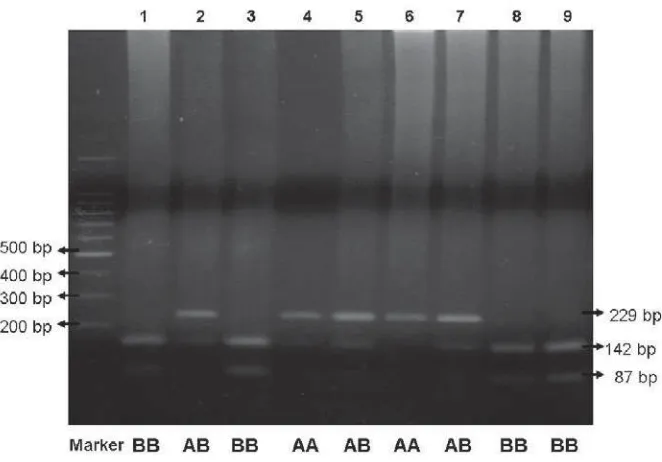
At genotype homozygote BB restriction enzyme Bgl II could recognize sequence of restriction site at 5’-gatct-3’, so that producing band of DNA that formed have double band (142 bp and 87 bp). Furthermore,
the genotype heterozygote AB is combination by 2 patterns genotype homozygote AA and BB, so that will producing three DNA bands (229 bp, 142 bp, 87 bp).
Figure 1. PCR product of 229 bp of TCR-β gene after being run on 1% agarose gel for 60 minutes 500 bp
400 bp 300 bp
229 bp
Distribution of TCR-β allele on NPC patients and control
At the Table 1 and Figure 3 we can see the genotype distribution and allele frequency of TCR-β gene on NPC group and control group. Tables 1 indicate that genotype distribution of TCR-ß in NPC population and control
Figure 2. Electrophoresis PCR-RFLP DNA TCR-β product in 2% gel agarose for 60 minutes, visualized with an UV illuminator and documented with a Polaroid camera.
Table 1. Comparison distribution genotype and allele frequency between NPC patients group and control group
Group Total AA AB BB Allele frequency A (%) B (%) NPC 102 13 39 50 31.86 68.14 Control 117 7 45 65 25.21 74.79
Here in after for allele frequency, at NPC group, B allele has high frequency (68.14%), whereas A allele
has low frequency (31.86%). At Non-NPC group, B allele has high frequency (74.79%), whereas A allele has low frequency (25.21%). Through chi-square test, the allele frequency of TCR-β in NPC group did not signifi cantly differ from control group (p>0.05). Nevertheless, distribution of genotype AA (A allele) is tending to increase on the NPC group.
Figure 3. Difference in allele frequency of TCR-β gene in patient NPC group and control group (p>0.05)
Distribution of TCR-β allele in China ethnic group and indigenous in Indonesia
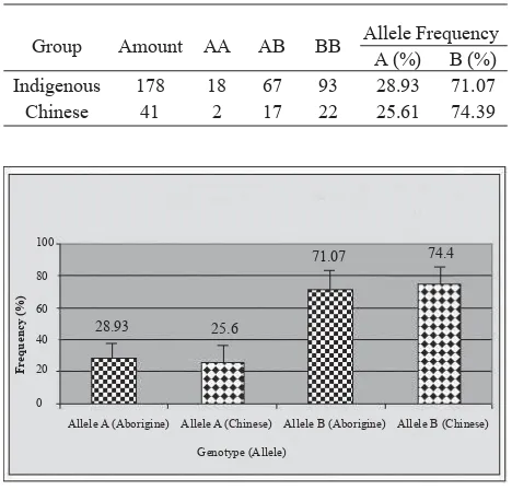
Table 2 and Figure 4 show the distribution of genotype and allele frequency TCR-β gene at Chinese ethnical group and Indonesian genuine population (Indigenous) group in Indonesia. Distribution of genotype TCR-ß at indigenous and Chinese in Indonesian population shows spreading pattern that does not fl atten. In the indigenous group, BB genotype has highest proportion (52.24%), followed by AB genotype with lower proportion (37.64%), and AA genotype with the lowest proportion (10.11%). In the Chinese group, BB genotype has highest proportion (53.65%), followed by AB genotype with lower proportion (41.46%), and AA genotype with lowest proportion (4.87%). Furthermore, for the allele frequency, at indigenous group, B allele has high frequency (71.07%), whereas A allele has low frequency (28.93%). At Chinese group, B allele has high frequency (74.39%), whereas A allele has low frequency(25.61%). As a whole in population indicate that B allele has high frequency (71.69%), whereas A allele has low frequency (28.31%). By chi-square test, allele frequency TCR-β in Chinese group was not signifi cantly different from indigenous group (p>0.05). This condition indicated that allele frequency of TCR-β gene at ethnical Chinese in Indonesia did not differ from the indigenous population in Indonesia (p>0.05).
Table 2. Comparison of genotype distribution and TCR-β allele at Chinese ethnic group and Indigenous group
Group Amount AA AB BB Allele Frequency
A (%) B (%)
Indigenous 178 18 67 93 28.93 71.07
Chinese 41 2 17 22 25.61 74.39
Figure 4. Difference in allele frequency of TCR-β gene in Chinese ethnic group and indigenous group in Indonesia (p>0.5).
DISCUSSION
Polymorphism of the TCR-β gene is shown by variation at span of TCR-β DNA that covers polymorphic restriction sites of Bgl II and located at 1.5 kilo base pairs,
100
80
60
40
20
0
Fr
equency (%)
Allele A (NPC) Allele A (Non-NPC) Allele B (NPC) Allele B (Non-NPC)
Genotype (Allele)
31.86
25.21
68.14
74.79
Allele A (Aborigine) Allele A (Chinese) Allele B (Aborigine) Allele B (Chinese) 100
80
60
40
20
0
Fr
equency (%)
Genotype (Allele)
28.93 25.6
71.07 74.4
Fr
equency (%)
Allele A (Aborigine) Allele A (Chinese) Allele B (Aborigine) Allele B (Chinese)
Genotype (Allele) 100
80
60
40
20
upstream direction to the end of 5’ Cβ2. Amplifi cation area of span Cβ1-Cβ2 are conducted with PCR method. Analysis polymorphism of TCR-β gene determine by undertaking PCR-RFLP. From RFLP analysis result that 3 genotypes TCR-β gene, where AA genotype are represented by single band DNA or wild type (229 bp), BB genotype represented double band DNA (142 bp and 87 bp), and AB genotype represented three band DNA (229 bp, 142 bp, and 87 bp).
At AA homozygote genotype restriction enzyme Bgl II did not recognize restriction sites sequence that changed from 5’-gatct-3’ to 5’-aatct 3’, so that the DNA showed a single band (229 bp). At BB homozygote genotype restriction enzyme Bgl II recognized the restriction sites at 5’-gatct-3’, so that produce two bands DNA (142 bp and 87 bp). Hereinafter, AB heterozygote genotype is combination from 2 patterns AA homozygote genotype and BB homozygote genotype, so that will produce three bands DNA (229 bp, 142 bp, 87 bp). In consequence, at PCR-RFLP method changing of endonuclease restriction sites will produce different fragment length DNA.17
Table 1 and Figure 3 indicate that distribution of genotype TCR-ß gene in NPC population and control shown that spreading pattern. By chi-square test, allele frequency of TCR-β gene between NPC group was not signifi cantly different with control group (p>0.05). But, distribution of A allele frequency TCR-β gene was tending to increase on NPC group. In this case, it can be anticipated that allotype TCR-β gene has infl uenced with susceptibilities of the individual to NPC, although the reality has been seen yet, where A allele frequency tend to increase at NPC patient and maybe predisposed at NPC pathogeneses. Involvement of A allele as predisposing factor of NPC will possibly be seen it reality if amount samples is improved two till three or conducted samples selection based staging of disease in tightens. Recognition EBV antigen by T cell through TCR also depend on antigen presentation by molecule HLA until HLA genotype class I and II on NPC patient also need to be considered, because HLA genotype modus certain to present EBV to T cell determine accuration and strong its cytotoxic response host to cell that infected by EBV.18
Table 2 and Figure 4 indicate that distribution of genotype TCR-ß gene at indigenous and Chinese groups in indonesia shows spreading pattern that not fl atten. By chi-square test, allele frequency of TCR-β gene between indigenous group was not signifi cantly different with Chinese group (p>0.05). This condition indicates that frequency of allele TCR-β gene in Chinese ethnic did not differ with indigenous groups in Indonesia. This is indicates that Indonesian people have the same chance
with Chinese ethnic in Indonesia to get NPC. This data indicates that maybe gene transfer and transmission have occurred between Chinese ethnic and indigenous from generation to generation.
Several other researches showed that, Chinese ethnic has a high incident of NPC compared to other ethnics, especially in South-East Asia. It is interesting to note the occurrence of NPC in Chinese migrant who has lived in Chinatown San Francisco United States for several generations. There is a signifi cant difference in NPC occurrence from Chinese immigrant compared to other populations such as Caucasians, Negroid, and Hispanics, where Chinese group shows a higher number of NPC cases.19 On the contrary, NPC cases in Chinese migrants that live in Chinatown showed a lower number compare to their brothers who live in China. Thus, it is possible that migrant groups still carry genes that are susceptible for NPC, but as a consequence of life style changes and eating habit during living in Chinatown, the trigger factors were suppressed so that the NPC does not develop.19
Other epidemiologic evidence occurs in the number NPC in Singapore, where the biggest percentage of NPC is Chinese clan society (18.5 per 100.000 residents), followed up by Malay clan (6.5 per 100.000) and the last is Hindustani clan (0.5 per 100.000).20 Furthermore, in Malaysia occurrence of NPC also many found in the Mongoloid race clan.21 That number is signifi cantly higher compared to that in European countries or North America with prevalence 1 per 100.000 per year.22 According to research result by Devi et al. in Sarawak-Malaysia, cases of NPC also have high prevalence, which is 13.5 per 100.000 people.23 Even though Korean, Japanese and north Chinese were included in Mongoloid race, not many were found to get NPC. Incidence of NPC in Asian countries is much higher compared to that in Europe or America. In UK, cases of NPC by age 0-14 year is 0.25 per 1.000.000 people, whereas by age 10-14 year is 0.8 per 1.000.000 people. Age estimating from England and Wales Cancer data indicates that 80% NPC patient at age 15-19 year, with number of occurrences 1-2 per 1.000.000 peoples.24
have an important role in response to EBV and NPC pathogenesis.11 In a research performed by Hirankarn et al.25 the association between HLA-E and genetic susceptibility to nasopharyngeal carcinogenesis was investigated by comparing the frequencies of HLA-E alleles in 100 Thai NPC patients and 100 healthy controls. HLA-E typing was performed by means of PCR–sequence-specifi c oligonucleotide probe method. The frequency of the HLA-E* 0103 allele and HLA-E 0103, 0103 genotype, but not others, was increased in NPC patients, compared to controls. This observation suggests a possible role for HLA-E in NPC development, possibly via natural killer cell or cytotoxic lymphocyte function.25
In conclusion, the distribution of TCR-β gene allele between NPC group and control groups showed no difference. The Distribution of TCR-β gene between ethnic Chinese and indigenous groups showed no difference. Polymorphisms TCR-β gene are not associated with NPC and ethnic groups present in the Indonesian population.
Acknowledgments
We thank to Irwan Ramli from Department of Radiotherapy and Umar Said Dharmabakti, Armiyanto, and Marlinda Adham from Department of ENT FMUI/ RSCM that has helped in levying samples research. Many thanks also handed to Dwi Ari Pujianto that helps in editing language of this article. This study was supported by HPTP project Directorate of Higher Education (DHE) Department of National Education (DNE).
REFERENCES
Mutirangura A. Molecular mechanisms of nasopharyngeal 1.
carcinoma development.Res Adv Res Updat Med. 2000; 1:18-27.
Mutirangura A, Tanunyutthawongese C, Pornthanakasem 2.
W, Kerekhanjanarong V, Sriuranpong V, Yenrudi S. et al. Genomic alteration in nasopharyngeal carcinoma: loss of heterozygosity and Epstein-Barr virus infection. Brit J Cancer. 1997; 76:770-6.
Roezin A, Adham M. Karsinoma nasofaring. Dalam: 3.
Soepardi EA, Iskandar N, Bashiruddin J, Restuti RD, editor. Buku Ajar Ilmu Kesehatan:Telinga Hidung Tenggorok Kepala dan Leher. Edisi ke 6. Jakarta: Balai Penerbit FKUI; 2007. p. 182-7.
Parkin DM, Freddy B, Ferlay J, Pisani P. CA: A Cancer 4.
Journal for Clinicians. available from http//:www.caonline. amcancersoc.org./cgi/contet/full/55/2/74. Diakses pada 24-9-2005.
Dutt MSN, Watkinson JC. The aetiology of nasopharyngeal 5.
carcinoma. Clin Otolaryngol. 2001; 26: 82-92.
Roezin A. Food and social background of nasopharyngeal 6.
cancer patient in Jakarta. ASEAN Otolaryngol Head & Neck Surg J. 1997; 1 : 21-7.
Yu MC, Yuan JM. Epidemiology of nasopharyngeal 7.
carcinoma. Semin Cancer Biol. 2002; 12:421-9.
Straten PT, Schrama D, Andersen MH, Becker JC. clonotypes 8.
in cancer. J Translat Med. 2004; 2:11. Available from: http:// www.Translation-Medicine.com/content/2/1/11.
Abbas AK, Lichtman AH, Pober JS.
9. Cellular and molecular
immunology. 4th ed. Philadelphia: WB. Saunders Company; 2000.
Chen Y, Chan SH. Polymorphism of T cell receptor genes in 10.
nasopharyngeal carcinoma. Int J Cancer. 1994; 56: 830-3. Berliner N, Dubby AD, Morton CC, Leder P, Seidman 11.
JG. Detection of a frequent restriction fragment length polymorphism in the human T cell antigen receptor beta chain locus. A diagnostic tool. J Clin Invest. 1985; 76: 1283-5. Knowless DM. Immunotype and antigen receptor gene 12.
rearrangement analysis in T cell neoplasia. Am J Pathol. 1989; 134:761-5.
Soeharso P, Summers KM, Cooksley WGE. Allotype 13.
distribution of human T cell receptor b and g chain genes in Caucasians, Asians and Australian Aborigines: Relevance to chronic hepatitis B. Human Genet. 1992; 89: 59-63. Sastroasmoro S, Ismael S. Dasar-dasar penelitian klinis.
14. Edisi ke-2. Jakarta: CV Sagung Seto; 2002.
Maniatis T, Fritsch EF, Sambrook J. Molecular cloning: A 15.
Laboratory manual. 2nd ed. New York: Cold Spring Harbor Laboratory Press; 1989.
Medis R. Statistical hand book for non-statistician. London: 16.
McGraw-Hill Book Co.: 1975.
Nussbaum RL, McInnes RR, Willard HF. Thompson & 17.
Thompson:Genetics in Medicine.6th ed. Philadelphia:WB Saunders Co.; 2001.
Munz C. Immune response and evasion in tha host-EBV 18.
interaction. In: Robertson ER, editor. Epstein-Barr Virus. England: Caister Academic-Press; 2005. p. 202-18. Parkin DM, Whelan SL, Ferlay J, Raymond L, Young J. 19.
Cancer Incidence in Five Continents. Vol. 7. Lyon: IARC Scient. Publ. ; 1997.
Armstrong MW, Armstrong MJ, Yu MC, Henderson BE. Salted 20.
fi sh and inhalant as risk factors for nasopharyngeal carcinoma in Malaysian Chinese. Cancer Res. 1983;43: 2967-70. See HS, Yap YY, Yip WK, Seow HF. Epstein-Barr virus 21.
latent membrane protein-1 (LMP-1) 30-bp deletion and Xho I-loss is associated with type III nasopharyngeal carcinoma in Malaysia. World J Surg Oncol. 2008; 6:18. Available from: http://www.wjso.com/content/6/1/18. Brennan B. Carcinoma nasopharyngeal. Orph J Rar Disease. 22.
2006; 1 (23):1-5.
Devi, BCR, Pisani P, Tang TS, Parkin DM. High incidence 23.
of nasopharyngeal carcinoma in native people of Sarawak, Borneo Island. Cancer Epidemiol Biomarkers Prev. 2004; 13 (3): 482-6.
Susworo, R. Kanker nasofaring: epidemiologi dan 24.
pengobatan Mutakhir. Cermin Dunia Kedokteran. 2004; 144: 16-9. [Indonesian]
Hirankarn N, Kimkong I, Mutirangura A. HLA-E 25.

