98 Wardhani et al. Med
J
Univ IndonTuberculosis Verrucosa Cutis
with Bilateral
Pulmonary
Tuberculosis
Tina Wardhani,
Adhi
Djuanda, Sri
Adi
Sularsito
Abstrak
Seorang laki-laki berusia 28 sejak 16 tahun lalu menderita bintil kecil, keras, kasar, dan tidak nyeri pada punggung kaki kanan. Dalam 2 tahun ini kelainan kulit menjadi tebal serta neluas dan pada paha kanan tinbul kelainan kulit serupa. Dalnn 6 bulan terakhir penderita nengeluh batuk-batuk kering, detrran, nudah lelah dan tidak nafsu makan. Pada peneriksaan fisik tatnpak penderita sakit dengan tubuh yang kurus. Terdapat petnbesaran kelenjar getah bening regional pada lipat paha kanan. Alat-alat dalam tidak nenuniukkan kelainan dan suhu nonnal. Pada peneriksaan kulit di punggung kaki kanan didapatkan jaringan hiperkeratosis berukuran 8 x 3 cm, nerah kecoklatan, dengan erosi, pus, darah, krusta, skuatna- Juga didapatijaringan hiperkeratosis berukuran 5 x 5 an dengan krusta dan skuatna sedikit proksitnal dari lesi pertana. Pada paha kanan bagian anterior terdapatjaringan hiperkeratosis berukuran 12
x
5 cm, serpiginosa, dengan erosi, pus,
darah, krusta dan skuatna. Pada diaskopi tidakdijunpai
applejellyJike
appearance. Petneriksaan laboratorium menunjukkan peningkatan laju endap darahyaitu 86 mny'jam dan tes fungsi hati dalatn batas nonnal. Pada petneriksaan foto toraks didapatkan ganbaran tuberkulosis paru bilateral disertai hasilpositif
pada petneriksaan basil tahan asam dalan sputmn. Tes tuberkulin dengan PPD 5 TUnenberi
hasil positif kuat. Hasil kultur jatnur dan nikrobakteriutn negatif. Hasil petneriksaan histopatologik tidak bertentangan dengatt tnberkulosLs kutis verukosa. Pengohatan dengan obat antituberkulosis yang terdiri atas rifampisin 450 ng/hari, pirazinanid 1OO0 mg/hari,dan
isoniazid jOO ng/hari nenunjukkan perbaikan klinis setelah 3 nûnggu. Setelah itu pasien tidak datang kenbali.Abstract
A rare case oftuberculosis verrucosa cutis with bilateral pultnonary tuberculosis itr a 28 year-old Indonesian nale is reported. He has been affected by this disease
for
16 years. The lesiotr began asa
slow growittg, ,to,, pruritic, rough, paùiless, warty lesion, at the dorsal aspect of his right foot. In tlrc last 2 years, the lesiott has thickened, and he noted a warty lesion on his right upper thigh. In thelast6nonthshesufferedfrondrycough,fever,generalnnlaise,andanorexia. Physicale.r.aninationrevealedanafebrile,chronically ill, lean individual, with nornalfunctions. Enlarged regional lytnph nodes of the right groin was fowtd. Cutaneous exatnination showed afinn,
slightly red-brown, hyperkeratotic plaque of 5 x 5cn
with crusts and scales proxitnal to thefirst
lesion.On
the anterior aspect of the right uppear thigh, there was a serpigitreous, hyperkeraatotic plaquel2
x 5 ctrr itr size, with erosions, pus, blood, crusts, and scales. An apple jelly-like appearance was not present on diascopic exanination. Sigttificant laboratory findings were and elevated blood sedimentation rate of 86 trurlh and nornal liver function tests. Chest x-rays revealed bilateral pulntonary tuberculosis and acid fast bacilli were found in the sputun. The tuberculitt test of PPD 5 TII was strongly positive. Tissue cultures of biopsy specinensfor
either fungi or mycobacteria were negative. Histopathologic exanination was sugestive of tuberculosis verrucosa cutis. The patient was given 450 nrg rifampicin, 10O0 mg pyrazinatnide, and 300 ng isoniazid daily. Afier 3 weeks of therapy, the skin lesion was altnost healed. The patient did not show upfor
a follow-up.Keywords
:
Tuberculosis verrucosa cutis, Bilateral pulmonary tuberculosis, Atttituberculous drugs.INTRODUCTION
Tuberculosis verrucosa
cutis
is
a verrucous
form of
skin tuberculosis in previously infected
and sensitizedindividuals
due to an exogenousinfection
ofMycobac-terium tuberculosis
ot Mycobacterium âovis.
It
is
aninoculation tuberculosis occuring
in
personswith
pre-vious
contactto Mycobacterium tuberculosis
and thushave acquired a certain
degreeof immunity
andsen-sitivity.l'2'3 The tuberculin test
is
therefore usually
positive
with hyperergic reaction.
The
inoculation usually occurs at sites of minor
wounds or
abrasions.l'2 Occasionally, the inoculation
canoccur by
thepatient's sputum. In low
socioecono-mic
areas,children
can become infected byplaying
andsitting on the sround contaminated
with
tuberculous
l-3 spurum.
Vol 3, No 2, April-June 1994
Tuberculosis
verrucosa
cutis usually
occurs
on the hands, often on theradial
border of the dorsum, andon the
fingers.
In
children,
the
predilection
sites
arethe lower extremities, which are most liable
to
betraumatized.l
The lesion
is
asymptomatic
and starts as asmall
papule
or
papulopustule
with
a
purple inflammatory
halo.
It
becomeshyperkeratotic
andwarty, with
slow
growing
development
of a
verrucous
plaque. The con-sistency isusually
firm,
but
the center can besoft;
pusand keratinous material can be
expressed
from
thefissures. There
is
a
serpigineous spreading
or
peri-pheral expansion
with cicatrix
on the central
lesion.4The
plaques
tend
to
be single, but
can
be multiple.
Lymph
nodes areusually not affected, however
enlar-gementof regional lymph
nodes can occur after secon-darybacterial infection.
1,2CASE REPORT
A 28
year-old Indonesian
man came to theDepartment
of Dermatovenereology
of Dr.
Cipto Mangunkusumo
Hospital,
Jakarta, onNovember
16,
1992History
At
the
age
of
12,
he noted a slow growth
of
a
nonpruritic,
rough,
and painlesswarty lesion
on the dorsal aspectof
theright foot.
He
did not recall
any trauma. He was treated at amedical
center on several occasionswithout
any success.In
the last2 years,the growth
hasthickened and he discovered another rough,
warty
lesion on hisright thigh.
Six months earlier, hLsuffered
from
adry
cough, fever, general malaise, and anorexia.He
wasnot
aware
of
any tubercular contacts
and has never been treatedfor
this ailment,
General conditions
Physical examination revealed
a chronically
ill,
leanindividual, with a blood
pressureof
l2Ol7OmmHg,
arespiration rate
of
2O/minute,
and apulse rate
of
g0/minute.
Thepatient
wasafebril with
normal functions.
No
organomegaly
or
palpable
masses
were
found.
There
were an enlargement
of
the regional
lymph
nodes of the right
groin, which
was elastic,mobile,
andpainful.
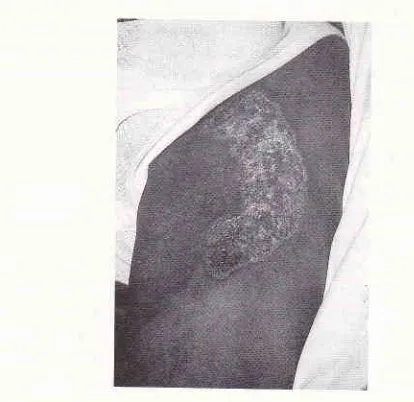
Cutaneous
examination
The lesion
was
firm,
slightly red-brown,
hyperkera-of
8by
3
cm
in
sions,
pus,, and scales on
th
eright
foot
earthis
lesion,w
erkeratotic
plaque
of
5
by 5
cm
in size,
with
crusts and
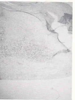
scales(Figure 2). On
theanterior
aspectof
theupper thigh,
there was aserpigineous, hyperkeratotic plaque
of l2
by
5cm,
with
erosions, pus,blood,
crusts, and scales.No
apple
jelly-like
appearancewas
present
on
dia-scopic examination.
Laboratory findings
Significant
laboratory
findings
were an elevatedblood
sedimentation rate
of
86/h and acid fast
bacilli
werefound
in
thesputum. Other laboratory
findings within
normal
limits.
[image:2.595.310.517.506.707.2] [image:2.595.49.272.511.698.2]Tuberculosis Verrucosa
Cutis
99Figure
2. A serpigineous hvperkeratotic plaque on the anterior part of the right upper thigh.Figure 1. 'lwo hyperkeratotic plaques on the dorsurn
of
100
Wardhani et al.Chest
x-ray
Bilateral pulmonary tuberculosis with
nopleural
invol-vement. Thoracic and lumbal
vertebrae showed
noabnormalities.
Microbiologic examination
Tissue
culture
ofbiopsy
specimenfor fungi,
Mycobac-terium tuberculosis,
andatypical mycobacterium
wereall
negative.Histopathologic examination
Skin biopsy of all lesions
revealed hyperkeratosis,
acanthosis
of
the
epidermis
with
Langhans
cells,
chronic
inflammatory infiltrates, with
someepitheloid
cells
in
thedermis (Figure
3 and 4).Tuberculin
test
Tuberculin
skin
testwith Purified
Proteins
Derivative
5
TU
showed
anerythematous
induration with
adia-meter
of
20 x
20mm.
Therapy
On
first
admission,
the patient was given
oral
clin-damycin
4x
150mg/day
andtopical
potasium perman-ganate U5000 dressingfor
one week to treat secondaryinfection. Antituberculous triple-drug
therapy
of
iso-niazid
300mg/day,
rifampicin
450 mglday, andpyrazi-namide
2 x
500 mg/day,
with vitamin
86
30mg/day,
was started on
Novembet 28,
1992.The patient
camein
for
afollow-up
onDesember2I,1992,3
weeksafter
the
start
of
thetreatment, feeling much better
and hadreturned
to
work. The skin lesion was almost
clear
(Figure
5 and6). He did not
show upfor
a subsequentfollow-up.
DISCUSSION
In the
Dr. Cipto Mangunkusumo Hospital, tuberculosis
verrucosa cutis ranks
second,
after
scrofuloderma,
among
skin tuberkulosis
(13%).4The
differential
diagnosis
for
this patient
werelupus
vulgaris
andchromomycosis. Lupus vulgaris
isan extremely chronic and
progressive
form
of
skin
tuberculosis occuring in individuals
with
ahigh
degreeof
tuberculin sensitivity.
It originates
from
a
tuber-culous
condition,
or aclinically
inapparenttuberculous
focus elsewhere
in
the body,
by
hematogenous,lym-phatic, or
contiguous
spread.The lesions
areusually
solitary, but two
ormore
sitesmay
beinvolved
simul-Med
J
Univ Indontaneously,
and in patients
with
pulmonary
tubercu-losis,
multiple foci
may develop.
In
about90
%of the
patients, the
head
and neck are involved,
although
lesions
involving
theextremities
can occuroccasional-ly.l
Thecharacteristic
lesion is areddish-brown
plaquewith
deeply embeddedperipheral
nodulesabout
I
mm
in
size
and
yellowish
(apple
jelly)
in
color.
If
thesenodules
are pressedwith
a glassslide (diascopic
exa-mination),
they show
as a sharphymarginated
yellow
brown
macules.'
In our patient, the
plaques
werehyperkeratotic,
with
onelesion having
a serpigineous spreadwithout
an applejelly
appearance on diascopy.Chromomycosis
is
achronic
cutaneous and sub-cutaneousinfection of
theskin,
causedby
the speciesPhialophora, Cladosporium, or
Fonsecaea,
which
forms
awart-like
lesion
on theskin.
It
usually
beginsby
the environment,
for
example,
workers working
without
shoes
or
protective
clothing on their lower
extremities
aremore
likely
to suffer
trauma
on their
feet and
legs.l
Onculture, all
species producesimiliar
heaped-up dark
colonies
with
short aerial hyphae,pro-ducing
agrey,
greenor brown, velvety
surfaceresem-bling
a mousepelt.
The
various^ causal agents can bedifferentiated
microscopically.r
Tissue culture
of
biopsy
specimen
from this patient
was negative
for
fungi.
Although
skin
cultures
for
Mycobacterium
tuberculosis
or atypical
mycobacteria were
negative,our patient's sputum
waspositive fot M.
tuberculosis.
Djuanda
reported thatmycobacterium culture
wasim-portant
in confirming the etiology, b]tt
only
found
positive results
in 21,7
%of
the cases.*Histologic
examination of tuberculosis
verrucosa cutis shows hyperkeratosis,papillomatosis,
andacan-thosis.
An
acute
inflammatory
infiltrate can be
ob-served
under the epidermis. Tuberculoid
structureswith
a moderate amount of necrosis areusually
presentin
themiddermis. Tubercle
bacilli,
are more numerouswhen
compared
to
lupus vulgaris.lo
In
chromomy-cosis, clusters
of brown,
thick-walled,
septatedfungal
cells can be
seen.8Histopathologic exàmination
of
biopsy specimen of
our
patient
was
suggestive
of
tuberculosis
verrucosa cutis.Stokes et al.,regarded tuberculosis verrucosa
cutis
as a
hyperergic reaction,
which
gives
apositive
skin
reaction
with highly
diluted tuberculin (1:
1, 000,000).On
the
other hand,
lupus vulgaris is
a hyperergic or
normergic reaction, which gives a positive
reaction
with moderatelly
dilutecltuberculin (1:100,000). Other
Vol 3, No 2, April-June 1994 Tube rculos is Ve rrucosa C utis
l0l
[image:4.595.71.196.84.251.2] [image:4.595.325.454.85.252.2]Figure3. Skin biopsy from a disral part of dorsutn pedis revealed hype rke ratosis, acanthosis wit h lttng lnns cells, chronic inflammatory inftltrates, and sone epitheloid cells in the dennis.
Figure 4. Figure 3
fron
close up.Figure 5 and 6. After 3 weeks of therapy,
all
lesiotrs either on the rlorsuttr pedis or on the upper thigh showing itrtprovetttent.anergic
reaction.4 Tuberculin skin
test(ppD
5TU) in
our
patient
wasstrongly positive
with
a diameterof
20x
20 mm
erythematous
induration. This confirmed
ahyperergic reaction.
The patient
was treated with
isoniazid
300 mg/
day,
rifampicin
450mg/day,
andpyrazinamide
2 x 5OOmg/day.
Thesedrugs
have potentbactericidal effects,
therefore rapid improvements
were noted.Lymph
nodes
are
not
affected
in
tuberculosis
verrucosa cutis. The enlargement of the
regional lymph
nodes
of
the
right
groin found
in
this patient
wasprobably
due to secondaryinfection.
It
respondedwell
to
antibiotics
given
one week.CONCLUSIONS
A
rare
caseof
tuberculosis verrucosa cutis
with
bila-teral pulmonary tuberculosis
in
a28
yeatold
Indone-sian
male is reported.
Theskin
diseaseimproved after
treatment
with
acombination of isoniazid
300mg/day,
rifampicin 450 mg/day, and
pyrazinamide
2
x
500mg/day
for
3 weeks.REFERENCES
L02
Wardhani et al.General Medicine, 3 rd ed. New York: Mc Graw
Hill,
1987;2 153-80.
2. Braun-Falco
O,
Plewig G,Wolff
HH, Winkelmann RK. Tubercolisis of the skin, 3 rd ed. Berlin : Springer Verlag,1990; 459-60.
3. Moschella SL. Disease of the mononuclear phagocytic
sys-tem.
In
: Dermatology,vol
l,
2 nd ed. Philadelphia : WBSauders, 1985;921-46.
4. Djuanda A. Tuberkulosis kutis. In : Ilmu Penyakit Kulit dan
Kelamin.
Edisi
kedua. Jakarta:
Fakultas Kedokteran Universitas Indonesia, 1993; 52-6O.5. Utz JP, Shadomy HJ. Deep fungal infection. In : Dermatol-ogy in general medicine, 3 rd ed. new
York:
Mc GrawHill,
1987;2248- 71.
Med
J
Univ Indon6. Lanfas G, Fisher BK, Contreras M. Tuberculous ulcer of the
skin. J Am Acad Dermatol. 1988; 19: lO67-72.
7. Wortman PD. Pulmonary and cutaneous tuberculosis. J Am Acad Dermatol. 1992; 2'l : 459-6O.
8. Allen HB, Rippon JW. Subcutaneous and systemic fungal infection.
In
:
Dermatology,2
nd ed. Philadelphia: WBSaunders, 1985:,l: 77 4-87.
9. Marcoval
I,
Servitje O, MorenoA.
Lupus vulgaris. J Am Acad Dermatol.l99l;
26:4O4-7.10. Lever
WF,
Schaumburg-I-everG.
Bacterial disease.In
: