92
Suryantoro Med J IndonesAn
Indonesian
Case
of
Compound
Heterozygote
for
Hemoglobin
E
and
Deletion C at Nucleotide
391
of the B-Globin
Gene
Purnomo Suryantoro
Abstrak
Hemoglobin E (Hbe), yang mengalami pergantian asam amino ke-26 dari asam glutamat menjadi
lisin
padap-globin, adalah tnutasi p-globin yang paling sering ditemukan di Asia Tenggara- Mutasi heteini
bersama dengan mutasi lain pada gen p-globin, menyebabktn B-thalasemia, dan memerlulcan transfusi darah berulang. Seorang anHbF dan HbAz/HbE tinggi, s"laren, j"np-globinnya dianalisis menggunakan sekuenser DNA automatik. Hasilnya menunjulckan bahwa satu alel mengalami mutasi G -+ A pada iukleotiila (nt) 232, yang menyebabkan lenggantian asam glutamat menjadi lisin pada codon
26, dan beraiibat HbE; pada alel'lainnya terjadi delesi C pada
nt
39i .lnporan
kasus ini melaporkan satu kasus heteroziSot EandaHbE dan delesi C pada nt. 391 pada gen p+halasemia, yang berakibat anemia berat'
Abstract
HemoglobinE (HbE), havin| the substitutionof glutamic acidwithlysineat the 26thamino acidresidue onp-globin, is the most common
mutaion of the p-globin
gei"
in South East Asia. A compound heteroqgote of this mutation with another mutation in the $-globin geneIead.s to
o'rrrr)r"h"^otyi,
disease known as hemoglobin Et\-thalnssemia d.isease, where repeated blood transfusions are needed- In an Ind.onesiangirl
showing severe anemia andhigi
levels of HbF and HbAz/HbE, the sequences of the p-globin gene were analyzed using an automatic DNA iequencer. The results showed one allele had G to A mutation at nucleotide (nt) 232 which resulted in thesubitiution of glutamic
acii
with lysine at codon 26 resulting in HbE; in the other allele del.etion C atnt
391 was identified. This is a case report of a compound heterozygote of HbE and a deletion of C at nt 39 t in the p+halassemia region which showed severe anemia.Keywords: Codon 26, nucleotide 232, codon 35, p-thalassemia.
The
mutation substituting glutamic acid
with lysine
atthe
26th
residue
of
p-globin
is
called
ashemoglobinopathy
E (HbE)
andis
theresult of
aG
toA mutation
at thenucleotide232 of the p-globin
gene.HbE
is the mostprevalent hemoglobin variant in
SouthEast Asia, where the frequency
is
about
28 million
people in
apopulation
of
338millions.t
HbE homozygotes
showmicrocytic hemolytic
anemia,while
heterozygotes
for
this
mutation show only
microcytic
red blood cells but no clinical anemia. In
contrast, a Hb E heterozygote can
complicate
p-thalas-semia
trait called
Hb E/p+halassemia,
a diseasewith
Department
of Child
Health, Facultyof
Medicine, Gadjah Mada (Jniversity, Yo gyakarta, Indonesiavariable clinical
picture ranging from a mild
anemiato
a severe anemianearly
as severe asthe
P-thalassemia maJor.In
B-thalassemia more thanfour
hundredmutations
onthe p-globin
gene have been reportedfrom all
over theworld
and themajor
types of mutations have beenclari-fied in each
specific region.
A
heterocompound
caseof
HbE
(391 delC)
was
firs
et al,2with-out
any
Yang
et al3had a more clear case
report of a heterocompound
caseof HbE
and 391 del C.The
second caseexhibited
aIIb
of
7
gldl,
andconsisted
of
HbF
487o,HbE
46Vo, andHbAz4.4Vo. This deletion mutations is
rare, and sincethen, no more
report have
beenpublished about this
compound heterozygote
for both HbE
and 391del
C.82
Mustafa and Samudro Med J IndonesExtracorporeal
Membrane Oxygenation
(ECMO)
:
New Technology
or
Just
A
New Tool
for
Developing Countries
?
Iqbal Mustafa,
Heru
SamudroAbstrak
Manajemen pasien dengan ventil.asi melcanik pada gagal napas akut dan
/
atau aù;Jt respiratory dishess syndrome di negara-negara berkembang biasanya dilalalkan oleh ahli anestesiologi.Di negara maju pun, gagal napas akut, terutama adult respiratory
distress syndromemempunyaimortalitasyang masihtinggi. Extracorporeal membrane oxygenator (ECMO) merupakaninovasi teknologi tinggi dalam bidang inænsive care medicine yang dimulai sejak 20 tahun lampau. Pada beberapa unit perawatan intensif di negara majA ECMO digunalcan pada gagal napas akut sebagai rescue therapy atau sèbagai terapi alternartf pada predil<si mortalitas tertentu.
Di
USA, ECMO telah merupakan terapi standar pada gagal napas neonatus. Hasil terapi ECMO berbeda-beda pada kelompok umur yang berbeda. Hasil terbaik didapat pada neonatus, yaitu dengan 70-907o berhasil dengan selamat, sedangkan pada anak dan dewasa,didapatitnortalitas 45-55Vo untukpasienyang diprediksi mempunyai mortalins sekinr 807o denganvenrtbsimekanik Apakahmungkin
dilakukan ECMO di negaraberkembang? ECMO tidak dapat disangkal sangat efektifuntuk terapi pada neonatus dengan gagal napas,
tetapi ECMO sangat membutuhkan tenaga. Biaya ECMO juga sangat tinggi, kira-kira 2 kali terapi perawatan intensif standar. Dengan mempertimbangkan cost benefit analysis, ECMO tampalotya lebih baik dilakukan di negara-negara berkembang hanya pada rumah sakit tertentu yang mempunyai cukup pengalaman operasi jantung terbuka, dan hanya dilakul<nn pada gagal napas neonatus.
Abstract
The management of patient's mechanical ventilation, in acute respiratory failure and
/
or adult respiratory distress syndrome in developing countries is generally done by anesthesiologist. Even in developed counties, patients with acute respiratory failure andparticularly adult respiratory distress syndrome have a very high mortality rate. Extracorporeal membrane orygenation ( ECMO) is an innovation of high technology in the intensive care medicine which emerged nvo decades ago, In certain centers in several develctped
countries, ECMO
for
acute respiratory failure is used as a rescue therapy or as an altemative therapy at a certain predicted mortality rate. In fact, in neonatal respiratory failure in the United States, ECMO is considered as a standard therapy. IJnfortunately, the result of ECMO is dffirent at dffirent age groups. The best results is in neonates, i.e, 70-90Vo surtival rote, whilefor
older ckiWren and adults the mortality rate is 45-55Vofor
patients with predicted mortality rate around 80Vo with mechanical ventilation. Wouldit
bepossible 1o start ECMO therapy in developing countries? ECMO has been unquestionably swccessful in treating a large nurnber af term infants
with
respiratory failure, but ECMO is very labor intensiye. The cost for ECMO is very high, it is about tv,ice as kiglt as standard intensive care treatment. Taking into considerations the cost benefit analysis and cost effictive analysis ECMO would be better carried out in developing countries only at certain hospitals with enough bypass or open heart surgery expertence (tr-2 selected centers), and is best done ontry in neonatal respiratory failure.Keyword.s: ECMO, Developing countries, Cost, Neonates
HISTORY
Extracorporeal membrane oxygenation
(ECMO) is
aform
of
invasive cardiopulmonary support that
canprovide temporary
physiologic
stabilization in
revers-ible circulatory
and/or-respiratory
failure.
Thehistory
of
ECMO application
in
clinical
situation
has
beencontroversial.
In
essence,ECMO
is
an
innovative
Intensive
Care Unit,
National
CardiacCentre Hospital,
Jalarta,
Indonesiaintensive
careunit
application
of
operating room
car-diac technology. The
useof
an
artificial lung for
ex-tended applications
was
not
considered
a serious
possibility
until
Kolf and
Clowes,
demonstrated that
the interposition
of
a
gas permeable membrane
be-tween the
blood
and gas
greatly
reduced
both blood
trauma
andembolic
accidents dueto direct gas blood
exposure
in
the heartlung
machines thenin uJe.l
Work
with
new fabrication and
membrane
materials
andimproved
design concepts
contributed
to
the
steadyVol 6, No 2, April - June 1997
METHOD
Case
An
11
year
old
Indonesian
girl
was
admitted
to
Dr.
Sardjito
General
Hospital
Yogyakarta, Indonesia,
be-causeof
anemia andpallor.
For
2 yearsbefore
admis-sion
she hadapparently
been weak comparedwith
herfriends
of
the same age.During
the last two months shecould not go
to
school
becauseof
intermittent
fever.
Therefore,
the doctor from
the
primary
health
centerreferred
herto
thehospital.
Physical examination revealed thalassemic
facies4 moderatesplenomegaly
andmild
hepatomegaly.
Con-junctivae
were anemic and sclera
slightly
icteric.
Hemoglobin concentration was
only 5.2 g/dl,
HbF
detected
by
thealkali-denaturation
method
was 5.2Vo,and
HbE+HbA2
by
cellulose
acetatemembrane
dif-fusion
method
was l2.8%o.Her father
andmother
hadmild
anemia(Hb
ll.2
and 9.04g/dl).
Her farher's
andmother's HbF
were 6.5 and 7 .2 Vo(normal0.4
Vo), andtotal
HbE+HbAz
were
5.7
and2i.lVo
respectively
(Table
1).Table 1. Blood examination of the case and her family members.
Hb
g/dl
PCV(Eo)
HbF(Vo)
HbE andHbA2 7o
Compound
Heterozygote
93Sequencing
For
the sequencing,
the p-globin
genewas
amplified
as three separate
fragments (fragment
I,
II,
andIII)
by
using three sets
of
primers (Table Z).Each of
the threefragments cover
anentire exon
and afew
nucleotides
of
flanking introns. The PCR mixture
of
20
pl
con-tained approximately 0.3-0.5
pg of
the case'sDNA,
5pmols
of
eachprimer,
0.I
unit of
Taq polymerase,
30pMol
of
eachdNTP
in
10mM
Tris
HCI (pH
8.4),
50mM KCI
and
1.5mM MgClz.
The thermal cycle
con-sisted
of
primary
denaturation
temperature at 94oCfor
6
minutes,
followed
by
30 cycles
of
denaturation
at 94oCfor
I
minute, combined
with
annealing
tempera-ture at6ToCfor
I
minute,
andextension
atT2oCfor
90 seconds. The last extension temperature wasprolonged
for
4minutes
(modified from Varrawalla
etal,
1991).6The
amplified
DNA
fragments were
separated by
electrophoresis on a 3Eo agatose
gel
andphotographed
after ethidium bromide staining.
The
amplified
DNA
fragments were
directly
sub-cloned
into
pTTBlue T-vector (Novagen,
Madison,
WI).
The
sequencesof
eachinserted
DNA
fragments
(from
9-10clones)
were determinedusing
anautomat-ic DNA
sequencer(model
373A;
Applied
Biosystem,
Foster
City, CA) with
Taq dyeprimer cycle
sequencingkit
(Applied
Biosystem, Foster
City, CA).
Table 2. Primer sequences used to amplify the three fragments of the p-globin gene
Fragment Sequence 5'-+3' Complementary
site*
Ll
r.2
II. 1
It.2 II.3 38 32 36 34 2t 6.5 7.2 5.9 4.2 5.2 5.7 27.1 20.9 24.2 12.8
Father
11.20Mother
9.04Brother
10.89Sister
9.82Case
5.18by Cellulose Acetate Membrane diffusion the HbE comigrated with
HbAz
Screening
for
B-globin mutation
A blood
sample wastaken before transfusion,
and theDNA
was extracted by the standardphenol/chloroform
method
for
further
B-globin investigation and
se-quenctng.Screen
was
conducted by
PCR
u
ry
mutation
_system(ARM
elsewhere.5
This
method
was
applied
to
analyse
the 5
most prevalent
single mutations
in
South
EastAsia
(codon
26 GAG
-+
AAG)
andAsian-Indians
:IVS-l
position
5(G-+C),
codon 15
(G+A),
IVS-2
position 654
(G-+T)
andcodon
30(G+C).
ACC TCA CCC TGT GGA GCC AC GAG AGA GTC AGT GCC TAT CA
AGA AAC TGC GCA TGT GGA GA
CCC CTT CCT ATG ACA TGA ACT TAA ATT CTG AGT CCA AGC TAG GC
TGC ACT GAC CTC CCA CAT TC
III
-5to
14346 to 327 286 to 305
670.o 647
1386 to 1405
l77l to 1752
*Numbering of the p-globin sequence is according to
Lawn et al (1930).
RESULTS
Using
ARMS
PCRmethod, mutant
(M)
andwild
type(W) primer
can
bind to mutant or
wild
type
specific
sequence
respectively
(Figure
l).
By
detecting
theamplified
fragment
of HbE
genefrom
each reaction,
normal, hetero
or
homozygotes can
be
identified.
Results
in
Figure
2 disclosed that themother
(I.2)
was94
Suryantoro(27.l%o). The
brother
(II.1) and sister
(II.2)
wereiden-tified
as heterozygotes. The caseitself(II.3)
hasproven
to be a
HbE heterozygote but
she hashigh
H.bF (5.27o)which is
phenotypical
of a
p-thalassemia genecarrier,
possibly inherited
from
her father.
PT
A
: 5' ACC TCA CCC TGT GGA GCC AC [image:4.595.53.287.85.478.2] [image:4.595.311.547.91.524.2] [image:4.595.314.545.241.441.2] [image:4.595.311.547.435.674.2]Pr
I
: 5' ACC AAC CTG CCC AGG GGCTI
Pr2
:5'
ACC AAC CTG CCC AGG GGC TCFigure
l.
The sequences of the primers usedfor
detecting HbEcases.
The Pr A, cited from Varawalla et al (1991) is used as forward primer
for both the reversal primer Pr I and Pr 2. The Pr
I
primer, a mutantprimer, will anneal only if mutant G to A at nt 232 exits and Pr 2, a wild
type primer, will anneal if the mutation does not exist.
Kb
l.æ0 1.107
0.984 0.615
.o.246
0.123
MARKER
Figure 2. Gel electrophoresis ofamplifiedproducts detecing the
existance of HbE gene using ARMS method.
Wild type primer will amplify normal sequence (W) and mutant primer
will
amplify mutant sequence (M). This figure shows that all familymembers are heterozygote except the mother, where only the mutated
type at Co26 (GAG
-+
AAG) band appears, therefore being ahomozygote. The father shows only a wild type band therefore there is
no mutation at Co26 on his side.
To
clarify
the
HbE/B-thalassemia disorder
state, herB-globin
gene
was
subjected
to
sequencing using
anautomatic
DNA sequencer.
Results
of
the
nucleotide
sequences
of
amplified
DNAs
encompassing
exon
3
(fragment
III)
showedidentical
sequencesto
those
of
wild-type gene
in
all
clones studied.
In particular, two
type
of DNA clones
were obtained
by
subcloning
the amplifiedfragment
I
and
fragment
II encompassing.exon
I
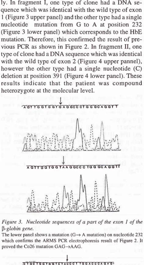
and2respective-Med J Indones
ly. In
fragment
I,
one
type
of
clone
had a
DNA
se-quencewhich
wasidentical
with the wild
type of
exonI
(Figure
3 upper panel) and the other type had asingle
nucleotide
mutation
from G
to
A
at
position 232
(Figure
3lower panel)
which corresponds
to
the HbEmutation. Therefore,
this confirmed
the result
of
pre-vious
PCR
asshown
in Figure
2.
In
fragment
II,
one typeof
clone had aDNA sequence
which
wasidentical
with the wild
type
of
exon
2(Figure 4 upper pannel),
however the other type had a single nucleotide
(C)
deletion
at position 391(Figure 4 lower panel).
Theseresults indicate
that the patient was
compound
heterozygote
at themolecular level.
ô-ÂE-GTT
I-ô-TTô-6T-0 C T
^ A q OCC C rs O GC A O O r I
Figure 3. Nucleotide sequences of a part of the exon I of the
$-globin gene.
The lower panel shows a mutation (G-+ A mutation) on nucleotide 232
which confirms the ARMS PCR electrophoresis result of Figure 2. It
proved the Co26 mutation GAG-+AAG.
Figure
4.
Nucleotide sequences of a part of the exon 2 of the p-globin gene.The lowerpanel shows the deletion C at the nucleotide 391. The Co35
(TAC) remains uncharged but a frameshift mutation in the subsequence
coding region will result.
I
Vol 6, No 2,
April
- June 1997DISCUSSION
In
dealing
with
single mutation
screening,
misincor-poration of nucleotides by the
TaqDNA
polymerase isa problem to
consider.
According
to Foecharoen et al7the
misincorporation
ratelnucleotide/cycle
in
the caseof
a239-bp product
in
30 cyclesof
PCRamplification
was estimated to be 2x
l}-a.In
thisreport,
at least threeclones
were shown
to
have the
samemutation,
andthereby the
possibility
of
themisincorporation
of
theTaq
polymerase
wasvirtually
eliminated.
We
detected
asingle p-globin
genemutation
from
G
to
A
at theposition 232 which
resultedin glutamic acid
being substituted
by
lysine
at codon 26.This mutation
not only
introduces
adifferent
amino acid
(glu+lys)
but
also
changes
the splicing pattern
of
the mRNA
because the
codon
24-27
are actually a rather goodfit
for
the
consensusdonor
signal
for splicing,
therefore
the mutation
activates
the "cryptic" splice site.
The resultant40%
abnormalmRNA
can notgive any
detec-tablep-globin protein, because
atotal
of
16nucleotides
are removed
from
thisexon.l'7 Furthermore,
instability
of
themRNA in
theerytroid
cell
and easy degradationin
thecytoplasmic celis'g
will
also reducethe
amountof
functional
mRNA,
therefore
synthesis
of
normal
B-globin is severely impaired.
Lie-Injo
et al2
reported thatfrom
36 Indonesian thalas-semia casesfrom
Jakarta, a HbE gene wasfound
in
13chromosomes. Furthermore,
from the
analysis
of
72 chromosomes (36 patients) they also described the mostprevalent mutations
as
IVS-l
nt5
(G-+C)
(32
chromosomes),
followed
by
IVS-2 nt5
(G+C)
(7
chromosomes),
IVS-1
ntl
(G-+T)
(6
chromosomes),codon
l5 (TGG-+TAG)
(4 chromosomes) and others (7chromosomes).
They described
I
chromosomehaving
deletion
C
atcodon 35
without
any
information
aboutthe paired
chromosomes.
In
the
sameyear the
samemutation was also
published in theMalayethnic
group.3In
this report, we
also
clarified
asingle deletion
C
atnucleotide
39 1.This
deletion introduces
astranslation
reading frameshift providing a different amino
acid
sequence
after the 35th
theamino acid
residue,before
a premature nonsensetermination
codon
atcodon
60(GTC
-+
TGA).
Therefore people
with
thecombina-tion
ofmutation at
codon 26 (GAG-+ AAG)
and codon35 (del C)
will
suffer from
severeimpairment
of
B-globin production
resulting
in
Bo-thalassemia.In
individual
with
a
mutation
at
codon
26
(GAG-+AAG)
and
35 (del C),
the
impairment
of
p-globin
gene expressionmight
activate the y
andp-tan-CompoundHeteroqgote
95dem
globin
gene, introducing the
HbF
and HbAz
production. The
ô
chain
isnormally
combined
with
anon ô chain designated as
lchain
resulting in
increasedHbF.
In
the erythrocyte this minor
component
com-poses about 2.57o (0.6-6Vo)
of
thetotal
hemoglobin.
Compared
with
a similar case describedby Yang et al3
the
HbF level
of
our case (5.2Vo)
is far
lower
thanthat
of
the
Malay
case(48.77o). Subsequently,
increasedamount
of HbAz
was demonstratedto
compensatefor
the
low
production
of HbA.
Our case
was a compound heterozygotefor
HbE
and amutation at
codon
35(del C) of
theglobin
gene.Using
the
cellulose
acetate membranediffusion
methods onecannot differentiate
theHbAz from
HbE,
becausethis
HbA2 comigrated
with HbE,
eventhough
from
Table
1 we
know
thateither HbE
and/orHbAz
level
maynot
be
abnormaly
increased.
Our
case
showed
a
lower
percentage
of HbE
andHbAz
(I2.8Vo) compared
with
those
of the
Malay
case(46.9Vo).However, this
resultsdid not affect the clinical
diagnosis.
Also,
the HbE
levels can
not identify
the genotype
(heterozygote,
homozygote
or
heterocompounds)
since there
is
noclear cut
off
between
the three
catagories.
Only
by
DNA
analysis one canclarify
the
genotype.In
Malay
patients
the most prevalent
B-thalassemiasare mutations
at
fVS-l
nt5
(G+C),
codons
4l-42
(-TCCT),
IVS-I
ntl
(G
+
T), IVS-2 nt654 (C
+
T)
exhibited by Yang
et
al (1988)' on
20,6,4,3
and,3chromosomes respectively.
This
data shows
close
similarity
with
those
by
Lie-Injo et
al
(1989)'and with
themu
cribed
in
theIndian
subcontinent.6
There
is
ant
similarity
with
those observed
in
the Chinese
community
where
thecommon mutations are
atpromoter site
e.g.-29(A-+G)
and -28
(A-+
G),followed
bycodon 17 (A
+
T),IVS-
1ntl
(G
+
T)
andIVS-I
nr5
(G
+
C).Lie-Injo et
al2
collected their
samples
from the
sur-rounding
areaof
the Jakarta
metropolitan
city
while
our case comes
from
an isolatedfamily
in
a remote area30
km from Yogyakarta, 600 km from
Jakarta. The
HbE
gene
in
our
case
possibly originated from
Thailand
where
the HbE carrier is prominent
with
afrequency
of
nearly
5o-60%o.toIt
is
possibly
true that
3000 years ago there were
tribal
movements from
north SEA
to the
south.We can
assumethat
theHbE
mutation
is
inherited
from
the Thai, other
B-thalas-semia
mutations
from
Asian-Indians,
while
thecodon
35 (delC)
is
a specificmutation
amongthe
96
SuryantoroMuslim-Thaill
since there
is no correlation
betweenreligion
andDNA
mutation.
lrÂ
[image:6.595.56.288.89.277.2]eaa f,
Figure 5. Geographic anthropology of the HbE and the $-thalns-semia gene disease and the spread in the South East Asia (SEA).
HbE carrier rate is high in the North Eastem ofThailand and spread to
the southem part of SEA nearly 3000 years ago. Other p-thalassemia
mutations have spread from India to SEA including Indonesia nearly
5000-6000 years ago. Some new mutations may be spesific to the
Malay-Indonesian ethnic group.
REFERENCES
l.
Boon WH. Thalassemia as community health problem in Souht East Asia.In:
SoemantriA,
Prabowo AP, editors.Proceedings
of
Hematologic Meeting; 1983 Sept 22-24; Yogyakarta. Semarang:Petra Satya, 1986.2. Lie Injo LE, Cai SP, Wahidiyat
I,
Muslichan S,Lim
ML,Evangelista L, e al. The p-thalassemia mutation in Indonesia
Med J Indones
and
their
linkage
to p
haplotype.
Am J
Hum
Genet 1989;45:971-5.3. Yang KG, Kutlar F, George E, Wilson JB, Kutlar A, Stoming TA, et al. Molecular characterization of p globin gene muta-tions in Malay patients with Hb E-p-thalassemia and
thalas-semia major. Br
J
Hematol 1989;72:73-80.4. Weatherall DJ, Clegg JB. Thalassemia intermedia.
In
:Weatherral
DJ, CIegg
JB,
editors. The
Thalassemiasyndromes. 3ro
ed. Oxford: Blackwell Scientific
Publ 198l;645-82.5. Newton CR, Graham A, Heptinstall LE, Powell SJ, Summers
C, Kalshekar
N,
et al. Analysisin
any point mutation inDNA.
The amplification refractory mutation
system (ARMS). Nucleic Acids Research 1989;17:2505-16. 6. Varawalla N, Old J, Sarkar R, Venkatesan R, Weatherall D.The spectrum of p-thalassemia mutation of the Indian
sub-continents : the basis for prenatal diagnosis. Br J Haematol 1991;78:242-7.
7. Fucharoen S, Fucharoen G, Fucharoen P, Fukumaki
Y. A
novel ochre mutation in the B-thalassemia gene of a Thai. JBiol Chem 1989 :264:'1 7 80-3.
8. GelehrterTD, Collins FS. Principle of Medical Genetics.
lst
ed. Baltimore: William
&
Wilkins Publ, 1990.9. Traeger J, Winichagon P, Wood WG. Instability
of
pE-mesenger
RNA during erythroid cell
maturation
in Hemoglobin E homozygote. J Clin Invest 1982;69:1050-5. 10. Fucharoen S, Winichagoon P. Thalassemiain
South EastAsia : Problem and strategy for prevention control. South
East Asia J Trop Med Publ Health 1992;23:64'7-55.
11. Laosombat
V,
Fucharoen SP, Panich-V, Fucharoen G, WongchanchailertM,
SriroongruengW,
et al. Molecular basisof
p-thalassemiain the
Southof
Thailand.Am
J