Clinical-pathologic factors, as predictor of lymph nodes metastasis in cervical
cancer stage IB and IIA
M.Farid Aziz*, Andrijono*, Laila Nuranna*, Sigit Purbadi*, Rukmini TjiptoMangunkusumo†, Budiningsih Siregar†, Santoso Cornain†, Abdul Bari Saifuddin*, Achmad Tjarta†, Bambang Sutrisna§
Abstrak
Tujuan penelitian ini adalah untuk mengidentifikasi faktor prediktor metastasis kelenjar getah bening (KGB) pada pasien dengan kanker serviks stadium IB dan IIA. Penelitian dilakukan dari bulan Mei 1996 sampai bulan Desember 2001. Ada 183 pasien kanker serviks dengan stadium menurut FIGO IB dan IIA menjalani operasi histerektomi radikal dan limfadenektomi. Dari pasien tersebut 158 pasien yang dapat dinilai, terdiri dari 43 pasien dengan metastasis KGB dan 115 tanpa metastasis KGB. Rancangan penelitian adalah kasus-kontrol. Kasus adalah pasien dengan metastasis KGB dan kontrol pasien tanpa metastasis KGB. Analisis multivariat dilakukan setelah analisis bivariat. Pada analisis bivariat umur < 39 tahun, diameter lesi >4 cm, stadium IIA > 4 cm, histopatologi dengan diferensiasi sedang dan buruk, invasi ke pembuluh darah dan limfa merupakan variabel yang independen terjadinya metastasis KGB dengan nilai p ≤ 0,05. Tetapi pada analisis multivariat yang muncul sebagai variabel independen adalah umur muda, paritas > 4, diameter lesi, histopatologi adenoskuamosa, dan invasi limfo-vaskular dengan nilai p ≤ 0,05. Kesimpulan: Usia muda, paritas > 4, stadium IIA > 4 cm, diameter lesi, histopatologi adenoskuamosa, invasi limfa-vaskular merupakan faktor risiko terjadinya metastasis dan dapat dipergunakan sebagai faktor prediktor metastasis KGB. (Med J Indones 2004; 13: 113-8)
Abstract
The aim of this study was to identify possible predictor factors of lymph node metastases in patients with cervical cancer stage IB and IIA. Study was conducted between May 1996 and December 2001. There were 183 patients of cervical cancer with FIGO Stage IB and IIA who were underwent radical hysterectomy and lymphadenectomy. From those 158 patients could be evaluated, consisting 43 patients with node metastases 115 patients without metastases. Research design was case control study. Case was patients with node metastases and control was those without node metastases. Multivariate analysis was made after bivariate analysis. On bivariate analysis age < 39 years, diameter of lesion > 4 cm, stage IIA > 4 cm, histopathology moderate and poor differentiation, blood and lymphatic vessel invasion were independent variables for node metastases with p value ≤ 0.05. However, on multivariate analysis younger age, parity ≥ 4, diameter of lesion, histopathology adenosquamous, and lymph vascular invasion (+) as independent factors for node metastases with p value ≤ 0.05. Conclusion: Younger age, parity ≥ 4, stage IIA > 4 cm, diameter of lesion, histopathology adenosquamous, and lymph vascular invasion (+) were risk factors for node metastases and can be used as predictors. (Med J Indones 2004; 13: 113-8)
Keywords: cervical cancer, radical hysterectomy, node metastases, case control study, predictor
The presence of lymph node metastasis is considered as the most important prognostic factor for cervical cancer stage IB and IIA.1,2Of 100 stage IB patients, 15 are likely to be node-positive and 45% of these patients would be cured. The majority of patients is
node-negative (85%), and of these 90% is cured.3Five year disease free interval decrease significantly from 95% with negative patients to 76% with node-positive (p<0.002). Therefore, the presence of positivity defines the high-risk group and nodal-negativity the low-risk group.
Detection node metastasis with CT-scan, MRI have high false negative especially for nodes seizing less than 2 cm,4 in fact 80% node metastases were less than 10 cm.5
Although many authors have reported risk factors to node metastasis, this study try to identify risk factors
*
Department of Obstetrics and Gynecology, Faculty of Medicine, University of Indonesia, Jakarta, Indonesia † Departmentof Pathological Anatomy, Faculty of Medicine,
University of Indonesia, Jakarta, Indonesia
§
to node metastasis and then make a formula that can predict probability of node metastasis.
METHODS
This study was conducted at Dr. Cipto Mangunkusumo Hospital Jakarta from May 1996 until December 2001.There were 183 patients with cervical cancer stage IB and IIA who fulfill inclusion criteria under-went radical hysterectomy and lymphadenectomy. Of those only 158 cases could be evaluated due to incompleteness of surgical procedure, lost of specimens, down stage, paraffin block damage. Stages were determined by physical, gynecological examination, chest x-rays, IVP, cystoscopy, rectoscopy. Ultra-sonography, CT-scan or MRI was optional. Peripheral blood test, blood chemistry and ECG was done prior surgery. The surgery was hysterectomy with or without removing one or both ovary and lympha-denectomy. All the specimens were processed and examined at the Department of Pathological Anatomy University of Indonesia.
Study design was a case-control study, patient with node metastasis as a case and who without node metastasis as a control. All clinical and pathological features were evaluated against node metastasis. A model of multivariate was made after bivariate analysis, and then develops a model to predict probability of node metastasis. Statistical program used was Stata Version 6 up-grade.
RESULTS
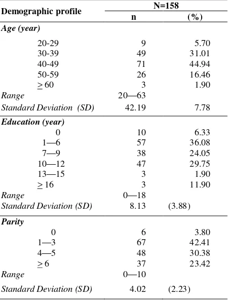
There were 43 patients of 158 with node metastasis. The mean age of the study population was 42.19 years with SD ±7.78 years, ranging between 20-63 years. The education range was 0-18 years, mean education 8.13 years with SD ± 3.88 years. The parity range was 0-10, mean parity 4.02 with SD ± 2.23 (Table 1).The diameter of the lesion ranging 10-70 mm, mean 33.84 mm with SD ± 13.37 mm. Most of the patients were
stage IB ≤ 4 cm (50.63%) followed by IB> 4 cm
(21.52%) IIA ≤ 4 cm and IIA > 4 cm (Table 2).
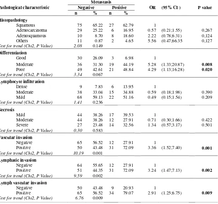
Bivariate analysis identified risk factors to node metastasis age < 39 years (p = 0.013) (Table 3), diameter of the lesion > 4 cm (p = 0.005), stage IIA
> 4 cm (p = 0.000) (Table 4), moderate (p = 0.008) and poor differentiation (p = 0.020), vascular invasion (p = 0.001), lymphatic invasion (p = 0.002), lymph-vascular invasion (p = 0.009) (Table 5). Multivariate analysis revealed that age (p = 0.024), parity ≥ 4 (p = found after radical hysterectomy should have adjuvant radiation, chemotherapy or combination of both. However radiation and surgery as a primary treatment yield the same result as shown by Landoni et al7 in their randomized study. Complication will increase if radiation or chemo radiation administered post radical surgery.8,9 If lymph node metastasis can be predicted prior surgery the treatment can be well planned and the complication can be avoided. The patient can choose that is suite for her condition. Currently morphological pathology used to predict lymph node metastasis.10 Sevin et al in univariate analysis found deep of invasion, diameter of lesion, lymph-vascular infiltration, volume of the tumor were significant predictor lymph node metastasis. Benedetti-Panici et al11 revealed pelvic lymph node metastasis 21% the lesion < 4 cm and 23% if the lesion > 4 cm.
Our study in multivariate analysis revealed younger
age, parity ≥ 4, diameter of lesion, histopathology
adenosquamous, and lymph vascular invasion as independent factors for lymph node metastasis with p
value ≤ 0.05. Based on this multivariate model and equation we can predict the probability lymph node metastasis (Form 1).
Form 1 showing the way to predict probability lymph
node metastasis. Conclusion: Younger age, parity ≥ 4,
Table 1. Distribution of patients with cervical cancer stage IB and IIA who underwent radical hysterectomy by dempgraphic profile
Demographic profile N=158
n (%)
Age (year)
20-29 9 5.70
30-39 49 31.01
40-49 71 44.94
50-59 26 16.46
> 60 3 1.90
Range 20—63
Standard Deviation (SD) 42.19 7.78
Education (year)
0 10 6.33 1—6 57 36.08
7—9 38 24.05
10—12 47 29.75
13—15 3 1.90
> 16 3 11.90
Range 0—18
Standard Deviation (SD) 8.13 (3.88)
Parity
0 6 3.80
1—3 67 42.41
4—5 48 30.38
> 6 37 23.42
Range 0—10
Standard Deviation (SD) 4.02 (2.23)
Table 2. Distribution of patients with cervical cancer stage IB and IIA who underwent radical hysterectomy by clinical characteristic
Clinical characteristic N=158
n (%)
Diameter of lesion
10—20 mm 35 22.15
21—30 mm 56 35.44
31—40 mm 35 22.15
41—50 mm 19 12.03
51—60 mm 9 5.70
> 61 mm 4 2.53
Range 10--70
Standard Deviation (SD) 33.84 (13.37)
Stage
Stage IB < 4 cm 80 50.63 Stage IB > 4 cm 34 21.52 Stage IIA < 4 cm 34 21.52 Stage IIA > 4 cm 10 6.33
Table 3. Odds ratio (OR) node metastasis of patients with cervical cancer stage IB and IIA by demographic profile (bivariate analysis)
Demographic profile
Metastasis
OR (95% CI ) P Value Negative Positive
n % n %
Age(year)
< 39 32 27.83 21 48.84 1
> 39 83 27.17 22 51.11 0.40 (0.19;0.84) 0.013
Test for trend (Chi2, P Value) 6.16 0.013
Education (year)
< 9 42 36.52 11 25.58 1
> 9 73 63.48 32 74.42 1.67 (0.76;3.69) 0.196 Test for trend (Chi2, P Value) 1.67 0.196
Parity
< 4 76 66.09 23 53.49 1
Table 4. Odds ratio (OR) node metastasis of patients with cervical cancer stage IB and IIA by clinical characteristic (bivariate analysis)
Clinical characteristic
Metastasis
OR (95% CI ) P value Negative Positive
n % n %
Diameter of lesion
< 40 mm 98 85.22 28 65.12 1
> 40 mm 17 14.78 15 34.88 3.09 (1.34;7.12) 0.005
Test for trend (Chi2, P Value) 7.78 0.005
Stage
Stage Ib < 4 cm 64 55.64 16 37.21 1
Stage Ib > 4 cm 22 19.33 12 27.91 2.18 (0.88;5.41) 0.840 Stage IIa < 4 cm 27 23.48 7 16.28 1.04 (0.38;2.82) 0.943 Stage IIa > 4 cm 2 1.74 8 18.60 16.0 (2.57;99.45) 0.000
Test for trend (Chi2, P Value) 6.64 0.010
Table 5 Odds ratio (OR) node metastasis of patients with cervical cancer stage IB and IIA by pathological characteristic
Pathological characteristic
Metastasis
OR (95% CI ) P value Negative Positive
n % n %
Histopathology
Squamous 75 65.22 27 62.79 1
Adenocarcinoma 29 25.22 6 16.95 0.57 (0.21;1.55) 0,267 Adenosquamous 10 8.70 8 18.60 2.22 (0.78;6.31) 0,124 Others 11 0.87 2 4.65 5.56 (0.47;66.35 0,127 Test for trend (Chi2, P Value) 2.08 0.149
Differentiation
Good 30 26.09 3 6.98 1
Moderate 36 31.30 19 44.19 5.28 (1.33;20.87) 0.008
Poor 49 42.61 21 48.84 4.29 (1.13;16.28) 0.020
Test for trend (Chi2, P Value) 3.34 0.067
Lymphocyte infiltration
Dense 9 7.83 6 13.95 1
Moderate 38 33.04 15 34.88 0.59 (0.18;1.98) 0.390 Mild 68 59.13 22 51.16 0.49 (0.15;1.54) 0.209 Test for trend (Chi2, P Value) 1.41 0.236
Necrosis
Mild 44 38.26 17 39.53 1
Moderate 44 38.26 12 27.91 0.71 (0.30;1.66) 0.422 Severe 27 23.48 14 32.56 1.34 (0.57;3.17) 0.501 Test for trend (Chi2, P Value) 0.30 0.583
Vascular invasion
Negative 65 56.52 12 27.91 1
Positive 50 43.48 31 72.09 3.36 (1.52;7.40) 0.001
Test for trend (Chi2, P Value) 10.19 0.001
Lymphatic invasion
Negative 64 55.65 12 27.91 1
Positive 51 44.35 31 72.09 3.24 (1.47;7.13) 0.002
Test for trend (Chi2, P Value) 9.59 0.002
Lymph vascular invasion
Negative 50 43.48 9 20.93 1
Positive 65 56.52 34 79.07 2.91 (1.25;6.75) 0.009
Table 6 Multivariate analysis Odds ratio (OR) node metastasis of patients with cervical cancer stage IB and IIA
Variable Coef. OR 95% CI P value
Lymph vascular (positive) 1.125 3.08 1.16 ; 8.18 0.024 Diameter of lesion ( mm) 0.080 1.08 1.04 ; 1.13 0.000
Age (year) -0.087 0.92 0.86 ; 0.98 0.012 Parity (> 4) 1.851 6.37 2.10 ; 19.26 0.001 Adenocarcinoma 0.315 1.37 0.43 ; 4.37 0.594
Adenosquamous 1.726 5.62 1.50 ; 20.99 0.010
Others 2.625 13.80 0.80 ; 236.78 0.070
Constanta -2.124
Form 1 Probability node metastasis in patient with cervical cancer stage IB and IIA
Variable
Individual
characteristic Index Score)
(IC) (I) (IC x I)
Lymph-vascular invasion Negative Positive (Negative=0; positive=1)
0 1,125
Diameter of lesion ( mm) 0,080
Age (year) -0,087
Histopathology Squamous Adenocarcinoma Adenosquamous Others
(squamous=0;adenoca=0;ad enosq=1;others=1)
0 0,315 1,726 2,625
Constanta -2,124
Total score
score
total
e
metastasis
node
1
1
Pr
( )CONCLUSION
On bivariate analysis of patients with cervical cancer stage IB and IIA who underwent radical hysterectomy, age < 39 years, diameter of the lesion > 4 cm, stage II > 4 cm, histopathology moderate and poor differentiation,
blood and lymphatic vessel invasion were independent variables for lymph node metastasis with p value ≤ 0.05.
However, on multivariate analysis younger age, parity
and lymph vascular invasion (+) as independent factors
for node metastases with p value ≤ 0.05. Model to predict the probability of lymph node metastasis can be made.
Acknowledgement
The authors would like to thank and highly appreciate to the trainees in gynecological oncology and staff of secretariat Division of Oncology Department of Obstetrics and Gynecology, University of Indonesia who have given devotion and care to the patients.
REFERENCES
1. Kamura T, Tsukamoto N, Tsunechi N, Saito T, Matsuyama T, Akazawa, Nakano H. Multivariate analysis of the histopathologic prognostic factors of cervical cancer in patients undergoing radical hysterectomy. Cancer, 1992, 69: 181-6
2. Sartori E, Fallo L, La Face B, Bianchi UA, Pecorelli S. Extended radical hysterectomy in early-stage carcinoma of the uterine cervix : failaring the radiology. Int J Gynecol Cancer 1995, 5: 143-7.
3. Thomas GM, Dembo AJ. is there a role for adjuvant pelvic radiation therapy after radical hysterectomy in early stage cervical cancer ? Int J Gynecol Oncol 1991, 1: 1-8 4. Kristensen GB, Kaern J, Abeler VM, Hagmar B, Trope
CG, Pettersen EO. No prognostic impact of flow-cytometric mesured DNA ploidy and s-phase fraction in
cancer of the uterine cervix: a prospective study of 465 patients. Gynecol Oncol, 1995,57:79-85.
5. Benedetti-Panici P, Maneschi F, Scambia G, Greggi S, Cutillo G, D’Andrea G, Rabitti C, Coronetta F, Capelli A, dan Mancuso S. Lymphatic spread of cervical cancer: an anatomical and pathological study based on 225 radical hysterectomies with systematic pelvic and aortic lynphadenectomy. Gynecol Oncol 1996, 62:19-24. 6. FIGO Annual report, 1998
7. Landoni F, Maneo A, Colombo A, Placa F, Milani R, Perego P, Favini G, Ferri L, Mangioni C. Randomized study of radical surgery versus radiotherapy for stage IB-IIA cervical cancer. Lancet 1997, 350, 535-40.
8. Zola P, Maggino T, Sacco M, Rumore A, Sinistrero G, Maggi R, Landoni F, Foglia G, Sartori E, De Toffoli J, Franchi M, Romagnolo C, Sismondi P. Prospective multicentre study on urologic complications after radicalsurgery with or without radiotherapy in the treatment of stage IB-IIA cervical cancer. Int J Gynecol Cancer 2000; 10: 59-66.
9. Choy D, Wong LC, Sham J, Ngan HYS, Ma HK. Dose-tumor response for carcinoma of cervix: an anlysis of 594 patients treated by radiotherapy. Gynecol Oncol 1993, 49: 311-7
10. Sevin BU, Nadji M, Lampe B, Lu Y, Hilsenbeck S, Koechli OR, Averrete HE. Prognostic factors of early stage cervical cancer treated by radical hysterectomy. Cancer 1995, 76:1978-86.