UNIVERSITI TEKNIKAL MALAYSIA MELAKA
This report submitted in accordance with requirement of the Universiti Teknikal Malaysia Melaka (UTeM) for the Bachelor Degree of Manufacturing Engineering
(Engineering Materials) (Hons.)
by
MOHD SAFUAN BIN ANUAR B050910116
870306-03-5193
FACULTY OF MANUFACTURING ENGINEERING 2012
DEVELOPMENT OF HYDROXYAPATITE/CHITOSAN
COMPOSITE POWDER VIA MECHANICAL MILLING
29 JUN 2012 29 JUN 2012
UNIVERSITI TEKNIKAL MALAYSIA MELAKA
BORANG PENGESAHAN STATUS LAPORAN PROJEK SARJANA MUDA
TAJUK: DEVELOPMENT OF HYDROXYAPATITE/CHITOSAN COMPOSITE POWDER
VIA MECHANICAL MILLING METHOD
SESI PENGAJIAN: 2011/12 Semester 2
Saya MOHD SAFUAN BIN ANUAR
mengaku membenarkan Laporan PSM ini disimpan di Perpustakaan Universiti Teknikal Malaysia Melaka (UTeM) dengan syarat-syarat kegunaan seperti berikut:
1. Laporan PSM adalah hak milik Universiti Teknikal Malaysia Melaka dan penulis. 2. Perpustakaan Universiti Teknikal Malaysia Melaka dibenarkan membuat salinan
untuk tujuan pengajian sahaja dengan izin penulis.
I hereby, declared this report entitled “Development of Hydroxyapatite/Chitosan Composite Powder via Mechanical Milling Method” is the results of my own
research except as cited in references.
Signature : ……….
Author’s Name : MOHD SAFUAN BIN ANUAR Date : 29TH JUN 2012
ABSTRAK
ABSTRACT
DEDICATION
Dedication to my beloved parents, family,
ACKNOWLEDGEMENT
First of all, in a humble way I wish to give all the Praise to Allah, the Almighty God for with His mercy has given me the strength and time to complete this report.
I am deeply indebted to Miss Adibah Haneem Binti Mohamad Dom, my supervisor, for her patience, supervision, encouragement and thoughtful guidance towards the completion of this report.
I am particularly grateful to Department of Material Engineering, Faculty of Manufacturing Engineering, Universiti Teknikal Malaysia Melaka (UTeM) for financial support.
TABLE OF CONTENT
2.1 Introduction of Hydroxyapatite 6
2.1.1 Hydroxyapatite Properties and Applications 7
2.1.2 Crystal Structure of Hydroxyapatite 9
2.2 Chitosan 11
2.3 Chitosan / Hydroxyapatite Composite 14
2.4 Biomaterials and Their Application 15
2.5 Bone 24
2.5.1 Structure and Composition of the Bone 25
2.6 Method of Preparation Composite 30
2.6.1 Mechanical milling 30
2.6.1.1 Planetary ball mills 30
3. METHODOLOGY 33
3.1 Experimental Procedures 33
3.2 Materials Preparation 35
3.2.1 Materials 35
3.3 Fabrication of HA/CS Composite 36
3.3.1 Powder Mixing 36
3.3.2 Sieve shaker 38
3.3.3 Forming 38
3.3.4 Sintering 39
3.4 Sample Preparation 40
3.5 Materials Characterization 41
3.5.1 X-ray Diffraction (XRD) 41
3.6 Mechanical Test 42
3.6.1 Vickers Micro Hardness Testing 42
3.6.2 Density Testing 43
3.6.3 Compression testing 44
4. RESULT AND DISCUSSION 46
4.1 Observation of Sample Preparation 46
4.2 Characterization Of composite powder 49
4.2.1 X-ray Diffraction Analysis 49
4.3 Mechanical Testing 51
4.3.1 Vickers Hardness Test (HV) 51
4.3.2 Density Testing 53
5. CONCLUSION AND RECOMMENDATIONS 58
5.1 Conclusion 58
5.2 Recommendations 60
REFERENCES
LIST OF TABLES
Table 2.1 Physical Properties of Calcium Phosphate 9 Table2.2 Examples of Substitutions in the Apatitic Structure 11 Table 2.3 Mechanical Properties of Some Metallic and Ceramic Materials 17
Table 2.4 Types and Properties of Biomaterials 17
Table 2.5 Composition of Bone 27
Table 2.6 Mechanical Properties of Human Compact Bone 29
Table 4.1 Diameter Samples Before and After Sintering 48 For Each wt. % of Chitosan
Table 4.2 Particles Size of HA/CS Composite 51
Table 4.3 Average Microhardness of HA/CS Composite 52 Before and After Sintering
Table 4.4 Result of Density Test of HA/CS with 53
Different CS Concentration
LIST OF FIGURES
Figure 2.1 Hydroxyapatite Structure Projected Down the 10 C Axis onto the Basal Plane
Figure 2.2 Structures of Chitin and Chitosan 12
Figure 2.3 Biomaterial Application of Chitosan 14
Figure 2.4 Classification of Polymer Composite Biomaterials 20 Figure 2.5 Range of Mechanical Properties of Some Biomaterials, 21
Cancellous and Trabecular bone
Figure 2.6 Various Applications of Different Polymer Composites 23
Figure 2.7 Organisation of Bone 26
Figure 2.8 Stress as a Function of Strain and Strain rate 28 For Human Compact Bone
Figure 2.9 (a) Fritsch Pulverisette P-5 four station ball mill 32 (b) Schematic depicting the ball motion inside the ball mill.
Figure 3.1 Process Flow Chart 34
Figure 3.2 (a) HA and (b) CS 35
Figure 3.3 Planatery Ball Milling 36
Figure 3.4 (a) HA-5 w.t% CS (b) HA-10 w.t% CS and (c) HA-20 w.t% CS 37
Figure 3.10 XRD Panalytical Xpert Pro MPD Pw 3040/60 42
Figure 3.11 HM-20 Series Micro-hardness Vickers 43
Figure 3.12 Densitimeter 44
Figure 4.1 Samples of CompoSite Powder Pellets Produced from 47 HA Powders with Different Ration of CS Addition
(a) HA-5 wt. %CS; (b) HA-10wt. %.CS ;( c) HA-20wt. %CS.
Figure 4.2 Pellets After Sintering Temperature 48 Figure 4.3 XRD Pattern of HA/CS composites (a) Before 50
(b) After Sintering Process
LIST OF ABBREVIATIONS
CHAPTER 1
INTRODUCTION
1.1
BackgroundThere is a necessity for replacing bone substance which has been lost due to traumatic or nontraumatic events (Suchanek et al., 1998). The implanted biomaterial
must have certain desired properties in order to achieve a satisfactory result and to have an appropriate host response at the hard tissue implantation site. The microstructural and mechanical properties of the bone must be thoroughly understood for the successful preparation of candidate hard tissue implant biomaterials in order to mimic the natural bone structure.
Biomaterials must be compatible with body in order to exhibit their function properly. The use of incompatible materials as medical implants in the body may induce unfavourable immune reactions, undesirable interactions with blood and other body fluids as well as damaging the genetic material at the chromosomal and DNA levels.
Bone consists of 69 wt. % calcium phosphate (mainly hydroxyapatite (HA)), 21% collagen, 9%water and 1% other constituents. It has a composite nature which is composed of mainly ceramic and polymeric components with a complex hierarchical microstructure difficult to imitate which gives most of the superior mechanical properties to bone. Extensive research has been conducted on bone substitute composite materials composed of mainly hydroxyapatite and a polymer (chitosan).
Hydroxyapatite (Ca 10 (PO 4) 6 (OH) 2) has excellent bioactivity, biocompatibility, non-toxicity and osteoconductivity properties but also has low toughness. Chitosan (CS), deacetylated form of chitin, is a natural polymer found in vast amounts in crustaceans. It is biocompatible and bioresorbable/biodegradable. It is non-toxic and easily soluble in dilute weak organic acids. The recent research on chitosan/hydroxyapatite composites which are partially biodegradable indicate that this behaviour of the composite may even be an advantage. New bone may intergrowth around the hydroxyapatite particles when the polymer matrix is resorbed. Extensive research on relatively new chitosan/hydroxyapatite composites has been conducted recently (Zhao et al., 2007).
1.2 Problem Statement
Metallic implants are widely used in many treatments and are fairly successful. However, they do not provide the optimum therapy due to their short comings such as stress shielding during post-healing, chronic inflammation caused by corrosion, and fatigue and loosening of the implant. As a result, a second surgery is often required to remove the metallic implant after healing, and it increases the risk of the operation and the expense to the patient. Degradable polymeric implants eliminate the need for a second operation and can prevent some of the problems associated with stress shielding during post-healing, and can also be used simultaneously to deliver therapeutic drugs to treat infections or growth factors to accelerate new bone growth. Chitosan (CS), was suggested as an alternative polymer for use in orthopedic applications to provide temporary mechanical support the regeneration of bone cell ingrowth due to its good biocompatible ( Vande et al., 2002), non-toxic,
biodegradable, and inherent wound healing characteristics (Lee et al., 2002).
According to Sabokbar et al., (2001) HA was used in various biomedical fields such
as dental material, bone substitute and hard tissue paste. HA can accelerate the formation of bone-like apatite on the surface of implant. CS can be utilized in combination with other bioactive inorganic ceramics, especially HA to further enhance tissue regenerative efficacy and osteoconductivity. Incorporation of HA with CS, the mineral component of bone, could improve the bioactivity and the bone bonding ability of the HA/CS composites (Wang et al., 2002). The aim of this work
1.3 Objective of Research
The main objectives of this research are:
i. To prepare composite biomaterials by using chitosan and hydroxyapatite powder through mechanical milling.
ii. To characterize and evaluate the physical and mechanical properties of the produced composite powder.
1.4 Scope of Research
In this study, HA (Merck, US) and CS (Sigma Aldrich, UK) powder were used in preparation of HA/CS composite. This research is focused on the effect of enhancement various component of chitosan on HA/CS composite. There are 3 samples of HA/CS were produced and the concentration of CS powder added to the HA powder are 5, 10 and 20 wt. %. HA and chitosan powder will be blended using ball milled machine in order to produce homogenous HA/CS composites powder. The HA powder is mechanically mixed with the chitosan powders for 5 hours in a planetary mill using Al2O3 balls and a jar, resulting in the preparation of the homogenous HA/CS powder mixture. Thereafter, the mixture of powder were sieved using sieve shaker 40 µm to obtain smaller HA/CS particles size. In fabricating process, for powder compaction, die pressing method is used with pressures of 3.5 tons to form HA/CS pellet with the diameter size of 13 mm and thickness is 6 mm. The density of the HA/CS pellet produced from die pressing is measured before undergo sintering process. Then samples were sintered in electrical furnace. The samples were sintered at 1000ᵒC heating temperature with heating rates of 5°Cminˉ ¹ and soaking time 60 minutes.
CHAPTER 2
LITERATURE REVIEW
This chapter discussed on structure composite material. The contents are focusing on introduction, structure, application and advantage from the study.
2.1 Introduction of Hydroxyapatite
According to Soley (2012), Hydroxyapatite is a mineral. It is a naturally occurring form of calcium apatite with the formula Ca5 (PO4)3(OH), but is usually written Ca10 (PO4)6(OH)2 to denote that the crystal unit cell comprises two entities. Hydroxyapatite is the hydroxyl endmember of the complex apatite group. The OH- ion can be replaced by fluoride, chloride or carbonate. It crystallizes in the hexagonal crystal system. It has a specific gravity of 3.08 and is 5 on the Mohs hardness scale. Pure hydroxylapatite powder is white. Naturally occurring apatites can however also have brown, yellow or green colorations, comparable to the discolorations of dental fluorosis.
Hydroxyapatite is the major constituent of the inorganic component of bone. Although similar to natural bone in many respects, synthetic HA vary from the natural bone in that they tend to contain larger and more uniform crystals with a more homogenous composition than those found in natural bone. Due to this more crystalline and organized structure and since hydroxyapatite is resorbed by foreign body giant cells, which stop ingesting once 2 to 10µm of hydroxyapatite has been consumed, synthetic HA tend to resorbd extremely slowly in the body, often taking many decades to resorb fully (Kurtz et al., 2006).
Hydroxyapatite can be found in teeth and bones within the human body. Thus, it is commonly used as a filler to replace amputated bone or as a coating to promote bone ingrowth into prosthetic implants. Although many other phases exist with similar or even identical chemical makeup, the body responds much differently to them. Coral skeletons can be transformed into hydroxyapatite by high temperatures; their porous structure allows relatively rapid ingrowth at the expense of initial mechanical strength. The high temperature also burns away any organic molecules such as proteins, preventing graft-versus-host disease (GVHD) and rejection.
2.1.1 Hydroxyapatite Properties and Applications
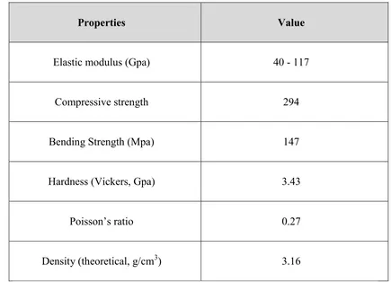
There is a wide variation of mechanical properties of synthetic calcium phosphate as given in Table 2.1. This variation of properties is the result of the variation in the structure of polycrystalline calcium phosphates, in turn the result of differences in manufacturing processes. Depending on the final firing conditions, the calcium phosphate can be calcium hydroxyapatite or β-whitlockite. In many instances, however both types of structure exist in the same final product.
mineral. Dentin (E=21GPa) and compact bone (E=12-18GPa) contain comparatively less mineral. The poisson’s ratio for the mineral or synthetic hydroxyapatite is about 0.27, which is close to that of bone (≈0.3) (Park et al., 1992)
Hydroxyapatite (HA) is a bioactive ceramic material with high bioaffinity, biocompatibility and osseoconductivity which is the main constituent of bones and teeth. Natural HA has the advantage that it inherits some properties of the raw material such as composition and structure. Properties of HA have found useful application in low-load bearing porous implants and coatings of metallic implants. Bioactivity and acting as a template for forming and growing of the surrounding bone tissues make HA an excellent choice for coating of the metallic implants.
Table 2.1 Physical Properties of Calcium Phosphate
Properties Value
Elastic modulus (Gpa) 40 - 117
Compressive strength 294
Bending Strength (Mpa) 147
Hardness (Vickers, Gpa) 3.43
Poisson’s ratio 0.27
Density (theoretical, g/cm3) 3.16
2.1.2 Crystal Structure of Hydroxyapatite