and sharing with colleagues.
Other uses, including reproduction and distribution, or selling or
licensing copies, or posting to personal, institutional or third party
websites are prohibited.
In most cases authors are permitted to post their version of the
article (e.g. in Word or Tex form) to their personal website or
institutional repository. Authors requiring further information
regarding Elsevier’s archiving and manuscript policies are
encouraged to visit:
Contents lists available atScienceDirect
Materials Science and Engineering B
j o u r n a l h o m e p a g e :w w w . e l s e v i e r . c o m / l o c a t e / m s e b
Electrochemical characterization of BSA/11-mercaptoundecanoic
acid on Au electrode
Teodora Ignat
∗, Mihaela Miu, Irina Kleps, Adina Bragaru, Monica Simion, Mihai Danila
Laboratory of Nanotechnology, IMT-Bucharest, Erou Iancu Nicolae 126A, 077190 Bucharest, Romania
a r t i c l e
i n f o
Article history: Received 9 June 2009
Received in revised form 2 November 2009 Accepted 9 November 2009
Recently, it has becoming increasingly important to control the organization of self-assembled mono-layers (SAMs) of functionalized thiols and to bind various proteins on gold/silicon substrates for their potential integration in nanoscale sensors/biosensors and optical devices. The biomolecule immobiliza-tion on the surfaces by covalent chemistry allows fabricaimmobiliza-tion of reproducible, protein-modified surfaces and became also a model to investigate the electrochemical response induced by protein binding. In this study, we report different nanostructured gold substrates and the adsorption of a protein, bovine serum albumin (BSA) on the 11-mercaptoundecanoic acid (MUA) layer for further biomedical applications. Nanostructured gold layers of 200 nm thickness have been prepared on both, flat and macroporous sili-con (macroPS) substrates. The X-ray diffraction analyses emphasized a dominant (1 1 1) crystallographic orientation of nanostructured Au substrates, which is preferred orientation for binding and detection of organic molecules on the gold surface. Impedance spectroscopy measurements performed in specific fre-quency ranges show that the binding of protein to a single monolayer of MUA can be easily detected. The impedance changes were also corroborated with cyclic voltammetry and Raman spectroscopy analysis for further development of the biosensor transducer for converting of the specific molecular recognition events into either an optical or electrical signal.
© 2009 Elsevier B.V. All rights reserved.
1. Introduction
Self-assembly strategy to exploit specific noncovalent interac-tion between a protein and a metal complex-based self-assembled monolayer (SAM) with interesting application in microarray tech-nology or in sensors for lactoferrin, has been recently reported in
[1]. However, utilization of noble metals, particularly functional gold nanoparticles, in biological and pharmaceutical fields remains attractive for ultrasensitive detection and imaging methods for bioreorganizing events, because they have great biocompatibil-ity, allow a variety of surface coatings and exhibit unique optical properties, as surface plasmon resonance absorption and resonance light scattering[2]. Moreover, gold is commonly used for fabrica-tion of working electrodes; therefore, defining surface properties of this material is important for electrochemical sensor development. The immobilization chemistries as thiol:gold provide the user with a variety of potential methods for coupling anchor molecules to the sensor surface. Modifying gold surfaces by SAM have been the subject of intense investigation over the past twenty years [3–7]. The thiols of similar chain length led to well-ordered monolayers. By synthesizing appropriate
alka-∗ Corresponding author. Tel.: +40 740088663; fax: +40 214908238. E-mail address:[email protected](T. Ignat).
nethiol molecules, gold surfaces can be rendered protein reactive, by incorporating aliphatic or reactive polar groups [8,9]. Cova-lent linkage of any protein to a carboxylic acid terminated SAM is most conveniently done by first converting the car-boxylic acid functions into N-hydroxysuccinimide esters (NHSs), in the presence of 1-ethyl-3-(3-dimethylaminopropyl)carbodiimide hydrochloride (EDC)[10,11]. Model protein, bovine serum albumin (BSA) was covalently bound to MUA through carbodiimide chem-istry. The BSA, also known as “Fraction V”, has been chosen in our experiments because it displays a strong affinity for various types of surfaces, has good stability and lack of effect in many biochem-ical reactions, as well as low cost since large quantities of it can be readily purified from bovine blood, a byproduct of the cattle industry.
Moreover, BSA has numerous biochemical applications includ-ing protein microarray technology[12], lysate microarray technol-ogy[13], ELISA (Enzyme-Linked Immunosorbent Assay)[14,15], immunoblots as a blocking agent during western blotting to reduce background and non-specific binding[16]and immunohis-tochemistry[17]. BSA is used in fundamental researches for the development of better therapeutics to convert globular proteins which is non-cytotoxic into fibrillar form that causes cell death[18]; it is also used as a nutrient in cell and microbial cultures.
The aim of this paper is to realize a new microstructured Au/Si substrate with appropriate structure for thiol binding and
56 T. Ignat et al. / Materials Science and Engineering B169 (2010) 55–61
improved properties to enhance Raman signal, and further to establish the conditions for covalent protein immobilization using surface chemistry of carboxyl containing thiols, such as 11-MUA. Deposition of thiols and proteins has been monitored by Raman spectroscopy and confirmed by cyclic voltammetry with potassium ferricyanide serving as a probe molecule.
2. Experimental
2.1. Fabrication of test structures
p-Type Si wafers with resistivity in the range of 10–18cm, provided bySi-MatSilicon Materials – Germany, have been used as substrate. The surface micropatterning has been achieved by an electrochemical porozification process using the single-tank (AMMT GBH) wet etching system for 4 in. diameter silicon wafers with programmable power supply and dedicated software for time-based current profiles as fabrication set-up. The process parameters were: 4% HF:DMF (dimethylformamide) electrolyte solution, 8 mA/cm2 current density and 8 min time, specific for fabrication of a macroPS layer[19].
Thin gold layers have been deposited using vacuum coating sys-tem (BOC Edwards Auto 500) on both flat silicon substrate and macroPS. The physical vacuum deposition (PVD) process is com-puter controlled and supplementary X-ray reflectivity analyses confirmed that the resulting metallic layer thickness is 200 nm.
MUA was self-assembled on all investigated substrates by their immersion in 2 mM MUA in ethanol solution. We have chosen MUA because it has a long alkane chain, which produces highly packed and ordered surface monolayer[20]with carboxylate binding sites. Analytically graded BSA, 1-ethyl-3-(3-dimethyl amino-propyl)carbodiimide hydrochloride (EDC), N-hydroxy succinimide (NHS), phosphate buffer solution (PBS, pH 7.4) were purchased from Sigma–Aldrich Co. Deionized water was used to prepare the solution.
For investigation the covalent immobilization of proteins to carboxyl end groups of 11-MUA self-assembled on gold we have used a common methodology for construction of biosen-sors [21,22]. (N-hydroxysulfosuccinimide) NHSS as catalyst has been used in conjunction with 1-(3-(dimethylamino)propyl)-3-ethylcarbodiimide hydrochloride (EDC) to convert 11-MUA carboxyl groups to Sulfo-NHS esters which react with primary amines from BSA. Freshly prepared 0.1 M EDC/0.1 M NHS and BSA (1 mg/l) in PBS (1%, pH 7) were poured over the 11-MUA/Au/PS samples and stirred for 1 h at 0◦C, being kept overnight at room temperature. The surface was rinsed with PBS buffer to remove unreacted protein molecules. This immobilization protocol has been applied on a high number of samples and has a very good reproducibility.
2.2. Investigation methods
Morphological characterizations were realized using Nova NanoSEM 630 system, a Field Emission Scanning Electron Micro-scopes (FE-SEM) – FEI Company, USA – with ultra-high resolution at high and low voltage in high vacuum: 1.6 nm at 1 kV.
The crystalline structure of the experimental samples was inves-tigated by different X-ray diffraction methods using a Rigaku Smartlabthin film 9 kW rotating anode equipped with an in-plane arm in–2angular scanning configuration using a Cu X-ray tube
and a multilayer mirror (parallel beam, Cu K˛X-ray wavelength of
K˛1=1.540609 Å ) and a flat graphite analyzer (one reflection). The electrochemical experiments were carried out using the advanced electrochemical system PARSTAT 2273 – Princeton Applied Research – and theModel K0264 Micro-Cell Kitin a
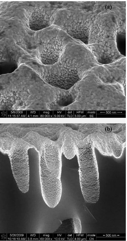
conven-Fig. 1.Plan view (a) and cross section (b) SEM images of PVD-evaporated Au thin film on macroPS substrate.
tional three electrodes cell configuration, with Pt counter electrode wire, reference Ag/AgCl (1 M in KCl) and the test structures elec-trode as working elecelec-trode. The electrolyte was 0.1 M KCl solution in either the presence or the absence of [Fe(CN)6]3−/4−redox probe. Raman measurements were carried out on High Resolution Raman Spectrometer, LabRAM HR 800–Horiba Jobin Yvon–with spectral resolution 0.35 cm−1/pixel for laser of 633 nm and diffrac-tion gratings of 1800 gr/mm. The spectrometer is equipped with a microscope and a 50×objective was used to focus the incident
laser beam to a spot size of about 2m on the sample surface and to record the scattered light from a very small focus area.
3. Results and discussion
3.1. SEM analysis and pore size distribution
Fig. 2.X-ray spectra of Au thin film deposited on flat Si substrate (a) and on macroPS/Si substrate (b).
3.2. X-ray analyses
Evaporated gold films, as well as sputter deposited films, are known to have a strong Au (1 1 1) character when deposited on a heated substrate. The crystalline structure of the gold film was investigated by wide angle X-ray diffraction methods (WAXRD).
The diffraction spectra of the gold on porous silicon (PS on Si (4 0 0)) presented inFig. 2a reveal Au (1 1 1) preferred orientation, a high Au crystalline phase content of the layer and a medium crystallite size of 23.16 nm (computed with the Hall–Williamson method[23,24]). The Au (1 1 1) comparatively with other gold ori-entation has the lowest energy, and therefore is the most stable; moreover, Au (1 1 1) comparatively with Au (1 0 0) has a higher density of atoms and favors the attachment of a higher number of atoms and bio-molecules on its surface. More pronounced Au (1 1 1) orientation has been obtained on Si (1 0 0) flat surfaces (Fig. 2b).
3.3. Raman measurements
Among biophysical and biochemical analyses, Raman spec-troscopy plays an important role in the identification and structural characterization of molecules. The wavelengths of the Raman emis-sion spectrum are characteristics of the chemical composition and structure of the light absorbing molecules in sample, while the intensity of the light scattering is dependent on the concentration of molecules in the sample.
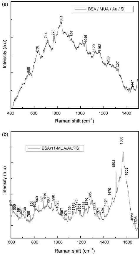
Fig. 3.Raman spectra obtained after 20 h of immersion of BSA for the functionalized substrates: MUA/Au/Si (a) and MUA/Au/macroPS/Si (b).
Although Raman spectroscopy is limited by extremely low scat-tering cross sections as compared to fluorescence, the surface enhanced Raman scattering (SERS) effect can provide cross sec-tions with several orders of magnitude higher than normal Raman spectroscopy[25]therefore allowing in some cases single molecule detection [26]. Raman amplification can be explained in terms of electromagnetic and chemical enhancement in the presence of metallic nanostructures[27]. In the first case scattering is enhanced by the local optical fields close to metallic particles after excitation of surface plasmon at resonance conditions.
Recently, the excellent SERS properties of nanostructured Au (1 1 1)/macroPS substrate have been demonstrated[28]. The recorded Raman spectra for SERS spectra of BSA/MUA/Au Si (flat and macroPS) samples are presented inFig. 3.
Bovine serum albumin consists of 583 amino acids. Its amino acid sequence contains 17 disulfide bonds and one free thiol, which are positioned in a repeating series of nine loop-like structures centered on eight sequential cys–cys pairs[29].
58 T. Ignat et al. / Materials Science and Engineering B169 (2010) 55–61
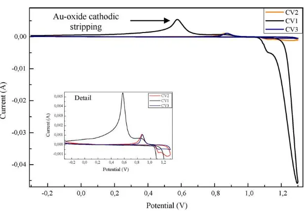
Fig. 4.The evolution of cyclic voltammograms of cleaning for a gold electrode.
The amide I bands corresponding to the ␣-helix, -sheet, and random coil conformations occur in the 1696–1665 cm−1, and 1666–1660 cm−1range, respectively. On the other hand, the amide III band positions of these conformations were localized in the 1310–1260 cm−1, 1242–1235 cm−1, and 1250–1240 cm−1regions, respectively. Amide III bands of BSA exist at 1270 cm−1. More-over, several weak signals appeared in the region of 1100 cm−1, 1300 cm−1; these peaks could belong to the signal of the secondary structure (␣-helix,-sheet), and phenyl stretching.
In our SERS spectra of labeled BSA the amide I band can be observed at 1663 cm−1, which could be supposed as a Raman shift toward a lower wavenumber, and the amide III band is localized at 1305 cm−1 spectra (Fig. 3). It is difficult to believe that the weak band at 1400 cm−1from the Raman is shifted to such lower wavenumbers in SERS. It would be more convincing to assume it to a SERS contribution derived from the large Raman band at 1334 cm−1 due to the Au/PS/Si surface micro/nanostructuration.
3.4. Electrochemical measurements
3.4.1. Electrochemical cleaning of gold surface electrode
The electrochemical cleaning of the gold surfaces was carried out in 0.1 M H2SO4 by means of cyclic voltammetry (CV), i.e., by sweeping the applied potential between−0.3 and 1.3 Vvs. Ag/AgCl
with scan rate of 30 mV/s. The cyclic voltammograms recorded for the cleaning regime of the gold electrode are presented inFig. 4.
The voltammograms show how a monolayer of oxygen is chemisorbed onto the Au electrode, in a relation of 1:1 with the sur-face gold atoms, followed by the electrochemical reduction of gold oxide. The stripping peak identified in graphs has a well-defined baseline, which can be easily integrated to obtain the experimen-tal charge density. Stable and reproducible Au-oxide formation and stripping peaks are indicative of a clean, well defined gold surface
[31].
3.4.2. CV study of MUA and BSA on the gold electrode
Redox process of protein at an electrode is not easy to explain only on the basis of its structural properties. Reductibility or oxid-ability of electroactive sites, presented in the respective molecule, may be affected by their position in the ordered structure, and also by adsorbability of the given macromolecular segment which is responsible for the exposure of the electroactive groups in the
redox processes. Therefore, only disulfide group of cystine and aro-matic ring of tyrosine and tryptophan in the corresponding class of proteins are electroactive[32].
The electrochemical analyses are known for their sensitivity in detection of new bonds creation or changes in original bonds with direct applications in a broad range of analyte recognition via formation of a covalent bond to the sensing layer.
The cyclic voltammograms recorded in 0.1 M KCl +
5 mM K3Fe(CN)6electrolyte on the MUA gold electrode/PS sample before and after interaction with protein (BSA) are presented in
Fig. 5. The potentials were swept between 0 and 0.6 V relative to [Fe(CN)6]3−/4−redox couple reduction/oxidation in 0.1 M KCl. As can be seen, the well-defined redox peaks commonly displayed on bare-Au[33]are drastically reduced for MUA-modified electrodes, since the recorded currents are at least one order of magnitude lower after functionalisation – the data recorded for substrate Au/PS/Si being an indication that the thiols are more densely packed on surface of plane gold comparing with nanostructurated ones. The corresponding CV curve in the first case there are no
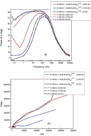
Fig. 6.Nyquist (a) and Bode (b) plots for SAM/Au/Si structures.
measurable pinholes and this provides substantial resistance to electron transfer [34], indicating that electron transfer occurs preferentially by through-film tunneling[35].
TheBSA/11-MUA/Auelectrode displays an increased redox cur-rent of sigmoid shape, probably due to reduction of the surface negative charge density that allowed [Fe(CN)6]3−/4−ions to pen-etrate the biomolecule layers and to reach the electrode surface
[36].
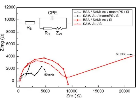
3.4.3. Impedance for Fe(CN)64−on modified Au electrode with BSA/MUA layer
The impedance biosensors measure the electrical impedance of an interface in AC – alternating current – steady state with con-stant DC – direct current – bias conditions; an important advantage is that, in order to promote biochemical events small voltages on the order of mV’s, who does not damage biomolecular probe layers, are necessary. As a result, monitoring impedance by electrochem-ical impedance spectroscopy (EIS) brings significant improvement
of detection over commonly used cyclic voltammetry (CV) and in addition enlarges the application area[37].
The changes in electrical properties of a sensor surface, as dielectric constant or resistance, which are recorded when a target biomolecule interacts with the probe-functionalized sur-face take place leading to modification of the electrode–solution interface impedance; since there is a direct consequence of the presence target molecule it is applicable scheme of detection to label-free biosensing. Moreover, impedance spectroscopy can be used with biosensors to detect an “electro-fingerprint”, a unique pattern of electrical changes as a function of the electrical frequency[38].
60 T. Ignat et al. / Materials Science and Engineering B169 (2010) 55–61
Fig. 7.Bode plot of the SAM/Au/PS/Si before and after the interaction with BSA protein.
force to charged molecules and also the probe–target binding ener-gies (in general, covalent bonds have ca. 1–3 eV enerener-gies, but not all the samples are covalently attached).
Our analyses have been performed using 5 mV rms AC amplitude and 5 DC bias voltages:±0.28,±0.18 V, and open circuit potential
(OCP) using a support electrolyte (0.1 M KCl) with and without the redox complex (5 mM Fe(CN)63−/4−).
The Nyquist (a) and Bode-phase (b) curves show the (Fig. 6) compartment of sandwich structures subjected to both nonfaradaic and faradaic processes induced by application of different DC biases. While the nonfaradaic processes appear normally when the electrolyte does not contain the redox species and there are only given by the interface capacitor change during the probe immobilization, the key parameter being theCsurf, the faradaic processes require the presence of redox species, alternately oxi-dized and reduced by the transfer of an electron to and from the metal electrode the charge transfer resistanceRctbecomes the key parameter.
The impedance changes observed in redox electrolyte for SAM/Au/PS/Si before and after the interaction with BSA protein are presented inFig. 7.
It could be observed that the impedance spectra obtained for two applied DC potentials present for the incomplete semicircles a rapidly shrink upon increasing overpotential. The experimental data were fitted to equivalent circuit models of the electrochemical interface using Zsim 3.21 software;Rs(CPE(RCTW)) is the equivalent circuit used to extract the electrical parameters. In this circuit,Rs represents the uncompensated electrolyte solution resistance, CPE is constant phase element – CPE = 1/(jωY)n– for interface capaci-tance, in a parallel grouping withRCTis charge transfer resistance series with the Warburg impedance – W – arising from diffusion of the electroactive species to the electrode happened at low fre-quencies.
Analyzing the response in impedance of both types of experi-mental samples the differences between the recorded values are evident.
The Nyquist curves demonstrate that at the higher frequen-cies an electron transfer-limited process is dominant, followed by a diffusionally limited electron transfer process at the lower fre-quencies [44]. On the other hand, using macroPS as metallic layer substrate determines increasing of electrode surface and conse-quently a higher number of active sites on, which finally leads to decreasing of charge transfer resistance, theRCTvalues being more than 3 times lower for Au/PS structures.
For example, the values obtained for Rs varies from 48
(BSA/SAM/Au/Si) to 10(BSA/SAM/Au/PS/Si), while theRCTvaries from 7585 to 1709. In the same time, the fractional power of CPE,
n, takes the valuesn= 0.8 (BSA/SAM/Au/Si) and respectivelyn= 0.97 (BSA/SAM/Au/PS/Si), confirming the voltammetric results.
3.5. Conclusions
The paper presents a comprehensive study of the nanostruc-tured gold deposited on macroPS or on flat Si and modified with MUA and BSA. Deposition of alkanethiols and proteins was moni-tored by Raman spectroscopy, and was further confirmed by cyclic voltammetry and impedance spectroscopy with potassium ferri-cyanide serving as PROBE molecule. Both investigated structures, nanostructured Au (1 1 1) deposited on Si (flat surface) and on macroPS/Si substrates are appropriate for self-assembly and fur-ther protein binding. Raman measurement results show a better enhancement signal when nanostructured gold is deposited on the Si substrate subjected to a previous microstructuration process (macroPS). Electrochemical characterization techniques relieve improved characteristics for nanostructured Au (1 1 1) deposited on Si (flat surface), both investigated structures being suitable for impedance biosensors.
Acknowledgment
The authors gratefully acknowledge the support of the Romanian Ministry of Education and Research through PNII-11023/71120 and IDEI-884 national programs. The authors are pleased to acknowledge Mr. Adrian Dinescu for SEM characteri-zation and Mr. Florin Comanescu for Raman measurements.
References
[1] N. Tuccitto, N. Giamblanco, A. Licciardello, G. Marletta, Chem. Commun. (2007) 2621–2623.
[2] A.P. Alivisatos, Nat. Biotechnol. 22 (2004) 47–52.
[3] A.S.N. Murthy, J. Sharma, Anal. Chim. Acta 363 (1998) 215–220. [4] M.Y. Tsai, J.C. Lin, J. Colloid Interf. Sci. 238 (2001) 259–266.
[5] M.K Calabretta, K.S. Matthews, V.L. Colvin, Bioconjugate Chem. 17 (2006) 1156–1161.
[6] J. Tkac, J.J. Davis, J. Electroanal. Chem. 621 (2008) 117–120.
[7] J. Pillay, B.O. Agboola, K.I. Ozoemena, Electrochem. Commun. 11 (2009) 1292–1296.
[8] J.C. Love, L.A. Estroff, J.K. Kriebel, R.G. Nuzzo, G.M. Whitesides, Chem. Rev. 105 (2005) 1103–1170.
[9] S. Balasubramanian, A. Revzin, A. Simoniana, Electroanalysis 18 (2006) 1885–1892.
[10] M.G. Kim, Y.B. Shin, J.M. Jung, H.S. Ro, B.H. Chung, J. Immunol. Methods 297 (2005) 125–132.
[11] Y.C Lin, Y. Tsao, W. -H. Tsai, T.S. Hung, K.S. Chen, S.C. Liao, Sens. Actuators. B: Chem. 133 (2008) 370–373.
[12] O. Gutmann, R. Kuehlewein, S. Reinbold, R. Niekrawietz, Lab Chip 5 (2005) 675–681.
[13] C. Mircean, I. Shmulevich, D. Cogdell, W. Choi, Y. Jia, I. Tabus, S.R. Hamilton, W. Zhang, Bioinformatics Advance 12 (2005) 1935–1942.
[14] H.J. Willison, K. Townson, J. Veitch, J. Boffey, N. Isaacs, S.M. Andersen, P.Z.C.-C. Ling, D.R. Bundle, Brain 127 (2004) 680–691.
[15] S. Mirapurkar, U.H. Nagvekar, N. Sivaprasad, J. Immunoassay Immunochem. 28 (2007) 119–126.
[16] Y. Studentsov, M. Schiffman, H.D. Strickler, G.Y.F. Ho, Y.Y.S. Pang, J. Schiller, R. Herrero, D. Burk, J. Clin. Microbiol. 40 (2002) 1755–1760.
[17] M. van Timmeren, S.J.L. Bakker, C.A. Stegeman, R.O.B. Gans, H. van Goor, Nephrol. Dial. Transpl. 20 (2005) 2349–2357.
[18] C.Y. Huang, C.M. Liang, C. Li Chu, S. Mei Liang, BMC Biotechnol. 9 (2009) 1–12. [19] P. Bettotti, L. Dal Negro, Z. Gaburro, L. Pavesi, A. Lui, M. Galli, M. Patrini, F.
Marabelli, J. Appl. Phys. 92 (2002) 6966–6972.
[20] N.K. Chaki, K. Vijayamohanan, Biosens. Bioelectron. 17 (2002) 1–12. [21] D.C. Kim, D.J. Kang, Sensors 8 (2008) 6605–6641.
[22] B.L. Frey, R.M. Corn, Anal. Chem. 68 (1996) 3187–3193.
[23] V. Pecharsky, P. Zavalij, Fundamentals of Powder Diffraction and Structural Characterization of Materials, second ed., Springer, 2008.
[24] I. Kleps, M. Danila, A. Angelescu, M. Miu, M. Simion, T. Ignat, A. Bragaru, L. Dumitru, G. Teodosiu, Mater. Sci. Eng. C 27 (2007) 1439–1443.
[25] A.I. Abdelrahman, A.M. Mohammad, T. Okajima, T. Ohsaka, J. Phys. Chem. B 110 (2006) 2798–2803.
[26] U. Landman, W.D. Luedtke, Faraday Discuss. 125 (2004) 1–22.
[28] I. Kleps, M. Miu, M. Simion, T. Ignat, A. Bragaru, F. Craciunoiu, M. Danila, J. Biomed. Nanotechnol. 5 (2009) 300–309.
[29] T.J. Peters, All about Albumin: Biochemistry, Genetics and Medical Applications, Academic Press Inc., Orlando, 1996.
[30] S. Cavalu, C. Pinzaru, N. Leopold, W. Kieefer, Biopolymers 62 (2001) 341–348. [31] H.O. Finklea, A.J. Bard, I. Rubinstein, Electroanal. Chem. A 19 (1996) 108–335. [32] J. Anzai, B. Guo, T. Osa, Bioelectrochem. Bioenerg. 40 (1996) 35–40.
[33] S. Mendoza, Langmuir 23 (2007) 582–588.