102
Tjahjadi et al MedJ
IndonesPathological
aspects
of breast
cancer
in
Indonesian
females,
applying
a
modified
WHO
classification.
A
joint
study
between
Indonesia and Japan
Gunawan
Tjahjadi*, Goi
Sakamotot,
u
Yoshiyuki ônnolt,
Santoso
Comain*,
S
Darwis#,
Setyawati Budiningsih-,
SadaoSuzu
dAbstrak
Dari
aspek patolagi diteliti kembali varian histologik karsinoma payudara dan dibandingkan dengan kasus-kasus dari Jepang. Dipakai kLasifikasi yang dinnjurkan oleh Japanese Breast Cancer Society, yang membagi lagi karsinoma mammae duktal invasif meniadi3 subtipe, yaitu: tipe papilotubule4 solid-tubuler dan skirus. Dari Indonesia ada 515 kasus yang ditangani oLeh Rumah Sakit Umum pusat Nasional Dr. Cipto Mangunkusumo (RSCM), berasal dari penelitian
ke-l
dnn ke-2, seclangkan dari Jepang ada 445 kasus yang rlitangani oleh Cancer Institute Hospital (CIH). Di RSCM clitemukan sebanyak 0.977o karsinoma noninvasif, 89.l4%o karsinoma duktal invasif dan 9.50Vo tipe khusus.Di
CIH ditemukan sebanyak 7.4Va karsinoma non-invasif, 80.47o knrsinoma duktal invasif dan 11.3Vo tipe khusus. Insirlen karsinoma tluktal noninvasif tti RSCM lebih rendah daripada CIH dengan perbedaan 6.437o.Di
antara subtipe karsi-noma rluktal invasif, tipe skirus-lah yang paLing banyak ditemukan pada kedua kelompok (RSCM dan CIH), dengan insiden 50.497o (RSCM) dan 43.6Vo (CIH). Subripe yang paling sedikit di RSCM ialah tipe papiLotubulet; sedangkan di CIH ialah tipe solid-tubuler Tipe khusus ditemukan sebanyak 5 varian di RSCM, sednngkan di CIH 7 varian. lnsiden karsinoma musinosum dan karsinoma lobuler invasif di RSCM lebih rendah claripada di CIH, sedangkan karsinoma moduler lebih tinggi. Insiden penyakit Paget juga lebih rendah di RSCM.Abstract
From the pathological aspect, we analyzed again the histological variants of breast cancer and compare them with cases from Japan. We usecl the classification recommended by the Japanese Breast Cancer Society, which divided the invasive cluctaL carcinoma
of
NOS (no otherwise specified) of the WHO clnssification into 3 subtypes, namely papiLlàtubulaa solid-tubular and scirrhous carcinoma. We had 515 cases from Inclonesia (lst batch and 2"t batch) treated at the
Dr Cipto Mangwtkusumo National Central General Hospital
(RSCM)ani
445 cases from Japut, treated at the Cancer Institute Hospital (CIH), The RSCM cases had 0.97Vo noninvasive carcinoma, 89.I4Vo invasive ductal carcinoma antl 9.50Vo of the specialtypes. The CIH cases had 7.4Vo noninvasive carcinoma, B0.4Vo invasive ductal carcinomaanl
I L3Vo of the speciaL types. The incidence of noninvasive ductal carcinoma was lower at RSCM, with a 6.43Vo dffirence.Among the subtypes of invasive ductal carcinoma, the scirhous carcinoma was the most common in both groups (RSCM and CIH), with an incirlence
of
50.49Vo (RSCM) and 43.67o (CIH). The least common at RSCM was the paPilLotubular type, while at the CIH it was the solicl-tubular type. Among the special types, only 5 variants were encountered at RSCM and 7 at CIH. The incidences of mucinous car-cinoma and int,asiye Iobular carcinomawere lower at RSCM, while the medullary carcinoma was higher. The incidence of Paget's disease was also lower at RSCM.Keywords: Pathological, breast cance4 WHO-classification, Japan-lndonesia study.
d' Department of Pathology, Faculty of Medicine, University of
Indonesia, Jakarta I 0430, Indonesia
t
Department of Pathology, Cancer Institute Hospital, Tokyo 170, Japan#
Department of Surgery, Faculty of Medicine, Universityof
I nrlonesia, Jakarta I 0430, Indonesia*
Department of Surgery Cancer Institute Hospital, Tokyo 170, Japan-
Department of Community Medicine, Faculty of Medicine' University of Indonesin, Jakarta 10320, Indonesia 1l Department of Preventive Medicine, School of Medicine,Nagoya University, Nagoya 466, Japan
n
Department of Nutrition, Faculty of Medicine, IJniversity of Indonesin, Jakarta 10430, Indonesia
INTRODUCTION
Breast cancer
is
a
worldwide
and serious
disease,which
create
a major
public
health problem.
There
aregeographic differences
in their
incidence rate,
be-ing higher
in
Northern America
and
Europe than in
Asian
countries
and Japan.Its
frequency is increasing
during the
last
15 years, especially among
women
with a high
socioeconomic
status,namely from
l2.I-16.6
to
2l
per
100,000
females in
Japan
and from
7I.7
to
91
per
100,000 females
in Northern America.
In
Singapore
the incidence
of
all
ethnic groups
Vol 8, No 2,
April -
June 1999the
data
collected from 13 Pâthology
Laboratories
spread
throughout Indonesia, showed that
breastcan-cer ranked the second among females,
with
18.037orelative frequency (ASCAR
=
agestandardized
can-cer
ratio
17.84Vo)
in
1988 and
l8.44Vo (ASCAR
17.46Vo)
in
1989.These differences have attracted investigators
to look
upon
several aspects
of
breast cancer, including
epidemiological,
clinical,
pathological,
and
nutri-tional
aspects.A Joint Study on etiology
andClinico-pathology
of
breast cancer
hasbeen conducted
since1
988, between Indonesia
and Japan.In
the
first
batch
of
a three years
study we
encoun-tered dissimilar pathological finding.
The Cipto
Man-gunkusumo
National Central
General
Hospital
(RSCM)
caseshad lower incidences
of
noninvasive
ductal carcinoma, mucinous carcinoma and invasive
lobular carcinoma than
casesfrom
CIH
(Cancer
In-stitute Hospital, Tokyo). Medullary carcinoma
wasmore frequent at the RSCM.
An
extended
study
wasdone to include nutritional analysis.
From tlre
patho-logical
aspects
we
analyzed again
tlie histological
variants of breast cancer and compare
with
the
casesfrom
Japan.MATERIALS
AND METHODS
Thble
1.
Histological classificationof
breast tumors (Japanese Breast Cancer Society, 1984)Malignant (Carcinoma)
l.
Noninvasivecalcinomaa.
Noninvasive ductal carcinomab.
Lobular carcinoma in situ2.
lnvasive carcinomaa.
Invasive ductal carcinomna
l.
Papillotubular crrcinomatr2.
Solid-tubullrcarcinomaa3.
Scirrhouscarcinomab.
Special typesbl.
Mucinous carcinomab2.
Medullalycarcinomab3.
Invasive lobular carcinomab4.
Adenoid cystic carcinomab5.
Squamous cell carcinomab6.
Spindel cell carcinoma b'l.
Apocrine carcinomnb8.
Carcinoma with cartilaginous and or osseous metaplasiab9.
Tubular carcinomah 10. Secretory clrcinoma
bl1.
Others3.
Paget's diseaseNote: See Appenclix
for
tlrc principles antl detaiLs oJ the classification.WHO classification
of
breast cancer t03Two hundred and twenty
six
casesof
breast cancer
were available.
Some
of these
casesthe diagnosis
of
breast cancer
were performed at another Hospital
without mentioning the subtypes. This occurred
in
11cases, so
for
histopathological analysis we had
215 cases.The classification
used that recommended by
the
JapaneseBreast Cancer Society,
which
divided
the invasive ductal carcinoma
of N.O.S (no otherwise
specified)
of
the
WHO
classification inro
subtypes,papillotubular, solid tubular
andscirrhous carcinoma.
RESULTS
The
casedistribution by histological type is shown
in
Table
2.Thble
2.
Case distribution of histological types of breast cancer (1992-199s)Histological types Number of
cases
7oNoninvasive carcinoma
a. Noninvasive ductal
carcinoma
I
0.46 b. Lobular carcinoma in situlnvasive carcinoma a. lnvasive ductal carcinoma
a
l.
Papillotubulara2. Solid-rubular a3. Scirrhous b. Special types
bl.
Mucinous carcinoma b2. Medullary carcinoma b3. Invasive lobular carcinoma b4. Squamous cell carcinomal5
66
lt3
4
l0
5
1
6.98 30.70 52.86
1.86 4.6s 2.33 0.46
Total
100.00The invasive
ductal carcinoma accounted
for
9O.24Voof
the cases,followed by the special types with
9 ,3Vo.Only
I
case
of noninvasive ductal carcinoma
wasfound (0.46Vo).In one
case
of
papillotubular
carci-noma
acoincidental finding with
alobular
carcinoma
in situ was
seen.When we compiled the
lstbatch
andthe
2ndbatch
of the study, the results
is shown
in
Ta-ble
3.Noninvasive carcinotra
a. Noninvasivc ductal carcinorra
b. Lobuiar carcinorna in situ
lnvasive carcinonra
a. Invasive ductal carcinoma
al.
Papillotubular a2. Solid-tubular a3. Scirrhous b. Spccial typesb1. Mucinous carcinomil b2. Medullary carcinoma b3. Invasivc lobular carcinoma b4. Adenoid cystic ccrcinorna b5. St.;unrnous ccll cârcinorna
Paget's disease
104
Tjalladi
et alTablc
3.
Histological types and case distribution of breastcan-cer of the whole study (1988-199-5)
Histological types Number
ol'cases
okMed
J
IndonesThe most common
was theinvasive
ductal carcinoma
with
89.14o/o.'|he
special
types
with
9.5OVo.Only
5cases
of noninvasive
ductal carcinoma were found,
comprised
only
0.97Vo.Among
tlie
special
types,
themedullary carcinoma was
the
most common, with
5.24Voincidence rate. When
we compare tlre whole
caseswith
casesfrom
Japan,the
result is
seenin
Ta-ble
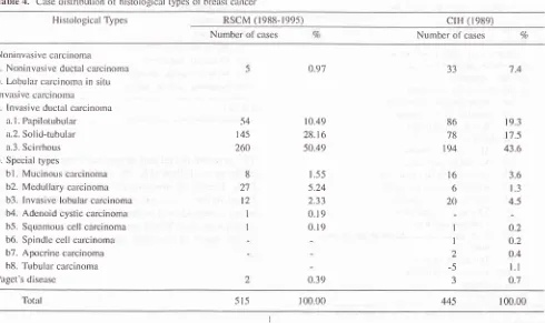
4.The
RSCM
cases had 0.91Vononinvasive
carcinoma,
89.147o
invasive ductal carcinoma,
and 9.5Oo/oof
thespecial types.
Tlie
CIH
caseshad
7 .4Vononinvasive
carcinoma,
80.4Vo
invasive
ductal
carcinoma,
andll.3t/o of
the
special
types. The incidence
of
nonin-vasive
ductal carcinoma is lower
at
the RSCM,
with
a 6.43o/adiftèrence.
Among
thespecial types
atthe
RSCM only
5variants
were encountered,
while at the
CIH,
there were
7variants.
Tlre
incidences
of
mucinous
carcinoma
andinvasive
lobular
carcinoma were
lower
atthe
RSCM,
while
the
medullary
carcinoma was higher.
The
inci-dence
of
Paget's disease
was also lower
at
theRSCM.
Tlre
ageincidences
of
the 226 çn5sssf
ths
Jndbatch
of
the study
arepresented
in
Table
5.0.97
54 145
260
8
2't t2
1
I
2
10.49 28.6 50.49
t.-55 5.24
Z.J J 0.19 0.19
0.39
Total
100.00Table
4.
Case distribLrtion of histologicirl types of brcast cancer 515Histological Types RSCM (1988-r99s)
crH
(r989)Number of cases Number of cases o/o
Noni nvz'rsive carcinoma
a. Noninvasive ductal carcinoma
b. Lobular calcinoma in sitr,r
Invasivc carcinorna
a. lnvasive ductal carcinoma
a.
l.
Papilotubulara.2. Solid-tubular
a.3. Scirrhous
b. Special types
bl.
Mucinous carcinoma b2. Medullary calcinoma b3. Invasive lobular carcinoma b4. Adenoid cystic carcinoma b5. Squamous cell carcinoma b6. Spindle cell calcinoma b7. Apocrine carcinoma h8. Tubulal carcinomaPaget's disease
54
145
260
8
27
l2
I I
0.97
10.49 28.16 50.49
r.55 s.24
2,33
0.1 9 0. 19
0.39
86 78
194
16
6 20
1
I
2 -5
-l
t9.3
17.5 43.6
3.6
t.3
4.5
0.2 o.2
o.4
t.l
0.774 JJ
[image:3.595.89.579.443.734.2]Vol B, No 2,
April -
June 1999Table
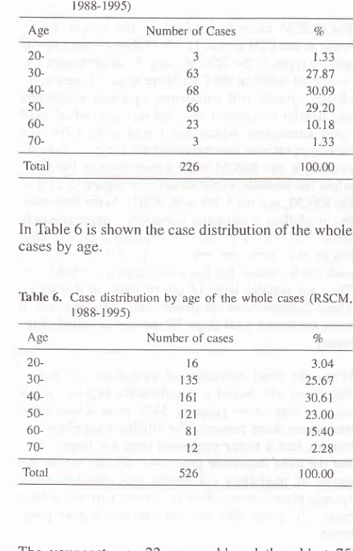
5.
Case distribution by age at operation/biopsy (RSCM, r988- r99-s)Age Number of Cases
WHO classfficatiort
of
breast cancer 105Both groups have the
samepeak incidence in the
5thdecade,
with
an incidence
of
30.6l%oat RSCM
and 38.3Voat
CIH.
In
the
4thdecade, the incidence
wasalmost
twice
compare
to
the
CIII
cases
(25.67Vo)against
13.2Vo).Also in the
3rd decade theRSCM
hadmore number of, cases,
with
a
3o/odifference.
The
casedistribution by
sizeof tumor of
the 2ndbatch
andcompare
to the
casesfrom
CIH
is shown
in
Table
8.The data were
available in
only
199 cases.Thble
8. Cases
distribution by size of tumor20- 30- 40- 50- 60-
70-3 63
68 66 23
3
1.33
27.87 30.09 29.20 10.r8 1.33
Tota 226 r 00.00
In Table 6 is shown
the casedistribution of the whole
cases
by
age.'lhble
6.
Case distribution by ageof
the whole cases (RSCM,r 988- r 99s)
Numbel of cases
No. of
cases
Vn No. of cases Size ofTumor
RSCM (1992-199s)
crH
(r989)Vo
TO TI '12
T3 ^t4
3 36 36 124
l.s0 18.09 18.09
62.31
22 t82
r9l
2l 30
4.9 40.8 42.8 4.7 6.7
Age
Totâl 526 r 00.00
Tl-re
youngest was 22 years
old
and
the
oldest
75years
old. The peak incidence was in 1le
Jth decade(30.617o),
followed
by
the
4thdecade
with
25.67Vo.Under
40
years
of age
there were
28.l1%o
of
the cases.Compare to
casesfrom CIH, the
casedistribu-tion
of
breast cancer by
ageis
seenin Table
7.Thble
7.
Case distribution of breast cancer cases by ageAse
RSCM (1988-1995)crH
(1989) No. ofcases
o/o No. of casesTotal
r99 l00o/o 446 t00%The RSCM T1 + T2 accounted
for
19.59Vo,while
atCIH it was
83.6Vo.The reserved occurred
for T3
andT4.
At
RSCM
it
was
80.4o/o,at
CIH
only
11.4Vo.DISCUSSION
In
this study we used the classification
proposed by
the
JapaneseBreast Cancer
Society, which is
essen-tially
the
same asthe WHO classification
with
a
mi-nor
modification.
The
invasive ductal
carcinoma
(WHO NOSAIST) = No otherwise
specifiedÆ.{ospe-cial type) which in reality
hasdifferent
pattern
of
in-filtration is
further
sub-classified
into
3 sub-types,
namely papillotubular, solid-tubular
and
scirrhous
carcinoma,
which
lraveprognostic significance. They
also reflect
the degree
of
differentiation,
which from
poor
to well is
in
an order
of
scirrhous ca, solid
caand papillotubular ca. The scirrhous carcinoma
hastlre poorest prognosis.
This
subclassification can
beused instead
of
the grading
system.
There were
sev-eral
grading systems proposed,8,e
but we
are still
looking
for
anInternationally
recognized breast
can-cer
grading
system.
Using
this classification
we
could avoid the universal problem
of
the grading
of
breast cancer.In the
second
batch
of the study we
found
only
Icases
of
noninvasive
ductal
carcinoma
(0.46V")
among 215
casesof breast cancer,
while in the batch
%
20- 30- 40- 50- 60-
70-l6
t35l6l
121 8l 123.04 2s.6'7
30.6 r
23.00 15.40 2,28
7a
20-
30-
40-
50-
60-
70-
80-16
135
l6l
12I 8l 123.04 25.67 30.6r 23.00 r5.40 2.28
2
59
l7l
t20 '74
t7
3
0.4 13.2
38.8 26.9 t6.6 3.8 0.7
[image:4.595.63.311.97.484.2]106
Tjahjatli
et aLof the
study,
we
encountered
4
cases ( I .33Vo)among
300
casesof
breast cancer.
When
we combined
the cases(Batch
1
&
Z,
RSCM)
and compared
with
the casesfrom
Japan
(CIH), we found that
the incidence
of noninvasive
ductal carcinoma was lower at
theRSCM,
with
a
6.437o
difference. The earlier
detec-tion of
breast cancer
could
beresponsible
for
the
dif-ference, especially
with mammography
screening
which has led to
increase
detection
of
ductal
carci-noma
in situ (noninvasive ductal carcinoma).ll
All our
casesof noninvasive ductal
carcinoma"/ductalcarcinoma
in
situ (DCIS) were
of the
comedo type.
In
our previous study, other
subtypes
were also
en-countered, such
asthe micropapillary, cribriform
andthe
solid
variety.l2 The
increasedprevalence
ofductal
carcinoma
in situ (DCIS) has produced
a
growing
awarenessof
the importance
of
its diverse pattern
andhave become
clinically significant
aspredictive
indi-cators
of
successfor
planned
local
excisions
of
small
DCIS
lessions.l3
Bellamy et alta in
areview
of
130 casesof
noninva-sive ductal carcinoma
of
the
breastconcluded that
thecomedo carcinoma DCIS
had an
occult
presentation
significantly more
likely
than other patterns to
in-volve multiple
quadrants
of
breast,irrespective
of
nu-clear
grade
or
necrosis.
Our
casesof DCIS were
de-tected at
early
stages(l
and
II).
In the second batch, the invasive ductal
carcinoma
accounted
for
90.24Voof the
cases,followed by
thespecial types
with 9.3Vo.ln
thefirst
batch
of
thestudy
we found
88.33Voof
invasive ductal carcinoma
and 9.67Voof the
special types. The
CIH
casesaccounted
for
80.4Voand
7l.3Vorespectively.
The
invasive
duc-tal carcinoma
was
the most common,
accounting
for
at least
80Voof
breast cancer.Among
the subtypes of invasive ductal
carcinoma,
the RSCM
cases
had
50.49Voof
the sciuhous
type
(Table
4),
followed
by the
solid-tubular
type of
28.l6Vo
and
the papilotubular type
of
10.49Vo.V/hile the
CIH
caseshad
43.6Voscirrhous type,
fol-lowed
by
the papilotubular type
of
19.3Voand
thesolid-tubular type was the least common. The
sub-classification
of
invasive
ductal carcinoma
into
3sub-types
has
prognosis significance. The
scirrhous
carcinoma
showsthe worst prognosis,
with
aten-year
survival rate of
61.2o/o.The papilotubular
carcinoma
shows
the most favorable prognosis, with
aten-year
survival
rate
of
77 .47a.The
ten-year
survival
rate
for
Med
J
Indonessolid-tubular
carcinoma
is
64.9Vo.The RSCM
cases
had
9.50Va
of the special
types,
while
at the
CIH it was
ll.3%o (Thble
4). Among
thespecial types at
the RSCM,
only 5 variants were
en-countered,
while
at theCIH,
there were 7 variants,
in-cluding spindle
cell
carcinoma, apocrine
carcinoma
and
tubular
carcinoma.
We had
one
caseof
adenoid
cystic carcinoma, which was found at the CIH. The
incidence
ratesof
mucinous
andinvasive lobular
car-cinoma at the RSCM were lower than at the CIH,
while the medullary
carcinoma was
higher
(5.24Vo at theRSCM,
against
7.37o at theCIH). In
the
literature,
the medullary carcinoma constitutes approximately
about 5 to
8Voof
invasive mammary
cancer.ls
His-tologically,
they
are
very poorly
differentiated or
high-grade
cancer,but
has a lessaggressive behavior.
They are usually large (4 cm
or
more
in
diameter).
These
features were consistent
with our
cases,which
were
presented
with
stageIII
disease
at initial
diag-nosis.
Using the strict
definition of medullary
carcinoma,
Ridolfi
et all6 found
a
significantly higher overall
survival rate
at l0
years of
84Vo,even when
nodal
metastases
were present.
The smaller medullary
car-cinoma had a better prognosis than the larger
ones,but the most important prognostic feature within
the
group
of medullary carcinoma was the density
of
lymphoplasmacytic
infiltrate. Those patients
with
areiatively
sparseinfiltrate
had
arelatively poor
prog-nosls.We had one case
of
adenoid
cystic carcinomain
a52-year
old woman in this
study,
presenting
with
a
cir-cumscribed lesion
of
6 cm
in
diameter. It
has
beenstated
that
this tumor is usually small, but may
be-come
aslarge as
8 cm in
diameter, and
grossly well
demarcated and
has agood prognosis.ls
The
two
casesof
Paget's
diseasein
our study were
55years and
60
years
old respectively,
similar to
thefindings
reported
in
theliterature, that Paget's
diseaseof
thenipple
tend
to
occur
in
anolder
agegroup. One
case
was
associatedwith
anin situ ductal
carcinoma.
Both
caseswere
of
stageI.
If we
compared the
casedistribution
of
breast cancer
by
age
among both groups (RSCM and CIH),
thenwe found that both
groups have
the
same
peak
inci-dence
in
the
5th decade,with
anincidence of
30.67VoVoL 8, No 2, ApriL
-
June 1999the
CIH
cases(25.67Vo
against
l3.2%o).Also
in
the3rd decade,
the RSCM had more number
of
cases,with a
3Va difference.
At
the
younger age
group,
breast
cancer occurred more
frequently
in
theIndone-sian females as compared
to
the
Japanesefemales.
Table 8 showed
the casedistribution by tumor
sizeof
the
2ndbatch
of
the
study.
At
the RSCM
Tl+T2
ac-counted
for
19.59Vo,and
in
the lst
batch
Tl+T2
ac-counted
for
l9%o,while
at theCIH, it
was83.6%. The
reversed occurred
for
T3
and
T4.
At
RSCM
it
wasB0.4Vo
(batch 2), or
SlVo
(batch
l),
while
at the
CIH
only
1l.4%a.
At
RSCM,
the
majority (more
than80Vo),
were
of T3+T4
size,while
atCIH
the
majority
(more than
807o)were
of
Tl+T2
size.
At
their
pres-entation, the
tumor
sizeof
breastcancer among
Indo-nesian
females was larger than that
among
Japanesefemales.
It
is well
establislred
that
the
size
of
an
invasive
mammary
cancer
is an
independent
prognostic
vari-able.lT'18
In
general,
the smaller the primary
tumor,
the
lower
the
chanceof axillary lymph
node
metasta-ses.
In
an analysis
of
200
breast cancers
treated
by
radical
mastectomy,
Fisher et alt9 found that
tumors
less
than
Icm
in
diameter
had a22Volikelihood of
ax-illary
metastases,and
tumors
with
more that 6cm
in
diameter had a
63Volikelihood
of
axillary
node
me-tastases.Sakamoto
et
al20in
a study
of
936
casesof
brcast cancer
surviving ten
years,
found that
tumors
between
2.1-5 cm in
size had 5I.ZVo
l0-year survival
rate, and
only
39.4Vofor tumors with
greaterthan
5.1cm
in
size.CONCLUSIONS
The
incidence
of
invasive
ductal
carcinoma
waslower
atthe
RSCM, with
a 6.43Vodifference. In
bothgroups
(RSCM
&
CIH), the
invasive
ductal
carci-noma
was
the most common,
accounting
for
at
least80o/o
of
breast cancer.
Among the
special
types,
theincidence
ratesof
mucinous and invasive
lobular
car-cinoma
at
the RSCM were lower than
at
the CIH,
while the medullary carcinoma
was
higher.At
younger age group (20-29 years),
breast cancer
had
already occurred
more
frequently
in
the
Indone-sian females
ascompared to the
Japanesefemales.
At
their
presentation,
the tumor
size
of
breast
cancer
among Indonesian females was larger than
among
Japanese
females.
Acknowledgments
The
authors
like to thank to the
nurses,Ms.
Ros
andWHO classification o.f breast
cancer
107Emi,
and
public health
nurses,Ms. July
and
Ms.
Er-Iaini
for
excellent epidemiological data collection.
We are
also indebted
to
SDP staffs
for helping in
dataprocesslng.
This work
was
suppofted by the
Ministry of
Educa-tion,
Science,
Sports and Culture
of
JapaneseGov-ernment, Grants
No.
0IO42OO1,
04042013
and06042006: and was
partially
supported
by the
Indo-nesian
Cancer Foundation.
This
collaborative
study
was
a
part
of
Special Cancer Research project
in
Monbusho Intenrational
Scientific
Research
Pro-gram,
with
the
approval
of the
Dean,
Faculty
of
Medicine, University
of
Indonesia,
No.
43831PT02.
H4.FKÆ/88.
RBFERENCES
l.
ElstonCW
Ellis
IO.
Prognostic tàctorsin
breast cancer.Short Course
XXth
International Congressof
the Interna-tional Academy of Pathology, Hongkong, October 1994.2.
Waterhouse J et al. Cancer incidence in five continents. In:International Agency for Research on Cancer, Vol.
III
IARC publication, 1 976.3.
Parkin DM, Laara E, Muir CS. Estimatesof
the worldwide frequencyof
sixteen major cancersin
1980.In J
CancerI 988; 4l
:
184-97.4.
NG EH, The Singapore breast screening project. Aprelimi-nary report. Proceeding APCC, page 274, Singapore 1995.
5.
Cornain S, Mangunkusumo R, NasarIM,
Prihaltono J. Tenmost frequent cancers in Indonesia: Pathology based cancer
registry
of
I 988- 1992. In: Cancer Regish'y in Indonesia. Na-tional Cancer Registry Center, Jakarta Coordinating Board,t992.
6.
PrihartonoJ,
MangunkusumoR,
PartoatrnodjoM,
Estab-lishing pathology based cancer registry: lndonesian
experi-ence. In: Sasaki R, Aoki K, editors. Epidemiology and
pre-vention
of
cancer. Proceedingsof
Monbusho, (Ministry ofEducation, Science and Culture) International Symposium on
Comparative Study
on
Etiology&
Preventionof
Cancer,Nagoya 1989. Nagoya: The University
of
Nagoya Press,1990:211-6.
'1
.
Japanese Breast Cancer Society. The General Rules for Clini-cal and PathologiClini-cal Recording of Breast Cancer. Jpn J Surg
1989; l9:613-32.
8.
Hartmann WH, Ozello L, Sobin LH, Stalsberg H.Histologi-cal typing
of
breast tumors.In:
International Histological classiflcation tumors, No.2 2nd ed. Ceneva: WHO, 198,l.9.
Dalton LW, PageDL,
DupontWD:
Hisrologic grading of breast carcinoma. A reproducibility study. Cancer 1994;72:2',165-70.
10. Elston CW Ellis IO. Pathological prognostic factors in breasr cancer. I. The value of histological grade in breast cancer: ex-perience tiom a large study with long-telrn follow-up.
108
Tjahjadi et olll.
Quinn CM, Ostrowski JL, Parkin GJS, HorganK,
BensonEA. Ductal carcinoma in situ of thc breast: the clinical
sig-nil'icance
of histologic classitication. Histopathology
1997;30:
ll3-19.
12. Tjahjadi G. Soetrisno E, Laihad PF. Pathology of malignant
breast tumors. In: Cornain S, Tjahjadi C, Marwoto W,
Se-tyawan S. eds. Malignant tumors
in
females. Jakarta:PA-FKUI: t986: 79-106 (in lndonesian).
13. Lennington WJ, Jensen
RA, Dalton
Lwl, Page DP
Ductalcarcinoma in sil-u
of the
breast. Hetcrogeneity of individuallesions. Canccr 1994:73: 118-24.
14. Bellarny COC, McDonald C, Salter DM, Cherry U, Anderson
TJ. Noninvasive ductal carcinoma
of
the breast: Therele-vance of histologic categorization. Hum Pathol
1993;24:16-23.
15. Calter D. Intelpretation ofthe bleast biopsies.2nd ed. New
York; Raven Press 1990: 160-4.
16. Ridolfi RL, Rosen PP, Port A, Kinne D, Mike V. Medullary
carcinoma
of the
breast. A clinicopathologic study withl0-year tbllow up. Cancer 199'1;40:136-5-8-5.
I7. Bedwani R..
Vann J. Rosnel D, Schmitz R, Murphy C.Man-agement and survival o1' t'emale patients with "minimâl"
breast cancer: as observed
in the
long-term and short-termsurvcys of thc American College o1'Surgeons. Cancer
l98l;
47:26'79-88.
18. Rosen PP, Groshen HF, Saigo PE, Kinne DW, Hellman S. A
long-terrn
lbllow up study
of survival in stageI
(TlNoMo)and stage
Il
(TlNoMo) breasl- carcinotna. J Clin Oncol 1989;7:355-66.
19. Fisher B, Slack NH, Bross ID and cooperating investigators.
Cancer ofthe breast: Size ol'neoplasm and prognosis. Cancer
1969:24: l07l-80.
20. Sakamoto G, Sugano H, Hartmann WH. Comparative
pal-ho-logical study of breast carcinoma among American and
Japa-nesc worlen. In: McQuire WL.ed. Breast ctncer. Nashville;
Plenum l93l:211-31.
APPENDIX
A.
Principles of classification
l.
Breast carcinoma is classified into three groups;nonin-vasive carcinoma, innonin-vasive ca[cinoma and Paget's dis-ease.
2.
Noninvasive
carcinomais
classifiedinto
noninvasiveductal carcinomâ and
lobular
carcinomain
situ;Inva-sive carcinoma into invasive ductal carcinoma and
spe-cial types.
3.
Invasive
ductal
carcinoma
is
tïrther
classified
intothree subgroups ; papilotubul ar carcinoma, sol id-ttrbul ar
carcinoma and scirrhous carcinoma.
B.
Definition
of histological
typesL
lnvasive carcinomaThis
group represents a carcinornawith
invasion.2a. Invasive ductal carcinonra
This is
classified
into
three subgroups; papilotubular carcinoma. solid-tubular carcinoma and scirrhouscar-Med
J Indones
cinoma. Classification
of
invasive cârcinomafollows
the
lule of
pledominancy when there are two or morelristological patterns.
In a case
where judgementof the
predominant
type
is difïicult, the
least differentiatedtype
slrouldbe
chosen asthe histological type, with
supplementary description
of
other histological types.Remark
l:
In practice, the predominant histological type should be
taken for the principal classification, and the secondary
histological
type
should be added as a supplementary description.Remalk 2:
The degree of diff'erentiation frorn poor to
well is
inor-der
of
scirrhous
carcinoma,solid-tubular
carcinornaand papilotubular carcinoma.
Remark 3:
A
lesionwith
slight
extraductal invasion should be sodescribed
for
easier correspondencewith the
WHO
cl assiflcation.
2.a\
.
Papilotubular carcinomaThis carcinoma is characterizedby papillary
pro-jection and
tubule forrnation, and may contain asolid
pattern inpart. Comedocarcinoma belongsto this type.
Remalk
l:
Cornedocarcinoma should be identified as that.
Rernark 2:
Papillary carcinoma
which
is
an
independenthistological type
in
theWHO
classiflcation hasbeen included in papilotubular carcinoma.
How-ever, there is still roorn fbr further considelation.
2.a2.
Solid-tubular carcinomaThis is a carcinoma characterized by a solid
clus-ter
of cancer
cellswith
expansive growthform-ing sharp borders.
Note:
Sincethis type
is
characterizedby
dis-tinct
bounderies,tumors
composedof
medullary and/orsolid nests are also
in-cluded, even
if
interstitial componentsof
strornal
tissue are present.Central
ne-crosis or
flbrosis
may also be evident.2.a3.
Scin'hous carcinomCancer
cells
of
this histologic type
showscat-ter-ed invasion into the stroma in small clusters or
in trabecular structures
with
accompanyingdes-moplasia of varying degrees. There are
two
sub-types. One is a pure scirrhous carcinoma which
has extrernely small amount
of
intraductalcom-ponent
and
extensive
stromal
invasion.
Theother derives
tiom
papilotubular or solid-tubularcarcinoma
with
a predominanceof diffuse
stlo-mal invasion.
Note:
The diftèrential
diagnosis betweenscir-rhous carcinoma and solid-tubular
carci-noma relies
on the
sizeof
cancer nestsand the f'ashion of
filtration
at the tumorInargin.
Scirrhous carcinoma
is
cotn-posed of small nests, grows
difiusely and
infrltrates into the
stromal tissue. On the
other hand,
solid-tubular
carcinoma haslarge nets,.grows expansively and is well