Serum biomarker proiles of interleukin-6, tumor necrosis
factor-alpha, matrix-metalloproteinase-2, and vascular endothelial growth
factor in endometriosis staging
Wachyu Hadisaputra, Sandhy Prayudhana
Department of Obstetrics and Gynecology, Faculty of Medicine, Universitas Indonesia, Cipto Mangunkusumo Hospital, Jakarta, Indonesia
Abstrak
Latar belakang: Penelitian ini bertujuan untuk membandingkan kadar serum penanda biologis: interleukin-6 (IL-6), tumor
necrosis factor-alpha (TNF-α), matrik-metaloproteinase-2 (MMP-2), dan vascular endothelial growth factor (VEGF) pada endometriosis stadium I-II dan stadium III-IV.
Metode: Penelitian ini adalah penelitian potong lintang pada empat puluh pasien endometriosis yang didiagnosis berdasarkan laparoskopi. Sampel serum diambil sebelum operasi, pemeriksaan penanda biologis dilakukan pada akhir penelitian dengan metode ELISA. Rerata kadar serum dibandingkan dengan menggunakan uji t tidak berpasangan. Variabel yang memiliki perbedaan rerata bermakna diuji dengan pemeriksaan ROC dan ditentukan titik potong optimal. Hasil: Kadar serum IL-6, TNF-α, dan MMP-2 tidak berbeda bermakna pada pasien endometriosis stadium I-II dan stadium III-IV dengan hasil rerata 1,58 ± 0,78 vs 1,55 ± 0,98 pg/mL; 1,5 ± 0,47 vs 1,49 ± 0,29 pg/mL; 152,04 ± 27,32
vs 140,98 ± 28,08 ng/mL. Hanya kadar VEGF yang memiliki perbedaan yang bermakna (289,76 ± 188,13 vs 581,29 ±
512,85 pg/mL (p < 0,05)). Perbedaan rerata VEGF memiliki nilai AUC 74,5%. Titik potong optimal VEGF ≥ 314,75 pg/ mL dengan sensitivitas 78,6% dan spesiisitas 69,2%.
Kesimpulan: Penelitian ini menunjukkan penanda biologis serum VEGF (tetapi tidak IL-6, TNF-α, dan MMP-2)
dapat digunakan untuk mengukur derajat keparahan endometriosis. Kadar VEGF dari 314,75 pg/mL merupakan
titik potong antara stadium yang lebih rendah dan lebih tinggi dari derajat keparahan. (Med J Indones. 2013;22:76-82)
Abstract
Background: The focus of this study was to compare serum biomarkers: interleukin-6 (IL-6), tumor necrosis factor-alpha
(TNF-α), matrix-metalloproteinase-2 (MMP-2) and vascular endothelial growth factor (VEGF) in endometriosis stage I-II and stage III-IV.
Methods: This is a cross-sectional study. Forty endometriosis patients were diagnosed using laparoscopy procedure. Serum sample was taken before the surgery. The serum biomarkers (IL-6, TNF-α, MMP-2, and VEGF) were analyzed with ELISA method at the end of research. Mean of serum biomarkers in endometrosis stage I-II and stage III-IV were compared using unpaired t-test. Variables that show signiicant mean difference were tested using ROC measurement and the optimal cut-off point was determined.
Results: There was no signiicant difference in mean serum biomarkers level of IL-6, TNF-α, and MMP-2 between endometriosis stage I-II and stage III-IV (1.58 ± 0.78 vs 1.55 ± 0.98 pg/mL, 1.5 ± 0.47 vs 1.49 ± 0.29 pg/mL, and 152.04 ± 27.32 vs 140.98 ± 28.08 ng/mL, respectively). On the other hand, the comparison of VEGF level in endometriosis stage I-II and stage III-IV demonstrated signiicant difference (289.76 ± 188.13 vs 581.29 ± 512.85 pg/mL(p < 0.05)). Mean difference of VEGF had AUC of 74.5%. Optimal cut-off point for VEGF was ≥ 314.75 pg/mL with sensitivity 78.6% and speciicity 69.2%.
Conclusion: This study showed that serum biomarkers of VEGF level (but not IL-6, TNF-α, and MMP-2) can be used to measure the degree of severity in endometriosis. VEGF level of 314.75 pg/mL represents the cut-off point between lower and higher stage of severity. (Med J Indones. 2013;22:76-82)
Keywords: Endometriosis, interleukin-6, matrix-metalloproteinase-2, TNF-α, vascular endothelial growth factor
Correspondence email to: [email protected]
Endometriosis is a chronic inlammatory disease related to immune system which is characterized by ectopic endometrial tissue (stroma and gland).1-3 The most common symptoms are infertility and dysmenorrhea.1,2
Endometriosis is a common gynecologic disease in reproductive age which has affected 25-50% infertile women.4 There is an increase in incidence of endometriosis in Indonesia as noted from one study in Soetomo Hospital, Surabaya, which show
Based on guidelines of European Society of Human Reproduction and Embryology in 2005, practice guidelines of American College of Obstetricians and Gynecologists in 2000 and committee opinion of American College of Obstetricians and Gynecologists in 2005, it is stated that the diagnosis of endometriosis can be determined by visualization of the lesion in laparoscopy.8 In undetermined cases, biopsy with histologic conirmation is recommended, but positive histologic indings were not needed to diagnose all cases. The surgeon is responsible to ind endometriosis lesion with his judgement during operation.2,8,9 Recently, it has been accepted that the gold standard for diagnosis of endometriosis is by lesion visualization with laparoscopy.2,9
Classiication of severity of endometriosis was done by examining lesion location at pelvic cavity. The American Society for Reproduction Medicine (ASRM) in 1996 divided endometriosis into 4 stages: minimal (I), mild (II), moderate (III), and severe (IV).2 This classiication is often narrowed into stage I-II and stage III-IV to reduce the subjectivity for classifying the severity.10
Changes in body immune system have important role in development of endometriosis. Local immune-inlammation in peritoneal cavity will activate immune cells, together with endometriosis implantation will produce cytokines, growth factors, and angiogenic molecules in high level.1,3,4,9,11 Systemic immune changes also occur in endometriosis, including increase of peripheral blood monocytes.12,13 These cells produce higher cytokines than peripheral monocytes in basal condition of healthy women. In endometriosis patients, these cytokines will attract and recruit more immune cells, promote implantation and growth of ectopic endometrium by inducing proliferation and angiogenesis on the surface of peritoneum, and help invasion of cells to the mesothelium.3,12,14
After iniltration of endometriosis cells, angiogenic process takes important role to sustain the implant and promote growth of endometriosis. VEGF has important role in the regulation of normal angiogenesis and pathogenic neovascularization.3,4,15 VEGF level in peritoneal luid was higher in endometriosis patients compared to control.16 However, some studies reported controversial results.16,17
The changes in cells and cytokines within peritoneal luid is undoubtedly due to endometriosis induced local inlammation. However, it is not clear yet that local immunologic inlammation can affect the peripheral cytokines, and whether the changes are signiicant
enough to be used as non-invasive diagnostic tools for endometriosis need further studies.
Many cytokines have been studied in relation with endometriosis, either from peritoneal cavity or from serum. Among them, IL-6 has shown robust result of increment in the peritoneal luid and serum of endometriosis women.2,12,18-20 TNF-α and MMP-2 levels also increase in endometriosis women.12,15,20-22 The purpose of this study is to evaluate whether severity of endometriosis is related to differences in serum biologic markers level.
METHODS
This cross-sectional study was performed from January-April 2012 at Raden Saleh Reproductive Health Clinics Dr. Cipto Mangunkusumo Hospital, Bunda Hospital, and YPK Maternal and Children Hospital in Jakarta. The inclusion criteria were women at reproductive age (18-42 year old) who had the complains related to endometriosis (infertility, dysmenorrhea, chronic pelvic pain, cervical tenderness, and rectovaginal nodule) with no history of pelvic inlammatory disease, cancer or autoimmune disease and had given informed consent to join the study. Meanwhile, those under hormonal treatment at least in last three months, leukocyte level > 12,000/ µL, erythrocyte sedimentation rate (ESR) > 30 mm/ hour, and/or quantitative C-reactive protein (CRP) level > 10 mg/L were excluded. The protocol of the study has been approved by Ethics Committee of Faculty of Medicine Universitas Indonesia with the letter number 46/PT02.FK/Etik/2012.
We use α 5% and β 20% to calculate minimal sample requirement for mean difference of two unpaired group. The minimal sample requirement was 38 subjects. Anamnesis, general physical examination including gynecologic examination and some laboratory tests were performed. Laparoscopy was used as gold standard to diagnose endometriosis. Endometriosis was classiied in accordance with ASRM classiication for endometriosis.
Education level of the subjects was recorded. The patient is classiied as having high education if she graduated with bachelor degree, moderate education if she had high school degree, and low education if she only inished junior high school degree or lower.
directly stored at Prodia Kramat laboratory at -20°C. The serum biomarkers (IL-6, TNF-α, MMP-2, and VEGF) were analyzed in duplo with enzyme-linked immuno sorbent assay (ELISA) method. The reagents used were from R&D products except for MMP-2 which was Biovendor product. Means of serum biomarkers in endometrosis stage I-II and stage III-IV were compared using unpaired t-test. P-value of < 0.05 were considered signiicant. Variables that show signiicant mean difference were tested using ROC measurement and the optimal cut-off point was determined.
RESULTS
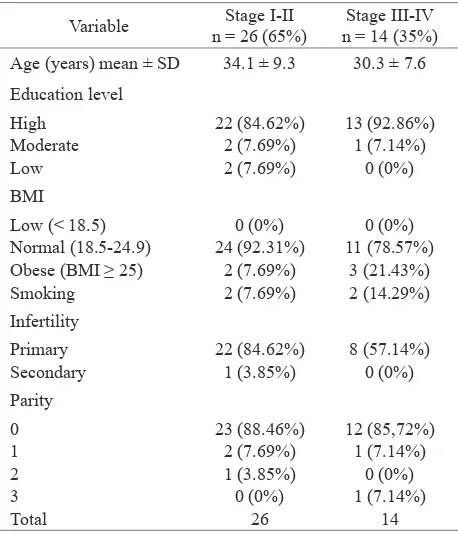
Forty patients with endometriosis were included in this study. Twenty six were classiied as endometriosis stage I-II and the remaining were classiied as stage III-IV. Table 1 shows the characteristic of study subjects.
Biologic markers
The results of statistical analysis of biologic markers are shown in table 2. There was no statistically signiicant difference of IL-6, TNF-α, and MMP-2 serum levels between stage I-II and III-IV. On the other hand, the comparison of mean VEGF serum level in stage I-II and stage III-IVdemonstrates signiicant difference (289.76 ± 188.13 vs 581.29 ± 512.85 pg/mL;p < 0.05).
Mean differences for biologic markers TNF-α and MMP-2 were shown in arithmetic difference (Δx) because they have normal distribution. Meanwhile, IL-6 and VEGF data had to be subjected to logarithmic transformation because they are not normally distributed therefore the mean differences were shown in geometric ratio. Area under curve (AUC) of receiver operating characteristic
Variable n = 26 (65%)Stage I-II n = 14 (35%)Stage III-IV Age (years) mean ± SD 34.1 ± 9.3 30.3 ± 7.6
Education level
High 22 (84.62%) 13 (92.86%) Moderate 2 (7.69%) 1 (7.14%)
Low 2 (7.69%) 0 (0%)
BMI
Low (< 18.5) 0 (0%) 0 (0%) Normal (18.5-24.9) 24 (92.31%) 11 (78.57%) Obese (BMI ≥ 25) 2 (7.69%) 3 (21.43%) Smoking 2 (7.69%) 2 (14.29%)
Infertility
Primary 22 (84.62%) 8 (57.14%) Secondary 1 (3.85%) 0 (0%) Parity
0 23 (88.46%) 12 (85,72%)
1 2 (7.69%) 1 (7.14%)
2 1 (3.85%) 0 (0%)
3 0 (0%) 1 (7.14%)
Total 26 14
Table 1.
Table 2.
Subject characteristics
Statistical analysis of serum biologic markers of endometriosis patients
(ROC) ≤ 0.5 has weak diagnostic value to differentiate stage I-II and stage III-IV.
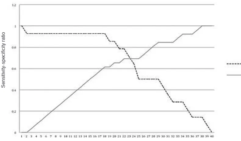
Cut-off point for serum VEGF level
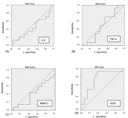
In bivariate analysis, only serum VEGF level had signiicant difference for endometriosis stage I-II vs stage III-IV. From ROC analysis, it is found that AUC of ROC 74.5% (p < 0.05) could be considered to have good diagnostic value (Figure 1). It is shown in table 3 several cut-off point for serum VEGF level to differentiate stage I-II and stage III-IV. Figure 2 demonstrates the optimal cut-off point in between
Variable Mean ± SD CI 95% p AUC
of ROC
p from
AUC IL-6 (pg/mL)
Std I-II 1.58 ± 0.78
Std III-IV 1.55 ± 0.98 -0.53-0.58 0.640 0.453 0.63 TNF-α (pg/mL)
Std I-II 1.50 ± 0.47
Std III-IV 1.49 ± 0.29 -0.27-0.29 0.078 0.499 0.989 MMP-2 (ng/mL)
Std I-II 152.04 ± 27.32
Std III-IV 140.98 ± 28.08 -7.44-29.57 0.857 0.402 0.314 VEGF (pg/mL)
Std I-II 289.76 ± 188.13
sensitivity and speciicity. The optimal value for serum VEGF level to differentiate stage I-II and stage III-IV is 314.75 pg/mL with sensitivity 78.6% and speciicity 69.2%.
DISCUSSION
Endometriosis is a subclinical chronic inlammation related to immune system.3,4,12,19 There has been several previous researchers who analyzed biologic marker level in endometriosis patients by comparing its increment to normal population. Biologic marker level can also be used to determine the progression of endometriosis. Based on Sampson’s theory of menstrual regurgitation, 10% women who had menstrual regurgitation will have endometrial cells which adhere to peritoneal wall. This adherence will in turn activate macrophage to trigger inlammatory response.1-4
This study was meant to see the progression of inlammatory response in advanced stage
(moderate-Figure 1.ROC graphics of serum biologic marker level. (A) IL-6,(B) TNF-α, (C) MMP-2, and (D) VEGF. Only VEGF has AUC more than diagonal line.
No Cut-off point
VEGF (pg/mL) Sensitivity Speciicity
1 56.20 1 0
2 66.65 0.929 0
...
18 285.60 0.929 0.615
19 289.95 0.857 0.615
20 295.85 0.857 0.654
21 305.00 0.786 0.654
22 314.75 0.786 0.692
23 323.95 0.714 0.692
24 346.80 0.643 0.692
25 368.05 0.500 0.692
26 372.95 0.500 0.731
...
40 2009.2 0 1
severe) of endometriosis in comparison with early (minimal-mild) stage. The results of this study show that there were no difference in IL-6, TNF-α, and Table 3.Several cut-off point for serum VEGF for differentiating
Figure 2. Sensitivity and speciicity curve of serum VEGF
MMP-2 serum level between stage I-II and stage III-IV. These biologic markers do not have diagnostic value to differentiate both conditions. The AUC-ROC of each serum biologic marker value, were < 50% with p-value > 0.05. This means their usage will have 50% of probability to differentiate both conditions.
IL-6 is a main indicator of acute inlammatory response. IL-6 has several biologic activities, including acute phase protein on hepatocyte, lymphocyte-B growth and differentiation, and lymphocyte-T activation. In fact, IL-6 and TNF-α are also secreted by peritoneal macrophage and ectopic endometriosis tissue.
The increase of peritoneal IL-6 and TNF-α level in patients with endometriosis has already been proven by many researchers. Maas23 performed in vitro study to examine angiogenesis process by injecting peritoneal luid of endometriotic women to chicken chorioalantoic membrane. The peritoneal luid was measured for cytokines of TNF-α, IL-1b, and IL-8.23 Only TNF-α showed signiicant positive correlation. Peritoneal luid of IL-6 and TNF-α also reported to increase proliferation from eutopic and ectopic endometrial tissue.23
MMP-2 system is assumed to have association with pathogenesis of endometriosis during implantation process. Matrix degradation is needed to ensure invasion of endometriosis implant. From in vitro experimental study, there was increment of MMP-2 expression from endometriosis cells of endometriosis women transplant.15 There is only one study that
compares the level of serum MMP-2 in endometriosis patients to control. Research performed by Huang22 had shown that there were increase in peritoneal luid and serum of MMP-2 in endometriosis patients. However, our study demonstrates different result from Huang by showing increment of MMP-2 in different stage of endometriosis.
Mihalyi19 performed a study to analyze six serum biologic markers (IL-6, IL-8, TNF-α, hs-CRP, CA-125, and CA-19-9) in attempt to ind the difference in these biologic markers level between endometriosis patients and control. It was found that only three serum biologic markers level (IL-6, IL-8 and CA-125) that increase in endometriosis patients compared to control.19 Serum IL-6 level had diagnostic value with AUC 70.5%, sensitivity 59% and speciicity 76.3% on optimal cut-off point.
Martinez24 and Othman25 reported mean serum IL-6 in endometriosis women stage I-II (28.4 and 5.39 pg/mL) was higher than stage III-IV (17.5 and 3.45 pg/mL). Only study conducted by Othman25 reported analytic statistical test to ind the mean difference between both endometriosis stages, which showed no signiicant difference between both conditions.
May had done systematic review of serum biologic markers in endometriosis patients compared to control. Studies that examined serum IL-6 and TNF-α still give conlicting result.12 Chae who conducted the study in Korea also reported no difference in serum TNF-α level of endometriosis women compared to control.26
Sensitivity
Speciicity
In pathogenesis of endometriosis, angiogenesis has important role. Angiogenesis is a complex process, including local degradation of basal membrane of native blood vessels and it is followed by migration and proliferation of endothelial cells. Angiogenesis is inluenced by angiogenic factors, one of which is VEGF.20 Stimulation of blood vessel growth by VEGF is already proven by in vitro and in vivo studies. McLaren reported that peritoneal luid VEGF level in endometriosis women was higher than control.27 Inlammation and neovascularization are also found in adjacent of ectopic endometrial tissue implant. Inlammatory leukocytes are found in this lesion and also in the peritoneal luid. Donnez had succeeded to show mRNA VEGF and expression of the protein in the endometriosis tissue.20 Study conducted by Cosin showed that peritoneal luid inluences VEGF secretion from neutrophils and ectopic endometrial tissue.28 Meanwhile, Estelles had proven positive correlation of peritoneal luid VEGF with ibrinolysis and metalloproteinase system with increment of urokinase plasminogen and MMP-3.29
The comparison of serum VEGF level in endometriosis and control was already examined by previous studies. In the systematic review done by May,12 at least four studies had already been conducted, all of which did not ind any increment of serum VEGF level compared to control. Only two studies showed the increment of VEGF level. Even though there was no study that differentiated endometriosis stage related to serum VEGF level.12
This study shows that there is signiicant difference of serum VEGF level in stage I-II and stage III-IV. Mean serum level of VEGF is 238.78 pg/mL in minimal-mild and 426.57 pg/mL in moderate-severe endometriosis. The difference ratio of mean is 1.81. Mean ratio was used rather than arithmetic ratio due to logarithmic transformation of data in the analysis. This difference has AUC of 74.5%. The cut-off point serum VEGF level is 314.75 with sensitivity 78.6% and speciicity 69.2%.
Manero30 found no difference of serum VEGF level in endometriosis patients who had no symptoms or minimal-mild dysmenorrhea compared to severe dysmenorrhea. Even though increment of serum VEGF had positive correlation with C-reactive protein.30 Study conducted by Nogueira found peritoneal luid VEGF level difference in endometriosis patients who had dyspareunia compared to patients who did not have the symptoms.31
The weakness of this study is that we did not consider the hormonal cycle of patient in the analysis. Estrogen
could affect the increment of serum MMP-2. Huang had shown positive correlation of serum 17b-E2 with serum MMP-2 level.22 Even though Othman showed that there was no difference in serum IL-6 and TNF-α level during proliferation phase and secretion phase of menstrual cycle.25 Our study also has limitation of internal validity of stress factor affecting the subjects before the surgery which could cause the increase of inlammatory factors.
In conclusion, this study shows that serum biomarkers of IL-6, TNF-α, and MMP-2 cannot be used to differentiate endometriosis stage I-II and stage III-IV. In contrast, VEGF level may be used to measure the degree of severity in endometriosis since higher VEGF level present in later stage of endometriosis. This result is in accordance with the pathophysiology of endometriosis.
Acknowledgment
We would like to thank dr. M Sopiyudin Dahlan, M.Epid as statistic consultant who wrote series of statistic books that used as statistic guidance analysis of this study.
REFERENCES
1. Jacoeb TZ. Faktor Imunoendokrinologis dan seluler lingkungan mikro zalir peritoneal yang berperan pada infertilitas idiopatik wanita [dissertation]. Mount Pleasant (MI): Universitas Indonesia; 1990. Indonesian.
2. Jacoeb TZ, Hadisaputra W. Penanganan endometriosis: panduan klinis dan algoritme. 1 ed. Jakarta: CV Sagung
pada gangguan folikulogenesis sebagai gambaran penurunan kualitas oosit pasien endometriosis yang infertil
[dissertation]. Mount Pleasant (MI): Universitas Airlangga; 2007. Indonesian.
6. Oepomo TD. Peran interleukin-6 serta interleukin-8 dalam zalir peritoneal penderita infertilitas disertai endometriosis
dalam proses apoptosis sel granulosa avarii yang patologis
[dissertation]. Mount Pleasant (MI): Universitas Airlangga; 2003. Indonesian.
7. Adiyono W. Dampak penambahan gonadotropin releasing hormon analog pada operasi laparoskopi terhadap manifestasi klinis, imunologis dan kualitas hidup
penderita endometriosis [dissertation]. Mount Pleasant (MI); 2003. Indonesian.
8. Stegmann BJ, Funk MJ, Sinaii N, et al. A logistic model for the prediction of endometriosis. Fertil Steril 2009;91(1):51-5.
9. Hadisaputra W. Endometriosis: tinjauan perangai
visualisasi Laparoskopi. Maj Obstet Ginekol Indones. 2007;31(3):180-4. Indonesian.
10. ASRM PC. Endometriosis and infertility. Fertil Steril 2004;81(5):1441-6.
11. Vignali M, Infantino M, Matrone R, et al. Endometriosis: novel etiopathogenetic concepts and clinical prespectives. Fertil Steril 2002;78(4):665-78.
12. May K, Conduit-Hulbert S, Villar J, Kirtley S, Kennedy S, Becker C. Peripheral biomarkers of endometriosis: a systematic review. Hum Reprod Update. 2010;16(6):651-74.
13. Gagne D, Rivard M, Page M, Shazand K, Hugo P, Gosselin D. Blood leukocyte subsets are modulated in patients with endometriosis. Fertil Steril 2003;80(1):43-53.
14. Gupta S, Goldberg JM, Aziz N, Goldberg E, Krajcir N, Agarwal A. Pathogenic mechanisms in endometriosis-associated infertility. Fertil Steril 2008;90(2):247-57. 15. Pitsos M, Kanakas N. The role of matrix metalloproteinases
in the pathogenesis of endometriosis. Reprod Sci. 2009;16(8):717-26.
16. Donnez J, Smoes P, Gillerot S, Casanas-Roux F, Nisolle M. Vascular endothelial growth factor (VEGF) in endometriosis. Hum Reprod. 1998;13(6):1686-90.
17. Laschke MW, Menger MD. In vitro and in vivo approaches
to study angiogenesis in the pathophysiology and therapy
of endometriosis. Hum Reprod Update. 2007;13(4):331-42. 18. Witz CA. Interleukin-6: another piece of the
endometriosis-cytokine puzzle. Fertil Steril 2000;73(2):212-4.
19. Mihalyi A, Gevaert O, Kyama C, et al. Non-invasive
diagnosis of endometriosis based on a combined
analysis of six plasma biomarkers. Hum Reprod. 2010;25(3):654-64.
20. Matalliotakis I, Goumenou A, Koumantakis G, et al. Serum
concentration of growth factors in women with and without
endometriosis: the action of anti-endometriosis medicines. Int Immunopharmacol. 2003;3(1):81-9.
21. Xavier P, Belo L, Beires J, et al. Serum levels of VEGF and TNF-alpha and their association with C-reactive protein in patients with endometriosis. Arch Gynecol Obstet. 2006;273(4):227-31.
22. Huang H-F, Hong L-H, Tan Y, Sheng J-Z. Matrix metalloproteinase 2 is associated with changes in steroid hormones in the sera and peritoneal luid of patients with endometriosis. Fertil Steril 2004;81(5):1235-9.
23. Maas JW, Groothuis PG, Dunselman GA, de Goeij AF, Struijker-Boudier HA, Evers JL. Development
of endometriosis-like lesions after transplantation of human endometrial fragments onto the chick
embryo chorioallantoic membrane. Hum Reprod. 2001;16(4):627-31.
24. Martinez S, Garrido N, Coperias J, et al. Serum
interleukin-6 levels are elevated in women with minimal
mild endometriosis. Hum Reprod. 2007;22(3):836-42. 25. Othman EE-DR, Hornung D, Hosam, et al. Serum cytokines
as biomarker for nonsurgical prediction of endometriosis. Eur J Obstet Gynecol. 2007;137:240-6.
26. Chae SJ, Kim H, Jee BC, Suh CS, Kim SH, Kim JG. Tumor
necrosis factor (TNF)-TNF receptor gene polymorphisms
and their serum levels in Korean women with endometriosis. Am J Reprod Immunol. 2008;60(5):432-9.
27. McLaren J. Vascular endothelial growth factor and endometriotic angiogenesis. Hum Reprod Update. 2000;6:45-55.
28. Cosin R, Estelles JG, Ramon L, et al. Inluence of peritoneal luid on the expression of angiogenic and proteolytic
factors in cultures of endometrial cells from women with
endometriosis. Hum Reprod. 2010;25(2):398-405.
29. Gilabert-Estelles J, Ramon LA, Espana F, et al. Expression
of angiogenic factors in endometriosis: relationship to
ibrinolytic and metalloproteinase systems. Hum Reprod. 2007;22(8):2120-7.
30. Garcia-Manero M, Santana GT, Alcazar JL. Relationship between microvascular density and expression of
vascular endothelial growth factor in patients with
ovarian endometriosis. J Womens Health (Larchmt). 2008;17(5):777-82.