166 Hadineg,oro Med J Indones
The kinetics of endotoxin and cytokines
in dengue
hemorrhagic
fever
Sri
Rezeki
H
Hadinegoro
Abstrak
Endotoksin serum dapaî ditemukan pada 63,3Vo di antara 120 kasus demam berdarah dengue (DBD). Proporsi endotoksemia maupun kadar endotoksin lebih tinggi pada sindrom syok daripada kelompok non-syok (berturut turut 13.9 dan 4.3 pg/ml, p=0'0008). Keadaan sindrom syok pada DBD mempunyai risiko 3 kali lebih besar untuk menderita endotoksemia daripada kelompok non-syok (OR 3.0; intervalkepercayaan 95Vo 1.29-7.05 ) Endotoksinmeningkatpada saat teriadi syokdanmenurunkembali apabila syok telah teratasi. Risiko endotoksemia untuk menjadi infeksi dengue berat 5.8 kali lebih tinggi daripada kelompok tanpa endotoksemia (OR 5.5; interval kepercayaan 95Vo 1.72-21.74). Tuntor nekrosis faktor alfa dan interleukin-6 merupakan jenis sitokinyang ditemukan pada kasus DBD-Sebagai mediator pro-inflamasi TNF-a meninglat pada fase awal penyakit dan menurun kembali pada fase penyembuhan, sedangkan IL-6 dapat ditemukan di dalam sirkulasi pada awalfase penyembuhan sebagai mekanisme umpan balik atas Peningkatan endotoksin dan TNF-a.
Abstract
Endotortnwas detected in 63lo among 120 dengue hemorrhagicfever (DHF) cases. The proportion of endotoxemia in shock group were higher than that non-shock as well as serum endotoxin level (13.9 and 4.3 pg/ml respectively, p=0.0008). Shock group has 3.0 times
isk
to have endotoxemia than that non-shock DHF group (OR 3.0; 1.29-7.05 95Vo CI). Serum end.otoin concentration increased at the time of shock and decrease after shock recovered. Patients with endotoxemia has 5.8 timeshigher
risk to develop severe dengue infectioncomparedtonon-endotoxemiagroup(OR5.5; 1.72-2I.7495VoCI).Pre-inflammatorymediatorTNF-acanbedetectedatthe beginningofrhediseaseanddisappearedonconvalescencesta7e,whilelL-6titerincreasedattheearly
convalescencephaseasafeed back mechanisû, to endotoxin andTNF-x.Keyw ords : endoloxin, cytokines, dengue hemorrhagic fever
Dengue hemorrhagic fever (DHF)
has been
alreadyknown
in
Indonesia since
two decades
ago.
As
anendemic
disease, dengueinfection
did not
appear as a healthproblem
at thattime,
butduring
the last 20 yearsmany
fatal
caseswere
reported.'
Government's
effort
has been succesful to decrease the mortality rate; how-ever,this
diseasethrough national
health proqram, themortality
rate in severalhospitals
is still high.'-'Bettercase management
is
needed, especially
for shock
syndrome.Shock
is
the
primary
evidence in
severeDHF,
other
vital
organ involvement are secondary
to
shock.6Hospital
datashow that
mortality of
shock
casesis
3to
10 times compared tonon
shock group. On theother
hand, the severity
of
shock
sy.ndrome dependson
thepathogenesis
of
the
disease.'
A
group
of scientist
hypothesized thatfour
pathways were activatedduring
the course
of
illness,
i.e.
complement, endothel,
platelets,
andcytokine pathway.
These fourpathways
work
synergistically. Complement and cytokines
promote hypovolemic shock via anaphylatoxin
sub-stance
(C3a, C5a) release.o
Cytokine
as
pre-inflam-matory mediators
of infectious
agents^ seemsto
play
arole in
the pathogenesis
of
DHF.e The
cytokine
production
is needed for
natural and specific
host
immunity
against
infection. Contrarily,
overproduc-tion of cytokine by monocytes
as the subsequenttarget
organin DHF
induced
cell damage.
Other
effects
of
increased vascular
permeability
andshock
areischemia, reperfusion,
and damage ofintes-tinal mucosa
integrity.l0'll Th"*" condifio;s
promote
bacterial/endotoxin translocation
from
intestinal
lumen
to lymphatic duct and
finally to
the
blood
cir-culation.l2il3'Endotoxin
as
an outér
capsul
of Gram
negative bacteria
has abiological activity
to stimulate
cardiovascular
and respiratory
system, complement,
coagulation,
metabolic, and cytokine
cascades.'"
Tumor necrosis factor-cx, interleukin-1, and
inter-Department of Child Health Faculty of Medicine Universityof
Vol 8, No 3, July - September 1999
leukin-6
are the most common cvtokinesthat
areac-tivated
by
endotoxemia.l5
Finaliy,
endotoxemia
will
induce severe
shock and hemorrhage
in DHF
cases.Concerning
the matter mentioned
above, the roles ofendotoxin and cytokine
in
DHF
should be further
studied.
METHODS
A
prospective, observational study was
conducted on
120 DHFpatients who were hospitalized
in Dr. Cipto
Mangunkusumo
Hospital Jakarta, during 26 month
period, from February 1993 through
April
1995. The diagnosis was establishedaccording to WHO
diagnos-tic
criteria, confirmed
by
hemaglutination
inhibition
and dengue
blot IgM/IgG
serological test. Intended
sample was taken from theDHF
cases who fulfilled theinclusions criteria.
The inclusion criterias were grade
I,
II, III,
andIV
of
DIIF
cases, no signs and symptomsof
bacterial infections,
and
all
patients signed the
informed consent.
Clinical
and laboratory
evaluation
Clinical
dataof DHF patients were recorded
onadmis-sion.
Clinical
condition and laboratory
examination
were followed
i.e. time of shock recovered,
complica-tion
of
shock (prolonged
or recurrent shock), time
of
defervesence
(fever
ceased), and gastrointestinal
bleeding.
Hemoglobin
content, hematocrit value,
WBC, differential count, and
platelet count were
ex-amined at the
Child
Health
Department Dr.Cipto
Mangunkusumo
hospital,
while
serum complement
C3, C4, and fibrinogen titers were examined
at the
Clinical
Pathology Department
of
the same hospital.
Examination of hemoglobin
content,hematocrit value,
andplatelet, count were repeated
every
6 hours until
the patients condition improved significantly.Limulus Amoebocyte Lysate (Endospecy) test
Serum
endotoxins were detected
by
chromogenic
limulus test,
i.e. limulus
amoebocyte lysate
(En-dospecy) test made by
Seigaku
Kogyo
Co.Ltd.Japan
with endotoxin
standardE.Coli ET.O11l:B4. The
pur-pose
of
pretreatment
of
serum specimen
using new
perchloric
acid was to minimize
the influence
of
en-dotoxin
inhibitor.
Besides endospecy test, an this studyalso
used
limulus toxicolor
test. Endospecy and
Toxicolor limulus
test was used simultaneously to
exclude the positive resultby p-glucan
as acomponent
of
mycosis, that could be
detected
by
toxicolor test
only.
Kinetics of endotoxin in
DHF
167All
glasses,tubes, and pipettes were heated
at 180
oCfor 4 hours by
autoclave to get pyrogen free, aswell
as the heparin solution and diluted water.Two milliliters
of
venous blood
were/poured
into
the heparini-zed
endotoxin free tube 30
IU/ml
(madeby Dalcon
com-pany), then centrifuged
at
1000xg
for
10 minutes.
Afterwards,
the procedure was done asfollowed,
50 Ul plasma
|
+ 50Ul
0.18 NaoHIncubated at37oC in 5 minutes
|
+ 50Ul
0.32 M perchloric acid Incubated at 37" Cin l0
minutes|
+ 100 Ul 0.18 M NaOH 150 Ul of aliquot|
+50Ut
0.2 M Tris-HCl buffer (pH8)100 Ul aliquot
|
+100 Ul Endospecy orToxicolor testIncubated at 37o C in 30 minutes
t
Diazotiation by 25Vo acetic acid
I
Read on spectrophotometer OD (optimal density) 545 nm
(Ref. K.Inada)
Control positive
and negative tests
were needed
for
each20 specimen
in
Endospecy and Toxicolor
test,respectively.
All
examinations
a were donein duplo.
Cytokine
AssayCytokine was examined
quantitatively by
atwo-
stepmonoclonal antibody method,
i.e
enzyme linked
im-muno-sorbent
assay(Elisa)
rnethod, madeby Endogen
lnc.Boston
MA,
USA,
used
for
TNF-g
detection;
while
detection
of il-6
concentration was done by
Genyme-kit,
BostonMA, USA.
The limitsof detection
of
these assays
was 3.0 pglml. The principle
of
cytokine
assay used was avidin-biotin amplifier in atwo-step sandwich method,
as describedbelow
:l.
Microplate
of 96wells covered by cytokine specific
monoclonal antibody
(TNF-crv and IL-6). This
an-tibody was called
aprimary antibody.
2.
Put
specimeninto
the wells of the microplate that
had been covered by primaryantibody.
3.
Then,
put
secondary monoclonal antibody
i.e.
biotinylated
anti-human cytokine (TNF-a
or IL-6)
to get the antigen-antibody complex.4.
Avidin
enzym
(HRP) which
has ahigh afinity to
biotin put
thereafter
to
get the antigen-antibody
eîzyme complex.
168 Hadinegoro
Endotoxin, TNF-c,
and IL-6 assays wereexamined
at theDepartment
of Bacteriology Iwate Medical Univer-sity, Morioka, Japan.Examination
of serum endotoxin,TNF-cr,
and
IL-6
titers were done every day during
hospitalization
(3
to
5
days).
^The
serum
specimen were collected and stored at -70uC before theexamina-tion
was done.Serological
test
Serological hemagglutination
inhibition (HI)
and Den-gue Blot IgM andIgG tests were taken admission
and on dischargefrom hospital. Serological
hemagglutina-tion
inhibition
were examined
at
the laboratory of
National Institute
of Health
Ministry of Health
of
In-donesia, while
Dengue
Blot IgM
andIgG tests were
done at the Prodia Laboratory, Jakarta.Research design
The designs
of
this study
were (1) cross sectional to
get the clinicalcharacteristics
and laboratory data, andthe endotoxemia prevalence.
(2) Follow-up study to
get thefigure of endotoxin, TNF-ct,
and IL-6 kinetics
during
the clinical course
of
the disease.
The sample
size was calculated
for
study
on endotoxemia
titer in
each stadium
of
the illness
by
estimation
of
the
en-dotoxemia proportion
of
non-shock
DHF
cases of
27Vo, Oddsratio 3.0, c, value
0.05,
and
B0.20. The
calculated sample size was
64.Statistics and analysis
Data were recorded
in
a the researchform and were
analyzed by EPI Infb 6 and SPSS programs. Oddsratio
was calculated
for
the different
endotoxemia
prevalence with
95Voconfidence
intervals,
x2 and p
value.
T
test was used
for
the difference
of
the two
independent means.
For proportion *2 o, Fisher Exact
test were used. Statistically significance
difference
was p<0,05.RESULTS
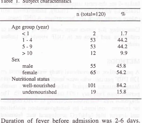
Subject characteristics
The mean age of the study
subjects was6.1
(SD
3.0) years,the youngest was 8 months
andthe oldest
was 13 yearsold. There were
45.8Vofemales and
54.2Vomales. Among
120patients,19 (l5.8Vo) were
under-nourished
(< l0
percentile
of NCHS).
Table
l.
Subject characteristicsMed J Indones
n (total=I20) 7o
Age group (year)
<l
1-4
5-9
>10
Sexmale female Nutritional status
well-nourished undernourished
2
53 53
12
55 65
r01
19
L7 44.2 44.2 9.9 45.8
54.2
84.2
I 5.8
Duration
of
fever
before admission was
2-6
days.Symptoms of vomiting, diarrhea,
andconvulsion
werecommon findings in young
children, while abdominal
pain wascommonly found in older children.
Sixty four
(53.3Vo) cases had evidenceof
shock. Seventy-eight
percent of cases showed petechi ae and I 47o amon g I 20 cases hadgastrointestinal bleeding.
Table 2. Clinical manifestations on admission
Clinical pictures n (Total=I20) Vo
Duration of fever (day) 2
3
4
5
6 Shock Hepatomegaly
Other signs
&
symptoms VomitingDiarrhoea Epigastric Pain Convulsion Bleeding manifestations
Positive Toumiquette Test Petechiae
Epistaxis Hematemesis Melena
t2
3841 J 3
64 87
42 28 t3
l0
77 94
21
t7
l9
10.0 31.1 34.2 2.5 2.5 53.3 72.5
35,0 23.3 61.5 8.3
64.2 78.3
17.5
14.2 15.8
Endotoxemia
[image:3.595.340.576.88.293.2] [image:3.595.342.577.394.673.2]Vol 8, No 3, JuIy - September 1999
Table 3. Endotoxin titre in DHF shock and non-shock
Grade of Illness Cases Mean Rank (pglml)
Kinetics of endotoxin in
DHF
169Table 5. Laboratory findings in endotoxaemia Endotoxemia (+)
Mean (957oCI)
Endotoxemia (-)
Mean (957oCI) Non-shock
Shock
56 64
4.3
13.9
Platelet (/pl)
Hemoconcentra-tion (Vo)
C3 (mg/dl)
C4 (mg/dl)
Fibrinogen (mgld[)
89500 (15000-348000) 24.6 (5.7-67.2) 20.6 (0.42.2)
t7.r (2.8-31.4) 95.5 (5.2-18s.8)
96500 ( 12000-323000)
15.7 (2.5-50.0) 34.7 (13.1-56.3)
23.O (9.'t-36.'7)
139 (52.7-225.3) Total
Mann-Vy'hitneyTest
z=-3,3656
pvalue=0.0008The endotoxin
meantiter in
thenon-shock group
was4.3 pglml, below
thecut off point
(9.8pg/ml) while in
the shock
group
was three timeshigher
than thatin
thenon-shock group
(13.9pg/ml).
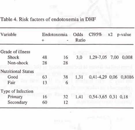
[n this study 76(63.3Vo)among 120
DHF
cases suffered
from
endotoxemia
(Table
4);
the proportion
of
endotoxemia
in
shockgroup
was
higher than that
in
non-shock group.
Therisk of shock group for having endotoxemia
was three times compared to thatof non-shock group
(Oddsratio
3.0,
1.29-7 .05of
95Voconfidence interval,
p value was0.008).
Table 4. Risk factors of endotoxemia in DHF
Variable Endotoxemia
Odds
Cl95Vox2
p-value+
-
RatioCytokine
assayCytokine examinations
i.e.
TNF-a
and
IL-6
titer
asshown
in Table
6. There was nosignificant difference
between
TNF-ct
andIL-6
meantiter
inendotoxemia or
without endotoxemia
group.Table 6. TNF-a and IL-6 titer in endotoxemia
Cytokine
EndotoxemiaEndotoxemia
z
p*(Mean
rank
(+)
G)pg/ml)
TNF-o 50.7
n=38
-l.1535 0.2487
50.0
n=36
-0.7192
0.47205,4
t20
58.
I
n=72Grade of illness Shock Non-shock
Nutritional Status
Good
Fair
Type of Infection
Primary Secondary
The
decreaseof C3
andC4 conçentration
wasclearly
shown
in endotoxemia group.
Thesefindings support
the high
level
of
hemoconcentration
in
endotoxemia
group. Hemoconcentration
in endotoxemia group
wasabove
20Vo,indicating
severe plasma leakage.
Con-sumption
of
fibrinogen
in
endotoxemia was higher
than
non-endotoxemia group,
while
theplatelet count
was
almost similar between
thetwo
groups.IL-6
54.5n=69
*Mann-Whitney Rank Test
The
follow-up
study showed that theduration of fever,
evidence
of shock, and gastrointestinal bleeding
during hospitalization were the important factors
of
prognosis. The
meanof the duration
of fever was
1.2(SD 0.9)
days,
in
shock
group
it
was
longer than in
non-shock group. The
meanof
the duration
of
shock was48.0 (SD 26.6) minutes,
while
in survivors
cases ofprolonged
andrecurrent
shockit
was 9 1.4 (SD26.3)
minutes.
Attention
should
bepaid to DHF
caseswho
had
gastrointestinal bleeding
onadmission,
since 737oamong
them continued bleeding
during
hospitaliza-tion.
The mean
of
the duration
of
gastrointestinal
bleeding
was2.1 (SD
1.0) days.Severe
dengue
infection
Severe dengue hemorrhagic
fever (shock group
andtheir
complications,
i.e. prolonged shock,
recurrent
shock,
gastrointestinal bleeding,
andencephalopathy)
occurred
in
32
(26,7Vo)among 120 studied
subjects.48 28
t6
28
3.0
1,29-7,05 7,00 0.00863
t3
l6 60
38
l,3l
632
l,4l t2o,4l-4,29 0,06 0.8086
[image:4.595.83.317.382.608.2]Hadinegoro
The median endotoxin
titer in
sêvereDHF
washigher
than that
in DHF, especially
in those with prolonged
shock,
recurrent shock,
or
gastrointestinal
haemorr-hage.
Median cndotoxin
titer
in
dengue
encephalo-pathy
wasbelow
thecut off point endotoxin level. The
cause
of
death
of 4
among
9
death patients
wereprolonged
andrecurrent shock which
havehigher
en-dotoxin titer
comparedto other
5 death patientswhich
were caused
by gastrointestinal bleeding
andencepha-lopathy.
Table 7. Endotoxin titer in severe DHF
Med J Indones
[image:5.595.341.575.93.204.2] [image:5.595.341.574.234.491.2] [image:5.595.80.314.235.382.2]Day
Figure
I.
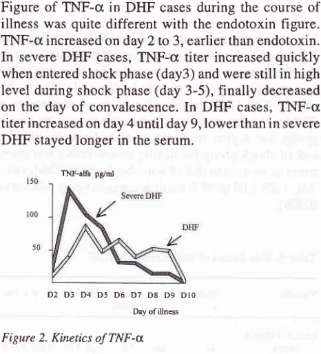
Kinetics of endotoxemiaFigure of TNF-c in DHF
casesduring
the course of
illness was quite different with
the endotoxin figure.
TNF-cr
increased on day 2 to 3,earlier
thanendotoxin.
In
severeDHF
cases,TNF-cr titer increased quickly
when entered shock phase
(day3)
andwere still in high
level during shock
phase(day
3-5), finally
decreasedon the day of convalescence. In
DHF
cases,TNF-c
titer
increased on day 4until
day 9,lower
thanin
severeDHF
stayedlonger in
the serum.)7 D8 D9 Dlo Day of illness
Figure 2. Kinetics of TNF-x
Interleukin-6 titer
at thebeginning
of theillness
didnot
increase
in DHF, while in
severe casesIL-6
increasedon day 5. The highest titer of
IL-6
was on day
6-7(phase
ofconvalescence)
and disappeared on day 9. So,IL-6 titer increased after shock recovered
andon
thetime of
convalescence.D2 D3 D4 D5 D6 D7 D8 D9 DIO
Day ofilbess
Figure 3. Kinetics of
Il-6
80
t35
Severe DHF
Cases (n=32)
Endotoxin (pe/ml)
Median Range
Prolonged Shock Recurrent Shock GI Bleeding Encephalopathy
Death
t5
l6
6
9
22.6
24.1 10.7
4.7 17.1
2.3-r80
1.6-1 0
0-r80
0-79 0-180
Endotoxemia
as asingle
dependentvariable proved to
be a
predictor
of
severeDHFas
shownin Table
8,with
an Odds ratio
of 5,8
(95Voconfidence
intervals:
1,7-21,7,
pvalue 0,0019).
Table 8. Proportion of severe dengue infection
Severe DHF
(+) C)
Endotoxemia
OR = 5.8 (Cl95Vo 1.72
-21.74)
x2 =9.60
p=0.00194Endotoxin
appearedin the
serumon day 3 of illness,
reaching the
peak level on day 6 (shock phase),
anddisappeared
after day 7
(convalescence phase).Com-pared
to the more moderate DHF
cases,endotoxin
in
severeDHF
appearedearlier
and had ahigher level.
96
44 48
40
28
(+)
(-)
120
84 32
D2 D3 D4 D5 D6 D? D8 D9
TNF-alfa pglol
[image:5.595.81.315.468.628.2]Vol 8, No 3, JuIy - September 1999
As
apre-inflammatory
mediator in
infections,
TNF-cr
titer
in
DHF
increased at thebegining of
the courseof
illness
and
decreasedimmediately
after the
infection
ceased.This picture
was more
clearly
seenin
severeDHF
than thatin DHF
group.
In DHF group
as aresult
of
immune response,
high
TNF-ct level
wasstill
seenin
the
convalescence phase.
This
study proved
that
TNF-a titer did
not correlate
significantly with
endo-toxemia; however
TNF-a
and
endotoxin
have quite
similar kinetic with
the pathogenesis and the courseof
illness
of
this
disease.Another cytokine
detected
in
DHF
was
IL-6.
Interleukin-6
titer in DHF
correlated
significantly
with
endotoxin and TNF-cr
titer.
This
correlation
was shown
in
the
follow-up
study
on
theIL-6
kinetic
level
during
hospitalization.
In
DHF
group,IL-6
did
not reachcut-off
point
eitherfollowing
endotoxin
or
TNF-cr
level. This figure
was
quite
dif-ferent compared to thatof severe DHF
group.In severe
DHF
group, endotoxin
aswell
as TNF-cr reachedhigh
level, followed by
increasedIL-6
titer.
As
there wasstatistically significant positive correlation
betweenIL-6
titer
andendotoxemia,
it was
suggestedthat
theincreased
IL-6
in severe
DHF
group was due
to
anegative
feedback
mechanismof endotoxin
andTNF-u
production.
DISCUSSION
In the last
five
years manyendotoxin
studies were donein
animal experiments, unfortunately there was
noavailable animal study
in
DHF.l0-t2 This study
was conducted toexplore
theroles of endotoxin in DHF by
systematic
clinical
and laboratory
observation.
Kinetics
of
endotoxin are characterized as
speciesspecific
in
animal
studies.Biological
respons and im-mune statusin
human ar-e the mostimportant
partsfor
endotoxin elimination.
l)
Endotoxin
is
a
polysaccharide component
of outer
capsul
Gram negative bacteria.
In
general, thereis
noendotoxemia
in viral
infection. Morales et
al proved
that endotoxin could
reach
the circulation
due
to
is-chaemia
or histological intestinal
mucosa
damagein
post
haemorrhagic shock
in
animal study.l0
Severalfactors,
i.e.,intestinal
trauma,burn,
shock, severemal-nutrition,
and prolonged parenterâlnutrition
also causeintestinal ischemia
to
promote bacterial/
endotoxin
translocation
from
gut
flora
to blood
circulation.
This
study
supported thehypothesis
ofbacterial/
endotoxin
translocation
in
DHF,
as
shown, endotoxin
titer
in-creasedthree times
in
shock compared
to
non-shock
group,
i.e.,
18.9
pg/ml
and
4.3
pglml
respectively.
Non-shock
group DHF
never reachedcut-off point
at9.8
pg/ml during
hospitalization.
Kinetics of endotoxin in DHF 171
The prevalence
of
endotoxemia
in DHF in this
studywas 63,3Vo,
it
was higher
in
shock compared
to
non-shock group. Usawattanakul
from Thailand
reported
that
endotoxemia occured in 43.5Vo
among
57 DHF
patient,
theprevalence
washigher
in
shock
comparedto
non-shock
group.'o Limulus
testswas
usedin
this
two
studies
were different.
This
study used
Limulus
Amoebocyte Lysate
(LAL)
test
which is more
sensi-tive
than
gelatin limulus
test
(semiquantitative)
usedby
Usa.
Plasmaendotoxin
concentration
is not
quite
similar to
endotoxin
tissueconcentration. Some
plas-ma protein such as transferrin,
HDL
(high
density
lipoprotein),
and
other
acute phaseprotein
will bind
endotoxin
in
the circulation. New-PCA
(perchloric
acid)
which contain hydroxy sodium
to
break
theconnection
betweenendotoxin
andplasma
qlotein,
to
get the true plasma
endotoxin concentration.'7
By
n"*
procedure
for
pre-treatment,
LAL test
is
more
ac-curate than
gelatin
limulus
test.DHF
cases can be divided into by shock and non-shock cases. Thetwo
groups havedifferent prognosis. In this
study, the risk to have endotoxemia
in
shock group wasthree times higher than non-shock group
(OR
3.095VoC11.29-7 .05).
This result
supportNimmannitya's
statement
that
shock
in
severe
DHF
is
primary
evidence
andother organ disturbance
is
secondaryto
shock.6
Hypovolemi"
sfrockin DHF
is
due tocomple-ment activation by release
of
anaphylatoxin mediators
(C3a, C5a).
Malasit
reported that
biological
effects
of
C5a
are
to
increase
capillary permeability,
increasechemotactic
effect
of
neutrophils and
monocytes,
stimulation
of monocytes
to
cytokines
release
par-ticularly TNF,
IL-1,
and
Il-6,
degranulation
of
mastcells followed
by
histamin
release,
stimulate
pros-taglandin
andfree radical
o^yg"n.8
Complement
con-centration
in this
study
was decrease dueto
activated
classic
and alternative pathways as reported by
Malasit.8
Shereported
hei
study on complàment
sys-tem
five
yearslater, proved that
in DHF,
SC5b-9
was activated.In
conclusion, complement activation
is themost important pathway
in
the pathogenesisof DHF.
Endotoxemia group
has
low
C3
concentration
com-pared
to
non-endotoxemia
group, but this
difference
did
not occurin
C4concentration. In
theearly
stageof
the disease
C3
and C4concentration
decreased dueto
classic and alternative pathways
activation
by
either
dengue
virus
or
immune
complex; after
endotoxin
entered the
circulation
C3 was
decreasedby
classicalpathway activation
due toendotoxin
release.Endotoxin
concentration
was detectedon
day threeof
172 Hadinegoro
5, and 6, and reached the
highest concentration
on day5,
and declined
on day
7 of
illness.
This
kinetics
followed
DHF
pathogenesis,
ie. febrile
phaseon
dayl-3,
shockphase
on
day3-7, and
convalescence phase after day 7ofillness.
So, endotoxin increased infebrile
phase and reached
the highest concentration on
shockphase, and
endotoxin concentration
as
well
asbody
temperature
decreasedon
convalescence
phase.This
result pointed
out that temperature was a goodclinical
parameter
for
follow-up
during
the courseof illness.
Evidence
of
endotoxemia correlated
with
gastrointes-tinal
hemorrhage.Endotoxin concentration
washigher
DHF
in
which gastrointestinal
haemorrhage occured
more than2
days.Disturbances of coagulation
systemand
decreaseof
platelet
are
the important
causesof
gastrointestinal
haemorrhagein
D[IF.
In this
study, nodiffererrce was observed between
platelet
concentra-tion
in
endotoxemia
or
no
endotoxin group; while
fibrinogen concentration showed significance
dif-ference
between
thetwo
groups (p=0,005).
The causeof
hemorrhagein
DHF
ismultifactorial,
as reportedby
Isarangkurà and
Funahara.ls'19
Bhu-urapravati
reported a massive haemorrhage of renal
cortical
tissue(Waterhouse-Friederichsen syndrome,
a
picture
of
severe
kidney
damagein
septic/
endotoxic
shock) in
the autopsy study,it
is suggested thatendotoxin played
a rolein fatal
severe dengueinfection
cases.'On
the
second
day
of
illness
65.5Voof
cases have increasedTNF-cr concentration, at29.5
pglml. Tumor
necrosis
factor
as
a
pro-inflammatory
mediator
produced
by
activated macrophage as a target organof
dengue
infection.'''^
IncreasedTNF-cl
concentration
occured either
in
shock
or
non-shock group.
In
the severeDHF
case(profound
shock
called
DI{F
gradeIV)
TNF-cr titer
was increaseduntil
theinflammation
process ceased. The concequences
of
increasedTNF-cr
titer
were
increased capillary
endothel permeability,
decreased tissueoxygen perfusion,
andfinally
patients
died
due tomultiorgan
failure."
Serum
TNF-a
concentration
increased on the secondday while endotoxin
increased
on the next day,
andserum
TNF-cr concentration reached highest
con-centration
on fourth
day
of illness, earlier
thanendo-toxemia
reached
the
highest concentration.
[t
is
suggestedTNF-cr
hasimportant role
since
thebegin-ning
of
the
illness
without
influence
of
endotoxin
stimulation.
This
result supported the opinion
that
production
of
cytokine occured at
thebeginning of
theillness stimuiated
by
immune complex
to
macro-phage.Med J Indones
Cytokine activation
stimulated
through cytokine
net-work,
aswell
asTNF-ct
stimulated
interferron, [L-1,
lL-6, or
TNF-a
(auto-induction
cycle).23
In
the
preliminary
study,
IL-l
as aspecific protein or
as an acuteinflammatory
mediator could not
be detectedin
DHF
patients.
Hanley et al
studied
56 children with
DHF
gradeI
toIV.'*
Hefound
TNF-cr
andIL-6
at thebeginning
of
illness
and disappearedon day 5
of
ill-ness.
At
the
same
time
TNF-cr
and
IL-6
can
bedetected
in
the urine
due
to
elimination
of
thesecytokine via kidney.
Interleukin-6,
also called asinter-feron-b,
is a mediator
which
stimulates
growth
factor
andB lymphocyte
differentiation,
alsogive
anegative
feed back to
TNF-c
andIL-1.
Thebiological function
of IL-6
is
stimulating
hepatocyteproduce
acute phaseprotein
and
IL-6
called
as anti-inflammatory
mediator.22
In viral infection,
thefunction
of
IL-6
was againstvirus activation via
RNA
andprotein
synthesisnon-specific
mechanism.
[n
this study
IL-6
titer
in-creased at median
titer
7.8pglml, in
shockgroup
IL-6
concentration was
higher thannon-shock group.
Therewas positive relation between endotoxin and
IL-6
levels.
IncreasedIL-6 titer in
shock group was
dueto
negatived feed
back
mechanism
to
endotoxemia
via
TNF-cr stimuli by
blockade endotoxin-induced
IL-1,
TNF-cr
andIL-6. IL-6
increasedslowly until
day 7of
illness
then
decreasedgradually.
This study
showedpositive correlation
between
IL-6
and endotoxin
aswell
asIL-6
andTNF-cr.
So,the aim
of
increasing
of
IL-6
concentration is to
reduce negativeeffect
ofTNF-c
and endotoxin. Examination ofcytokine
serumcon-centration
at
one time
does
not give
a true
value,because
cytokines have short
half
life
and
will
beeliminated
via kidney
immediately.24 Serial
examina-tion
of
cytokine
serum
concentration
will
show
thekinetics
of
the
truevalue
of
cytokine.2sOur
previous
experience showed thatabout 1/3
ofDHF
cases
suffered
from
shocksyndrome and
1/3of
shock caseswere
supposedto
have
recurrent
&
prolonged
shock,
aswell
asmassive gastrointestinal bleeding.)
Other
complication ie.
encephalopathy
occured only
in
5-7VoDIIF
cases.In this
study, recurrent
shock,prolonged shock, gastrointestinal bleeding more
than2
days,encephalopathy, and
all
death cases,were
all
found in
severe dengueinfection.
The
endotoxin titer
of severe
DHF
was 20.9pglml,
almostfour time higher
thanDHF
(5.4pg/rnl).
Endotoxin titer
increased at thetime
of
shock
and
very high
titer
showed
in
severe dengueinfection.
The
risk
to
develop
severeDHF in
endotoxemia group was 5,8 times
higher
than that
in
Vol 8, No 3, JuIy - September 1999
CONCLUSIONS
Endotoxemia
occurred
in
dengue hemorrhagic fever
cases
(63,3Vo),
in
shock
grotp
(75Vo)as
well
as
in
non-shock
group (507o);endotoxin titer in
shockgroup
(13,9
pg/ml)
wassignificantly
higher
than thatin
non-shock group (4,3
pglml).
The risk
to
develop
en-dotoxemia
in
shockgroup
was threetimes higher
thanthat
in
non-shock
DHF
group. TNF-cr
level
increasedduring
theeally
courseof illness
and thekinetics
weresimilar
with
the
pathogenesisof
the disease.In DHF,
no correlation was found
between increased
TNF-a
titer
andendotoxin
titer. Endotoxin titer in DHF
has apositive relation with
hematocrit
aswell
aswith IL-6
titer.
On
theother
hand,IL-6 titer
had
apositive
rela-tion with
TNF-a
titer.
It
is
suggestedthat
increasedIL-6
titer
related
with
feed back
mechanism
of
in-creased
endotoxin
and
TNF-cr
titer. The
laboratory
parameters
i.e.
platelet
count, C3, C4,
andfibrinogen
titer in
endotoxemia
DHF
cases werelower
than thosein non
endotoxemia
caseswhile the
hematocrit
titer
was higher. The
risk to
develop a
severe course
of
illness
in
endotoxemiaDHF
cases was 5.8 timeshigher
than that
in
non endotoxemia
cases.Acknowledgments
I
thank
Prof.
Masao
Yoshida
andDr.
Katsuya
InadaPhD
from
Department
of Bacteriology
Iwate
Univer-sity Morioka
Japan,
for
their
support
and
guidanceduring my endotoxin
andcytokine
assaystudy.
Also
thanks
to Dr. Riady Wirawan
for helping
with
thecomplement
and
fibrinogen
assays,Drh.
SuharyonoWuryadi
MPH
for
the
hemagglutination
inhibition
serological
dengue test, and ProdiaLaboratory
Jakartafor IgM
andIgG
dengueantibody examination.
REFERENCES
1.
Sumarmo. Demam berdarah (Dengue) pada anak.Tjo-kronegoro A, ed. Jakarta, UI Press 1983.
2.
SurosoT.
Perkembangan demam berdarah denguedi
In-donesia. Disampaikan pada Seminar Demam Berdarah Den-gue. Jakarta, Departemen Kesehatan 8 Juni 1991.3.
Azhali MS. Demam berdarah dengue: pengalaman di BagianIlmu Kesehatan Anak RS Hasan Sadikin Bandung. Cermin Dunia Kedok Terapi. 1992;81: 62-5.
4.
SachroADB.
Demam berdarah dengue: pengalaman di Bagian Ilmu Kesehatan Anak RS Karyadi Semarang. Cer-min Dunia Kedok 1992; 81: 66-9.5.
Hadinegoro SR, Nathin AN. The changing pattern of clinical manifestationsin
dengue haemorrhagic fever: a ten-year observations. Disampaikan pada The Intemational Sym-posiumon
Dengue Fever&
Dengue Haemorrhagic Fever. Bangkok, 1-3 Oktober 1990.Kinetics of endotoxin in
DHF
1736.
Nimmannitya S. Clinical manifestations of dengue/dengue haemonhagic fever. In: Thongchaeron P, ed. Monograph on dengue/dengue haemorrhagic fever.New Delhi:
World Health Organization Regional Ofhce for South-East Asia.-Regional Publication 1993; 22: 48-54.7.
Kurane I, Rothman AL, Livingstone PG, Green S, Nimman-nitya S,Innis FA. Immunopathologic mechanisms of dengue hemorrhagic fever and dengue shock syndrome. Arch VirolSuppl 1994; 9:59-64.
8.
MalasitP,
Mongkolsapaya, NimmannityaS,
Suvatte S.Complement in dengue haemorrhagic fever/ dengue shock syndrome. In: Proc. the
XIIIth
Intemational Congress for Tropical Medicine and Malaria. Pattaya, 29 November-4 December 1992.9.
Bhakdi S, Kazatchkine MD. Pathogenesisof
dengue: analtemative hypothesis. The International Symposium on Dengue Fever & Dengue Hemorrhagic Fever. Bangkok,
l-3
October 1990,602-7.
10. Morales J, Kibsey P, Thomas PD, Poznansky MJ, Hamilton SM. The effect
of
ischemia and ischemia reperfusion on bacterial translocation,lipid
peroxidation, ancl gut histol-ogy:study on hemorrhagic shock in pigs. J Trauma 1992;33:22t-7.
1 1. Pasquale MD, Cipoile JH, Cerra FB. Bacterial translocation:
myth versus reality. In: Reinhart K, Eyrich K, Sprung C, ed.
Sepsis current perspective in pathophysiology and therapy;
1 st.ed. Berlin, Budapest: Sprin ger-Verla g, 199 4: 84- I 06. 12. Deitch EA, MaWJ, Ma L, Berg R, Specian RD.
Endotoxin-induced bacterial translocation:
a
studyof mechanisms.
Surgery 1989; 106: 292-300.13. Edminton CE, Condon RE. Bacterial translocation. Surg Gynaec & Obstet
l99l;173:72-83.
14. Van Deventer SJH, Buller HR, Ten Cate JW, Sturk A, Pauw W. Endotoxaemia an early predictor on septicemia in febrile patients. Lancet 1988; 605-8.
15. Suffredini AF. Endotoxin administration to human. Dalam:
Lamy M,
Thijs
L. eds.
Mediators
of
sepsis. Berlin: Springer Verlag, 1992: 13-20.16. Usawathanakul
W,
NimmannityaS,
Sarabenjawong K, Tharavanij S. Endotoxin and dengue haemorrhagic fever. Southeast Asian J Trop Med PubHlth
1987; 8:8-12. 17. InadaK,
Endo S, TakahashiK,
SuzukiM,
Yoshida M.Establishment of a new perchloric acid treatment method to allow determination of the total endotoxin content in human plasmaby thelimulus test and clinical application. Microbiol
Immunol 1991; 35: 303-14.
18. Isarangkura PB, Pongpanich B, Pintadit P, Planichyakam. Haemostatic dearrangement in dengue haemonhagic fever. South East Asian J Trop Med Pub Hlth 1989; 20:325-30. 19. Funahara
Y,
liadinegoro SR, NathinMA,
TamaelaLA,
Dharma S, Sumarmo. One of
the
cause of recurrent shock in DHF. The Intemational on Dengue Fever/ Dengue Hemor-rhagic Fever. Bangkokl-3
October 1990.20. Bhamarapravati N. Pathogenesis
of
dengue hemorrhagic fever. The Intemational Symposium on Dengue and Dengue Hemorrhagic Fever. Bangkok,l-3
October 1990.174 Hadinegoro
Health Organization Regional Office for South-East Asia.-Regional Publication 1993; 22: 80- 103.
22.
Abbas
AB,
Lichtman AH,
PoberJS, ed.
Cytokinesmediators. In: Cellular and molecular immunology. lst ed.
Philadelphia: WB Saunders 1991; 259-81.
23. Brenner
MK.
The cytokinenetwork. In:
Galvani DW,Cawley
JC,
ed. Cytokine therapy.lst
ed. Cambridge: Cambridge University Press 1992: 177-86.J Indones
24. Hanley PDO. Potential pathogenic roles
of
acute inflam-matory cytokines andHLA
statusin
DHF. Cermin Dunia Kedok 1992;8l:
17.2434.25. Hadinegoro SR. Plasma endotoxin study on dengue hemor-'
rhagic fever cases. In: Harun SR, Inada K. eds, Proceedings
on one day seminar on endotoxin. Jakarta: University
of