Vol 8, No 4, October - December 1999 Glioblastoma multiforme in young patients 233
Presentation
of
glioblastoma
multiforme
in
young
patients:
a retrospective
analysis
from a
regional cancer center of
Northern
India
D.K.
Parida
Abstrak
Glioblastoma multifurme (GBM) yang lazimnya terjadi pada usia dekade ke 5 dan ke 6, juga dapat mengenai pasien anak. Pembedahan masih merupakan cara penanganan yang utama. Namun, kira-kira 50Eo tumor yang mengenai batang otak merupakan glioblastoma multifurme, di mana hanya radioterapi yang bisa diberikan. Dalam tulisan
ini
dilaporkan analisal1
penderita GBM antaratahun 1992-],996. IJsiapenderitaberkisar antara6 bulansampail5 tahun.
Sembilanpenderitatelahmengalamioperasi. Delapan pendertta mendapat radioterapi pasca bedah, dan delapan lainnya mendapat kemoterapi. Lamanyafollow-up berkisar antara 2-24 bulan dengan median 10,5 bulan. Pada waktu evaluasi, Iima penderita tidak menunjukkan tanda-tanda sakit, tiga penderita dengan status penyakit yang stabil dan dua dengan penyakit progresif. Dapat disimpulkan bahwa pendekatan terapi multimodal memberikanhasiL yang lebih baik dibandinglan dengan terapi tunggal dan pasien dengan tingkat penyakit yang masih dapat dioperasi menunjukkan waktu bebas penyakit yang lebih panjang.
Abstract
Glioblastoma multiforme (GBM) though commonly occurs in
fifih
to sixth decade of life, also found in paediatic patients. Surgery remains the mainstay of the management. However around 50Vo of the brainstem space occupying lesions are glioblastoma muLtiforme, wlrcre radiotherapy is the only therapeutic modality to be offered. We analysed II
patients of GBM between 1992 to 1996. The ageof
the patients were between 6 months and I5 years. Nine patients had undergone surgery, eight patients were ffeated with post-operative radiotherapy and eight patients had received chemotherapy. The
follow
up period ranged from 2-24 months with a median of 10.5 nxonths. At the time of analysis five patients were without any evidence of disease, there had stable and two patients had progressive disease. It may be concluded that multimodaltherapeutic approachoffereda betterdisease control than single modality and the surgically operable group showed a prolonged disease free survival.Keywords
:
G lioblastoma multifurme, radiotherapy, chemotherapy, brain tumorBrain
tumors
are the mostcommon solid malignancies
occuring
in children.
Out
of all cancers,
brain tumors
are
second
to
leukemia
in
young
patients.
Glioblas-toma multiforme
(GBM)
though
not
common in
children, accounts for
lTVoof them.l
The mean agefor
GBM
is around 54 yearswith
a malepredilection.
Themanagement
of
GBM in pediatric
patients
is a
chal-lenge
for the
neurosurgeon, radiotherapist
aswell
aspediatric oncologist. To
achieve
tumor control
at thecost of
growing
brain is a very critical cquation.In
spiteof
the
combination
of
various
therapeutic modalities,
the outcome
has
not
changed
significantly over
theperiod
of time. However
with
the advent oftechnologi-cal
innovations, improved neurosurgical techniques
D e p ar tment of Radio therapy,
ALI India Institute of Medical Sciences, New Delhi-110029, India
combined
with
newer
anaesthetic approach
and stereotacticradiotherapy,
thequality of
life
and diseasefree survival
have definitely changed.METHODS
This analysis was
carried out
with an
aim to
know
theexact incidence
of
GBM occuring in young patients,
their
prognostic factors
and clinical course.A total of
I I
patients were analysed
from
January 1992
tlll
December
1996.All
the patients hadhistopathological
proof
of
GBM.
All
the patients
were
evaluated
in
acombined
pediatric
cancer
clinic
and
they were
seen234 Parida
neurological
status
were recorded
very
carefully.
Simultaneously,
pre-anaesthetic
check-up were
alsodone.
Our institutional policy
is to
deliver 50 Gy
in
28-30
fractions to
the
whole brain followed by
boost
to
the tumor bed
with
a
margin
of
3 cm
till
66
Gy.
Medical
decompression
was done wheneverrequired.
Only two
patients
could tolerate the
full
course of
radiotherapy. Five
patients
received a
dose
between40-60
Gy
and one patient received 36
Gy.
Chemotherapy
was
apart
of
the
treatment protocol.
Eight
patients received chemotherapy
following
surgery
sisted
of
either
or
/m2,D11,
etoposid
2mglmz,
Dl)
and
3
weeks.A minimum of
threecycles
wereinstituted.
At
the endthe patients were followed
up at
an interval
of
six
weeks
for
thefirst
year, 2months
for
second year and 3 months there after. Change ofperformance
status andsymptoms were
carefully
noted during
follow
upvisits. Imaging.
studies were performed
at aninterval
of
six months or whenever required.
RESULTS
The
ageof
the
patients
varied
from 6
months
to
15years.
Majority
of
the patients were between l0-15
years
of
age.The
youngestpatient
wasof
6months
of
age at thetime of
diagnosis. There wasonly
onefemale
patient
in
this
series.
The most common
anatomical
sites were temporal (4), parieto occipital (3),
brain
stem
(2) followed by
temporoparietal
(1)
and
fourth
ventricle
(l).
Majority
of
the patients
in
this
seriesprcsented
with the
symptoms
of
vomiting,
headache,seizures.
Three patients complained
of
weakness
in
thcir extremities
and one presentedwith
doublevision.
All
these f'eatures are elaborated intable
I . In this seriesnine
patients had undergone
surgical
decompression andtwo
hadonly
biopsy.
VP
shuntwas given
in five
patients.
Eight patients had received post
operative
radiotherapy
which
was delivered
by
tele-cobalt
unit
with
parallel opposed
port.
The detailed
treatment
profile
is
given
in
table
2.
The radiotherapy
doseranged
fiom
36-66 Gy (Table
3). Thefollow
upperiod
ranged between
2-24
monrhs
with
a median
of
10.5months.
At
thetime
of
analysis
two
patients were lost
to fbllow
up. Out
of
nine
evaluable patients, five
patients were
without any
disease
and rest
of
the patients werehaving
disease outof whom two
patientswere
with
progressive
disease(Table 4).
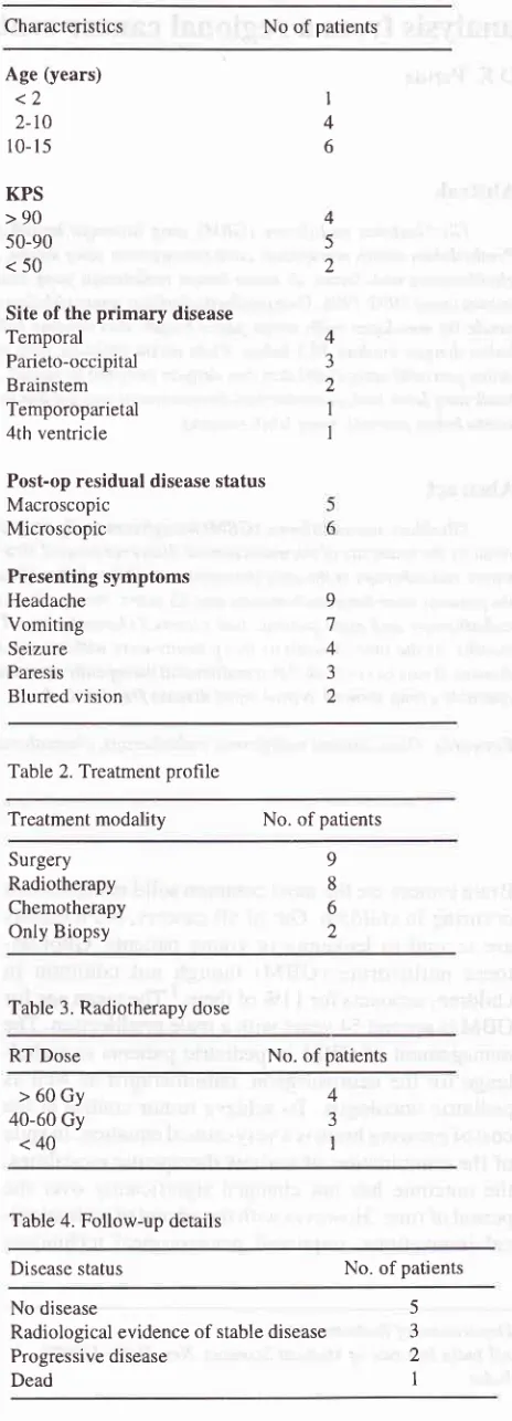
Table 1. Pre-treatment patient characteristic
Med J Indones
Characteristics No of patients
Age (years)
<2
2-t0 10-
l5
KPS
>90
50-90
<50
Site of the
primary
disease TemporalParieto-occipital Brainstem Temporoparietal 4th ventricle
Post-op residual disease status Macroscopic Microscopic Presenting symptoms Headache Vomiting Seizure Paresis Blurred vision I 4 6 4 5 2 4 J 2 1 I 9 7 4 3 2
Table 2. Treatment prohle
Treatment modality No. of patients
Surgery Radiotherapy Chemotherapy Only Biopsy 9 8 6 2
Table 3. Radiotherapy dose
RT Dose No. of patients
>60Gy
40-60 Gy
<40
4
3
I
Table 4. Follow-up details
Disease status No. of patients
No disease
Radiological evidence of stable disease Progressive disease
Dead
5 3
2
[image:2.595.341.573.103.746.2]VoL 8, No 4, October - December 1999
DISCUSSION
GBM
in paediatric patients
pose amajor
challengefor
the
oncologist. Beside surgery, radiotherapy
is
themain
^adjuvant
modality available
for
the
manage-ment.' Radiation
planning
is
the sole most important
aspect
for
theradiotherapist.
At
thetime of planning
it
should
bekept
in mind
that
thebrain
of
thechild
is
in
growing state
and
the
child
will
not
co-operate
for
positioning.
Computerised
tomographic (CT)
scan isthe major
diagnostic
tool
for
the
investigation
of
childhood brain tumors. But
it
has gotlittle limitations
in
case thetumor
is close to the baseof
skull
andin
thebrain
stem,where magnetic
resonanceimaging
(MRI)
is
abetter
option.
Contrast
enhancement studiesgive
better
sensitivity.
Our experience
showsthat
MRI
hasshown
to
be
more
effective
and sensitive
in
demonstrating
a
greater anatomic
details
for
the
in-filtrating
type
of
tumors.
The
clinical
symptoms
depend on the
anatomical location of
thelesion
and theage
of
thepatient.
GBM
situatedin
theposterior
fossa or cerebral hemisphere have a poorprognosis.
A
max-imum survival rate
of
2)Vo.can be
expected
in
thechildren
with
these
disease.'
Patients
who
undergosurgical debulking
live
longer
than those
without it.
This
difference
actually
depends
on the amount of
surgical
resection.3'4'sTh"
iole
of
chemotherapy in
paediatric
patientsis
still
investigational. Recently
one largetrial
ofAmerican
Children's
Cancer StudyGroup
concluded
that there was amarked
survival
advantageat 2 years and 5 years
in
the groupwho
receivedCT
in
addition
to
surgery and radiotherapy.
Randomised
studies have established that
addition
ofpost
operative
radiotherapy prolong
thesurvival.u
Albright
et al havereported
that most
of
the brainstem tumors are of
highgrade
nature.l Surgery
remains
the
mainstay
of
treatment.
It
helps
to
establish
the
diagnosis, tumor
debulking improves
the symptom
and increases
thesensitivity to
other modalities
of
treatment leading to
prolonged survival. Many
retrospective studies
have established therole of
theextent
of
surgical debulking
on the
ultimate survival period. Prognostically
it
is
thepost-operative residual tumor volume
which
isimpor-iunt
thutr the pre-operative
volume.3'4'5 Some other
prognostic factors
like
age Karnofsky
PerformanceScale
(KPS),
duration
of
symptoms
andlevel of
con-sciousness have already been establishedin
adults.We
have
tried
to
corelate
some
of
the
features
in
our
analysis.
It
was
found
that
theyounger
the ageof
thepatient,
the less was the tolerance tovarious modalities
of
treatment.
And in
patients
with
good performance
status,
the
response
was better. The patients
who
presented
with
symptoms
with
shorter duration,
tolerated
the treatmentbetter
and also respondedwell.
Glioblastoma multiforme inyoung
patients
235
Besides
slrf"ety,
radiotherapy
is animportant adjuvant
modality,'"'"'"
but in
caseof
brainstem
GBM it
is
theonly option. Brain Tumor Study Group (BTSG)
hasalready proved that the whole
brain irradiation with
atotal
dose
of
55-60
Gy
increases
the survival
over
surgery alone.
All
the patients
in
the
study
group
received
whole brain
radiotherapy
followed by
boost
to the
tumor
till
66 Gy.It
was observed that thepatients
who
received
higher dose, survived better.
As the
survival in
thesepatients
is limited,
achieving tumor
control
becomesmore
important
than the late effects
of
radiotherapy
on brain.
At
the
present
time, with
advent
of
newer technology,
availability
of
better
anaesthetic drugs,
delivering
radiotherapy to
thebrain
of
theyoung patients
hasbecome much easier.
Somecenters
are alsotrying different protocols like
hyper-fractionation,
interstitial
brachytherapy
andstereotac-tic
radiotherapy along
with
or
without
radiosensitisersare
being
tried
to improve upon
thelocal
control.
Therole
of
chemotherapy
ls stitÎ
investigational.T'8
How-ever, the study
by
the American Children's
CancerStudy Group
showed asurvival
advantage whengiven
along
with
surgery and radiotherapy.
Due
to
lack
of
therapeutic
specificity, intrinsic
cellular
resistance, in-toleranceof normal
tissues todrug
toxicity,
therole
of
chemotherapy
in
children
has
not
shown much
im-provement
in
patient survival.
In
conclusion
theincidence
of GBM in children
is not
so
common.
Due
to
less tolerance
to
various
therapeutic
modalities,
limited
survival and lack
of
proper
randomisedtrials
optimum combination
of
dif-ferent modalities
arenot possible. The tumor
behaves more aggressively than the adult ones. Surgery remains themainstay
of
treatment andradiotherapy
happensto
be themain adjuvant
therapy.REFERENCES
1. Albright AL,
Price RA, Guthkelch AN. Brainstem gliomasof
children: a clinicopathological study. Cancer 1983;52:2313-19.
2.
Ammirati M,Vick
N, LiaoY
et al. Effectof
the extent ofsurgical resection on survival and quality of life in patients
with
supratentorialGBM
and anaplastic astrocytoma. Neurosurg 1987 ;21 :2Ol -6.3.
Devaux BC, O'Fallon JR, Kelly PJ. Resection, biopsy andsurvival in malignant glial neoplasms: a retrospective study
of
clinical parameters, therapy and outcome. J Neurosurg 1993;78:767-75.4.
Curran tùy'J Jr, Scott CB, HortonJ
et al. Does extentof
236
5.
Parida
Simpson JR, Horton J, Scott C et al. Influence of location
and extent of surgical resection on survival of patients with GBM: results of three consecutive Radiotherapy Oncology (RTOG)
Clinical
Trials.Int J
Radioth OncolBiol
Phys1993 ;26:239. Walker MD, Green SB, Petal B D. Randomi sed
comparisons of radiotherapy and nitrosoureas for the
treat-ment
of
malignantglioma after
surgeryNEJM
1980;303:1323-29.
Deutsch
M,
Green SB, StrikeTA
et al. Resultsof
a ran-domisedtrial
comparing BCNU plus Radiotherapy,strep-tozotocin plus radiotherapy, BCNU plus hyperfractionated
radiotherapy and
BCNU
following
misonidazole plusradiotherapy
in
the post operative treatmentof
malignant glioma. Int J Radioth Oncol Biol Phys 1989;16:1389-96.Med J Indones
7.
WalkerMD,
Alexander E, Hunt WE et al. Evaluationof
BCNU 8clor radiotherapy
in
the treatmentof
malignant glioma; a co-operative clinical trial. J Neurosurg 1978;49:.333-43.
Liv
H, Davis R, Vestnys P, Resser K, Levin V. Correlationof
survival
and diagnosisin
supratentorial malignant gliomas. J Neuro Oncol 1984;2:268-73.Wood JR, Green SB, Shapiro WR. The prognostic impor-tance
of
tumor sizein
malignant gliomas: a computed tomographic scan studyby
the brain Tumor Cooperative Group. J Clin Oncol 1988;6:338-45.8.