O R I G I N A L A R T I C L E
Sergey ShabalaÆ Yuda Hariadi
Effects of magnesium availability on the activity of plasma membrane ion
transporters and light-induced responses from broad bean leaf mesophyll
Received: 12 September 2004 / Accepted: 14 October 2004 / Published online: 12 January 2005
Springer-Verlag 2005
Abstract Considering the physiological significance of Mg homeostasis in plants, surprisingly little is known about the molecular and ionic mechanisms mediating Mg transport across the plasma membrane and the impact of Mg availability on transport processes at the plasmalemma. In this study, a non-invasive ion-selec-tive microelectrode technique (MIFE) was used to characterize the effects of Mg availability on the activity of plasma membrane H+, K+, Ca2+, and Mg2+ transporters in the mesophyll cells of broad bean (Vicia fabaL.) plants. Based on the stoichiometry of ion-flux changes and results of pharmacological experiments, we suggest that at least two mechanisms are involved in Mg2+ uptake across the plasma membrane of bean mesophyll cells. One of them is a non-selective cation channel, also permeable to K+ and Ca2+. The other mechanism, operating at con-centrations below 30 lM, was speculated to be an H+/
Mg+ exchanger. Experiments performed on leaves grown at different levels of Mg availability (from deficient to excessive) showed that Mg availability has a significant impact on the activity of plasma-mem-brane transporters for Ca2+, K+, and H+. We discuss the physiological significance of Mg-induced changes in leaf electrophysiological responses to light and the ionic mechanisms underlying this process.
Keywords LightÆ MagnesiumÆIon transport Æ
Plasma membraneÆVicia faba
Abbreviations: ROS: Reactive oxygen species ÆNSCC:
Non-selective cation channel
Introduction
Magnesium is an essential nutrient that plays a key role in plant photosynthesis. In addition to being a central atom of the chlorophyll molecule, Mg is essential for the functioning of many enzymes, including RNA polyme-rases, ATPases, protein kinases, phosphatases, gluta-thione synthase, and carboxylases (Marschner 1995; Shaul 2002). Insertion of Mg2+ into the porphyrin structure during chlorophyll formation is catalyzed by Mg2+-chelatase. Many key chloroplast enzymes are strongly affected by small variations in Mg levels (Shaul
2002). Thus, Mg plays a fundamental role in both the ‘light’ and ‘dark’ reactions of photosynthesis. Both Mg deficiency and Mg oversupply have detrimental effects on plant photosynthesis. There are numerous reports that the rate of photosynthesis is severely reduced in leaves of Mg-deficient plants (Fischer 1997; Sun and Payn 1999; Ridolfi and Garrec 2000). Several factors, including structure and function of chloroplasts (Lavon and Goldsmith1999), impaired export of carbohydrates from source to sink sites (Cakmak et al. 1994), and production of reactive oxygen species (ROS) (Cakmak
1994), have been implicated in photosynthetic inhibition under Mg-deficiency conditions. Increased concentra-tions of free Mg may also impair photosynthesis via multiple pathways such as inhibition of K+ transport from the cytosol to the stroma, possible interference with Mg homeostasis inside the chloroplast, and im-paired regulation of transport events across the tono-plast (Shaul2002).
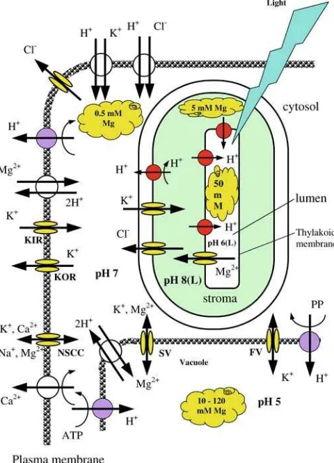
Magnesium control over membrane-transport pro-cesses in plant cells is rather complex. To date, the best studied are effects of Mg on transport activity in tono-plast and chlorotono-plast membranes. Physiological levels of Mg significantly affect activity of both slow (SV) and fast (FV) activating vacuolar ion channels (Allen and Sanders 1997; Tikhonova et al. 1997; Pottosin et al.
1997; Pottosin and Muniz 2002), thus playing an important role in maintaining the cytosolic homeostasis
S. Shabala (&)ÆY. Hariadi
School of Agricultural Science,
University of Tasmania, Private Bag 254, Hobart, TAS, 7001, Australia
E-mail: [email protected] Fax: +61-3-62262642
and in the regulation of cell turgor (Bruggemann et al.
1999; Pei et al.1999; Shaul 2002). In addition to regu-lation of vacuolar ion channels, Mg acts as an allosteric activator of vacuolar inorganic pyrophosphatase (with at least two Mg-binding sites known; Fraichard et al.
1996) and thus can modulate the activity of both the V-ATPase and the VPPase (Shaul2002). This is crucial for cellular pH homeostasis and turgor regulation, in both photosynthetically competent mesophyll cells and sto-mata guard cells. Also important is the role of Mg2+in membrane-transport processes in chloroplasts, where free Mg contributes to the regulation of photosynthetic enzyme activity (Shaul 2002). Additionally, proton pumping from the stroma into thylakoids lumen results in significant (DpH 2–3; Remis et al.1986) acidification of the thylakoid lumen. This massive light-driven transport of H+into the thylakoid lumen is electrically balanced by the counterflow of other ions (Hinnah and Wagner 1998) and, particularly, Mg2+ (Pottosin and Scho¨nknecht 1996). No details of specific transporters mediating Mg2+ transport across the chloroplast enve-lope are known to date.
Even more rudimentary is our knowledge of specific molecular and ionic mechanisms mediating Mg trans-port across the plasma membrane. It is not currently known through which transport proteins Mg enters the root symplasm and is loaded and unloaded from the xylem and phloem (Shaul 2002). The impact of Mg availability on the activity of other plasma membrane transporters also remains to be described. Despite recent advances in molecular biology studies of Mg2+ trans-port in yeast and bacterial cells (Gardner 2003), very little is known about the specific Mg transporters at the plasma membrane in higher plant cells. The aim of this study was to characterize the effects of Mg availability on the activity of major plasma-membrane transporters (those for K+, H+, Ca+) in photosynthesizing leaf tis-sues, and to relate Mg-induced ion-flux changes in re-sponse to light/dark fluctuations with cell photosynthetic performance and leaf expansion growth. Using the non-invasive ion-flux measuring (MIFE) technique, we were able to quantify (to our knowledge, for the first time) net Mg fluxes across the plasma membrane of leaf mesophyll cells and their responses to light fluctuations. Our data suggest that Mg availability significantly modifies transport of all major ions (H+, Ca2+, and K+) and provides some clues about mecha-nisms underlying Mg2+ transport across the plasma membrane.
Materials and methods
Plant material
Broad beans (Vicia fabaL cv. Coles Dwarf, Cresswell’s Seeds, New Norfolk, Australia) were grown hydropon-ically from seeds in a 70% : 30% (v/v) sterilized sand: perlite mix in a glasshouse as described in our previous
publication (Hariadi and Shabala 2004). Plants were grown at three different magnesium concentrations: deficient (1 ppm), optimal (50 ppm), and excessive (200 ppm), or 0.04, 2.0, and 8.0 mM, respectively. These are designated as Mg1, Mg50, and Mg200, respectively. For the yield data, see Hariadi and Shabala (2004). Our preference for parts per million rather than SI units is explained by an attempt to make our results comparable with the bulk of literature data (Tisdale et al. 1993). Plants were used for measurements after 4 weeks. Four hours before measurements, the fifth leaf was excised, transferred to the laboratory, and kept at room tem-perature (22C) under 0.45 W m 2illumination (Sylva-nia TLD 58 W cool fluorescence tubes, Australia), with the petiole immersed in distilled water. All measure-ments were made at the same time of the day, between 2 and 6 p.m.
Non-invasive ion-flux measurements
The leaf epidermis was gently removed, and mesophyll segments of about 5 mm·7 mm in size were cut and left floating in an aerated nutrient solution (0.1 mM CaCl2
+ 0.2 mM KCl) essentially for about 1.5–2 h as de-scribed by Shabala et al. (2000). One hour prior to the measurements, mesophyll segments were immobilized in a Perspex holder and then placed into a measuring chamber, filled with the appropriate solution (see details below). Dim green microscope light (1 W m 2) was used as background illumination during segment preparation and throughout the experiments.
Net fluxes of H+, K+, Ca2+, and Mg2+ were mea-sured using the non-invasive ion-selective vibrating microelectrode (MIFE) technique (University of Tas-mania, Hobart), generally as described in our previous publications (Shabala et al.1997; Shabala2000; Shabala and Lew 2002). Specific details of the liquid ionic ex-changes (LIX) and backfilling solutions used are given in Table1. All LIX were purchased from Fluka. The ref-erence electrode was a glass capillary of about 30lm
diameter, filled with 1 M KCl made up in 2% agar. Prior to measurements, electrode tips were positioned 50lm above the leaf surface, with their tips aligned and
separated by 1–3lm. During measurements, electrodes
were moved back and forth in a square-wave manner by a computerized stepper motor between two positions, 50 and 80lm above the leaf surface, with 0.1 Hz
Table 1 Liquid ion exchangers and backfilling solutions used for preparation of ion-selective microelectrodes
Potassium 60031 500 mol m 3KCl
Calcium 21048 500 mol m 3CaCl2
frequency. Net ion fluxes were calculated from measured differences in electrochemical potential for each ion be-tween these two positions as described earlier (Shabala et al.1997; Newman2001).
Nutrient uptake kinetics experiments
Mesophyll segments from severely Mg-deficient leaves (Mg1) were used for these experiments. Leaf samples were pre-incubated in 500 ll of measuring solution
(so-called ‘‘buffer’’ solution, 0.1 mM CaCl2+ 0.2 mM KCl,
pH 5.6) until steady fluxes were reached (usually 1–1.5 h after removal of epidermis). Steady-state fluxes were measured for about 5 min, and then an appropriate amount of Mg was added to the measuring solution, and transient ion flux kinetics recorded. To minimize the time required to reach the unstirred layer conditions (a prerequisite for MIFE measurements; Newman 2001), the chamber volume was made very small (1 ml), and Mg was added as a double-stock Mg solution made up in 500ll of the ‘‘buffer’’. With such experimental design,
the final solution concentration was reached at 20–25 s after Mg addition. Consequently, only the first 30–40 s were rejected during the data analysis, significantly improving the time resolution of experiments compared with our previous reports (Shabala2000; Shabala et al.
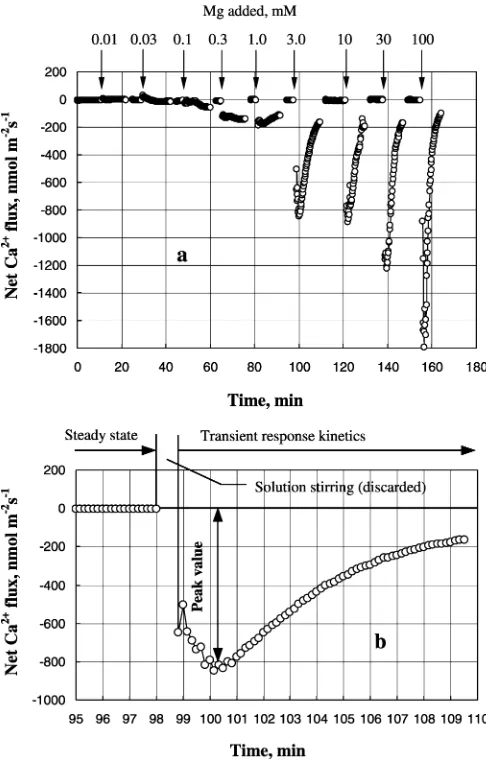
2000,2003). Altogether, nine different magnesium con-centrations were used (Fig. 1), covering the range of Mg in the measuring solution from 10lM to 100 mM.
For each segment, transient flux responses were measured for about 15–20 min. Then, a new chamber containing a fresh segment, pre-incubated in a ‘‘buffer’’ (i.e. Mg-free) solution, was put onto the microscope stage. The steady-state fluxes were measured, and the procedure repeated again, this time adding a different amount of Mg. Four to six segments were measured for each Mg treatment; each of these segments was cut from a different leaf.
Light-induced transients
Leaves were excised from bean plants grown in Mg1, Mg50, and Mg200 solutions (Hariadi and Shabala2004) and prepared and immobilized as described above. A mesophyll segment was transferred to a measurement chamber containing 5 ml measurement solutions (in mM: 0.1 CaCl2+ 0.2 KCl + 0.2 MgCl2) and left for 20–
30 min to adapt to light conditions (dim green micro-scope light of about 1 W m 2). After measuring steady-state (dark) ion fluxes for 5–10 min, the light treatment was applied (white light of 80lmol m 2s 1; fibreoptics
cool light KL 1500 LCD, Schott, Germany). Transient light responses were measured for 20–30 min or until the steady state was reached. This process was repeated for different mesophyll leaf segments, grown at different Mg levels, from deficient (1 ppm; Mg1) to excessive (200 ppm; Mg200) (see Hariadi and Shabala 2004 for
more details) in a random sequence. A total of 8–12 segments were measured for each treatment.
The above protocol was used to measure light-in-duced kinetics of H+, K+, and Ca2+ fluxes from leaf segments growth at various Mg supplies. Measurements of Mg2+fluxes, however, were significantly complicated by the confounding effects of Ca2+ on Mg2+ LIX selectivity. As all commercially available Mg LIX are very sensitive to Ca2+ (Fluka 1996 Selectophore cata-logue), there was a great danger of misinterpreting measured Mg2+flux in response to light treatment when Ca2+ was present in the measuring solution. To avoid this problem, Ca2+was omitted from the solution dur-ing Mg2+flux measurements, Although some Ca2+may have leaked from the tissue, its overall concentration
was too small to significantly affect Mg2+ flux mea-surements (<8 lM Ca2+compared with 200lM Mg2+
in the measuring solution). It should be noted that omitting Ca2+ from the measuring solution caused no significant changes in either H+of K+flux responses to light (data not shown), making light-induced Mg2+ transients comparable with the rest of the data.
Results
Nutrient uptake kinetics
Each cell possesses a sophisticated network of ion transporters, with numerous interactions and feedbacks between them. This significantly complicates the inter-pretation of long-term kinetics of ions in response to a particular stimulus. One of the objectives of this study was to characterize the effects of Mg availability on nutrient uptake kinetics of various plasma-membrane transporters in bean mesophyll cells. The experimental design allowed this to happen, with a (relatively) rapid equilibration of ions in solution taking place (see ‘‘Materials and methods’’). As a result, the time required to achieve the unstirred layer conditions was minimized to just 30–40 s (Fig. 1). Consequently, it became possi-ble to measure a ‘‘peak flux value’’ of the ion of interest (Fig.1b). This is a significant improvement compared with previous publications, where measurements were either made in larger chambers (Shabala 2000; Shabala et al. 2000) or the solution was replaced gradually (Garnett et al. 2003). In those studies, 2–3 min imme-diately after the treatment were usually discarded from the analysis.
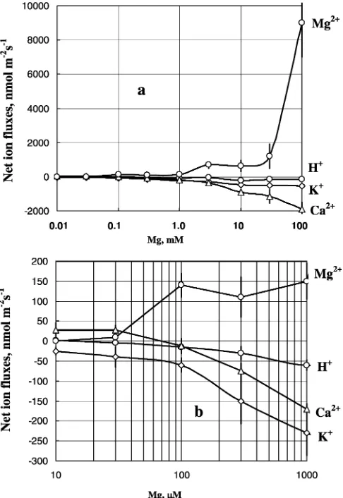
Generally, fluxes of all ions across the plasma mem-brane of bean mesophyll cells were affected by the addition of Mg to the bulk solution. In general, the higher the amount of added Mg, the larger the magni-tude of flux changes (Fig.2a). The most affected were Mg fluxes, followed by Ca2+. K+flux was next, and H+ flux had the lowest sensitivity to added Mg. When the peak magnitude (see Fig.1b) of ion flux change was plotted versus Mg concentration in the bulk solution, several specific regions were present in the dose response curves:
1. Addition of 10–30lM of external Mg caused a very
small (<10 nmol m 2s 1) uptake of Mg. Net H+ flux ‘‘mirrored’’ changes in net Mg2+flux (H+efflux of about the same magnitude; Fig.2b), with stoichi-ometry between 1:1 and 2:1 H+/Mg2+. Much more significant were changes in Ca2+ and K+ fluxes. At least an order of magnitude higher net Ca2+ influx and net K+ efflux were observed (Fig.2b). Calcu-lated stoichiometry values between Mg and these ions were as high as 25–30 (for 10lM Mg, Fig.3).
2. Concentration range 30–1,000 lM Mg. For these
concentrations, there was almost a linear increase in net Mg2+ uptake accompanied by a progressive
ef-flux of K+ and Ca2+ (Fig.2) Stoichiometry values approach some ‘‘reasonable’’ physiological values (Fig.3).
3. A ‘‘plateau’’ region in a 3–30 mM range. The mag-nitude of Mg2+flux responses remains steady; so are the changes in net H+ and K+ fluxes. Ca2+ shows progressive leakage. For this region, there is almost a 1:1 stoichiometry between Ca, K and Mg flux chan-ges (Fig. 3).
4. Further dramatic increase in Mg2+ uptake (‘‘low affinity’’ range in Epstein’s terms), accompanied by significant (although not very dramatic) Ca2+efflux. Both H+ and K+ fluxes are not responding any further to increasing Mg2+ concentrations.
Pharmacology and effect of leaf ‘‘growth history’’
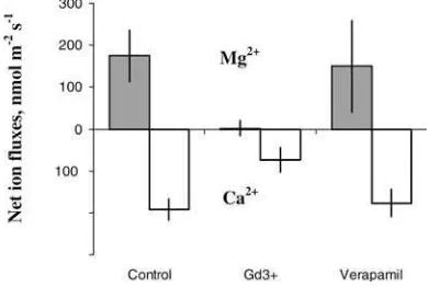
In order to provide some insights into the underlying ionic mechanisms of the above responses, a series of pharmacological experiments were undertaken. Leaf
incubation in 50lM GdCl3, a known blocker of
non-selective cation channels (NSCC), led to a significant reduction in the magnitude of both Mg2+and Ca2+flux responses (Fig.4). At the same time, 20lM verapamil
(specific Ca2+ channel blocker) had no effect on either Ca2+ or Mg2+ flux kinetics. Leaf incubation in 1 mM vanadate significantly (P=0.05) suppressed both H+ and Mg2+ flux responses (Fig.5a, b).
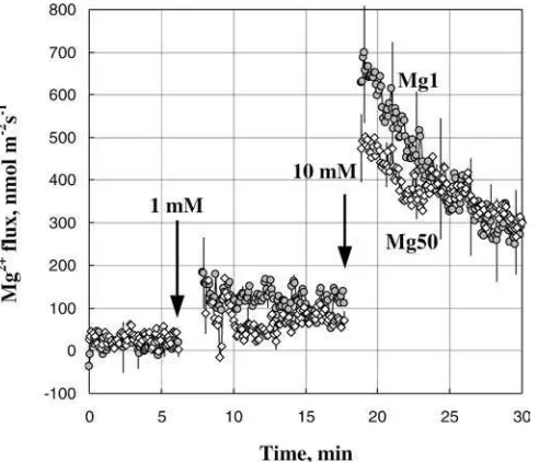
When Mg2+ uptake kinetics was compared between leaves from Mg-deficient (Mg1) plants and those grown at optimal (Mg50) supply, Mg1 plants were generally more responsive to supplemental Mg (Fig.6).
Steady-state fluxes
With the only exception of the data in Fig. 6, the
majority of the above nutrient uptake kinetics experi- ments were undertaken using leaves from Mg-deficient (Mg1) plants. The second question addressed in this study was to what extent Mg availability during plant growth would affect the activity of plasma-membrane transporters in steady-state conditions. Consequently, steady-state fluxes of K+, Ca2+, H+, and Mg2+ were measured from dark-adapted leaves of bean plants, grown for 4 weeks in deficient (Mg1), optimal (Mg50), and excessive (Mg200) conditions. Results are shown in Figs.7 and 8 (time intervals 0–2 min). Several distinct features were observed.
There was a pronounced effect of Mg availability during leaf growth on the magnitude of steady-state H+-flux (Fig.7a). H+ flux values were significantly more negative (net efflux) for Mg-sufficient variants, with the increase in H+ efflux almost directly propor-tional to Mg availability during plant growth. Calcium
Fig. 4 Effect of specific channel blockers (50lM Gd3+and 20lM verapamil) on the magnitude of net Ca2+ and Mg2+ fluxes in response to added Mg (1 mM). Data are average ± SEM (n=4–5)
Fig. 5 Effect of 1 mM sodium orthovanadate on net Mg2+(a) and H+ (b) flux responses from bean leaf mesophyll to added Mg (30lM at 3 min). One typical example (out of four) is shown. (c) Correlation between net Mg2+and H+flux changes in response to 30lM Mg2+from control (no inhibitors) leaf samples
fluxes essentially ‘‘mirrored’’ those of Mg. Higher Mg supply resulted in higher net Ca2+influx in the dark (0, 10 and 35 nmol m 2s 1, respectively; Fig.7b). Both H+- and Ca2+-flux differences were statistically signifi-cant at the P= 0.05 level. Net magnesium fluxes were near-zero for Mg1 and Mg50, with a significant (P=0.001) Mg2+efflux for excessive (Mg200) treatment (Fig.8a). Finally, there were near-zero net K+fluxes for Mg1 and Mg50 leaves, while a large K+efflux was ob-served for Mg200 treatment (Fig. 8b). In general, K+ data largely ‘‘mirrored’’ the Mg2+-flux data.
Light-induced transients
The onset of illumination triggers a cascade of electrical events in thylakoid and plasma membranes of green plant tissues. As can be seen from our data, Mg avail-ability has a significant impact on the activity of plasma-membrane transporters (not only for Mg2+ but also those for H+, K+, and Ca2+ ions) upon illumination (Figs.7,8). The highest magnitude of light-induced H+ -flux response was for Mg200 treatment (Fig. 7a). After stabilization, Mg50 (optimal) plants retained H+efflux, while in two other treatments, fluxes shifted to near zero. Light-induced Ca2+-flux kinetics was generally con-sistent with previous observations (Shabala and New-man 1999). The onset of illumination caused immediate and rapid influx of Ca2+ (Fig.7b), peaking within 1– 2 min after the light treatment, and then gradually decreasing over the next 20 min. As for the effect of Mg availability on Ca2+flux transients, there was a signifi-cant (P=0.05) difference in the temporal characteristics of light-induced Ca2+ uptake between treatments. In plants grown under optimal Mg supply (Mg50), Ca2+
influx peaked at about 1 min after onset of illumination, while in Mg1 and Mg200 treatments it was delayed for an additional 1–1.5 min (Fig.7b). Also, Mg-deficient (Mg1) plants retained Ca2+ influx for much longer compared with the other two treatments.
The onset of illumination caused rapid and prolonged net Mg2+uptake for all treatments (Fig.8a). Regardless of Mg availability during plant growth, light-induced Mg2+ influx peaked at about 2 –2.5 min after light exposure, and gradually wound down. As a general rule, net Mg2+fluxes in the light were significantly (P= 0.05) higher than those in the dark for each of the treatments. The magnitude of Mg2+-flux response was proportional to the level of Mg available during growth (Fig.8a). Light-induced K+-flux kinetics was also consistent with our previous observations (Shabala and Newman1999). Net K+ efflux was measured after some lag (of about 1 min).
There was a significant difference in light-induced K+-flux responses as a function of Mg availability
Fig. 6 Comparison of transient Mg2+ flux kinetics from Mg-deficient (Mg1, closed circles) and Mg-sufficient (Mg50, open diamonds) leaves. Data are average ± SEM (n= 4)
during plant growth. Mg-deficient plants showed the biggest magnitude of response (Fig.8b), followed by excessive (Mg200) plants. The smallest K+-flux changes were measured from leaves grown in the optimal Mg50 conditions.
Discussion
Our data represent the first real-time measurements of Mg2+ flux across the plasma membrane of leaf meso-phyll cells and their changes in response to light.
Nutrient uptake kinetics
The pathway(s) of Mg2+ entry through the plasma membrane of plant cells have not been clearly deter-mined (Shaul 2002). From our data, it is likely that at least two mechanisms are involved in Mg2+ uptake across the plasma membrane of bean mesophyll cells. At
low Mg2+ concentrations (<30lM), there was no
apparent correlation between the uptake of Mg2+ and Ca2+ (Fig.2). Both Mg2+ and Ca2+ ions were trans-ported in the same direction, but whilst Mg2+ uptake increased proportionally with Mg2+ concentration, Ca2+-flux responses remained more or less unchanged. Thus, it is unreasonable to suggest that any causal relationship exists between transport of Ca2+and Mg2+ ions at low (<30lM) Mg2+concentration. In contrast,
a high correlation (R2=0.8) between H+ and Mg2+ transport, ‘‘physiologically reasonable’’ stoichiometry (between 1:1 and 1:2) and the fact that both Mg2+and H+fluxes were suppressed by vanadate (Fig.5) suggests that in this concentration range, transport of Mg2+and H+ are coupled (Fig.9). There are very few molecules known to transport Mg2+ in eukaryotes (Haynes et al.
2002; Shaul 2002; Gardner 2003). Recent work by Li et al. (2001) suggested that among the ten members of the AtMGT family of transporters that are involved in Mg2+ acquisition from the soil and/or in Mg2+ trans-port in arabidopsis, at least one (AtMGTl) was localized at the plasma membrane.
The stoichiometry of this transporter is not clear. Although, due to the small magnitude of net Mg and H+ fluxes at low Mg concentration in our experiments, as well as because of the masking activity of H+-ATPase pump, the unequivocal deduction of the porter mecha-nism from our data is not possible. Our data (Figs.2,5) does allow us to speculate (at least qualitatively) that the Mg2+/H+ exchanger may be involved in Mg2+ trans-port across the plasma membrane of bean mesophyll cells, with stoichiometry between 1:1 and 1:2 (Fig.5c). So far, Mg2+/H+exchangers have been reported only at the tonoplast inArabidopsis(AtMHX, Shaul et al.1999) and Hevea(Amalou et al. 1994) vacuoles, with 1:2 and 1:3 stoichiometry, respectively. AtMHX is the first Mg2+ transporter to be cloned from a multicellular organism, and it shares limited sequence homology (36% identity) with NCX1, a mammalian plasma membrane Na+/Ca2+exchanger (Nicoll et al., 1990) as well as with the Mg2+permeable channel inParamecium
(Haynes et al.2002).
At higher Mg concentrations (>30 lM), there was
apparent competition between Ca2+and Mg2+for up-take. Judging by K+data and stoichiometry between all three cations (Ca, K, and Mg) it is likely that such a transporter could be a NSCC (Fig. 6). This is further supported by pharmacological experiments (Fig.4), which showed that both Mg2+ and Ca2+ fluxes were strongly inhibited by Gd3+, a known blocker of NSCC. At the same time, neither Ca2+ nor Mg2+ fluxes were affected by verapamil, a specific Ca2+channel blocker. In general, an almost electroneutral exchange of Mg2+ for K+ and Ca2+ could be observed in the 1–100 mM range of Mg concentration (Fig.3). Some observed deviation from electroneutrality may be explained by the fact that a small fraction of ion fluxes may originate from the cell wall, thus masking the activity of plasma-membrane transporters.
Importantly, the fraction of K+ tends to decrease with the increase in Mg concentration, which is expected under the condition of saturation of divalent cation binding sites within the NSCC. The molecular identity of the suggested Mg2+-permeable NSCC channel re-mains unknown. It has been shown in some species (such as wheat) that one possible route of Mg2+entry may be through putative homologs of the rca channel (White et al. 2000). The rca channel is defined as a calcium channel, but is also permeable to a wide variety of monovalent and divalent cations, including Ca2+, Mg2+, K+, and Na+ (Pineros and Tester1997; White et al. 2000). As this channel opens upon plasma mem-brane depolarization (Pineros and Tester 1997), light-induced Ca2+ uptake (Fig. 7) leading to rapid initial
depolarization of the plasma membrane (Spalding et al.
1992; Elzenga et al. 1995; Shabala and Newman 1999) may be responsible for the observed Mg2+ influx (Fig.8). As repolarization occurs (several minutes after light exposure; Shabala and Newman1999), this channel will be closed, and Mg influx will gradually decrease (Fig8a).
Steady-state fluxes
The observed dependence of H+efflux as a function of Mg availability during plant growth (Fig.4a; steady-state flux values before light treatment) is consistent with the role of Mg as the activator of plasma membrane H+-ATPase (Shaul 2002). Yazaki et al. (1988) showed that about 90% of cytosolic ATP is complexed to Mg. It may be suggested that, despite the large storage capacity of the vacuole (between 10 and 120 mM of total Mg; Dietz et al. 1992; Shaul 2002) and a strict Mg homeo-stasis (Gardner 2003), cytosolic-free Mg2+ levels differ significantly between Mg-deficient and Mg-sufficient leaves. Some additional support for this fact might be found in Fig.6, showing that plants grown at sufficient (Mg50) magnesium supply showed smaller responses to supplemental Mg2+ compared with deficient Mg1 plants. Assuming that under these conditions Mg2+ transport is mediated by the NSCC, reduced ability to take up Mg2+in Mg50 plants (Fig.6) may be indicative of higher cytosolic free Mg2+ concentrations. Even higher cytosolic free Mg2+ levels are expected to be present in ‘‘excessive’’ Mg200 plants, explaining the observed significant net Mg2+ efflux in steady-state conditions from Mg200 leaves (Fig.8a).
Light-induced transients
Mg availability had a significant impact on light-induced changes in net ion fluxes of Mg2+, H+, K+, and Ca2+ across the plasma membrane of bean mesophyll cells (Figs.4,5). Interpretation of the H+data is complicated by the fact that H+ fluxes were measured from the ac-tively photosynthesizing mesophyll tissues. Upon illu-mination, the cytosolic CO2 pool is quickly depleted,
leading to a significant influx of CO2 from external
media into the cell (Hansen et al.1993). That shifts the equilibrium between HCO3 and CO2 in the apoplastic
solution towards CO2 formation and results in
signifi-cant alkalinization of the measuring solution (Yin et al.,
1996), which is interpreted by the MIFE technique as an apparent influx (Shabala and Newman 1999). As a re-sult, activation of plasma membrane H+pump, known to occur in response to light (Elzenga 1997), is masked by this phenomenon. Nonetheless, steady-state H+flux values in the light (20 min after onset of illumination) showed the largest efflux for Mg50 leaves. This is con-sistent with the role of Mg as the activator of plasma membrane H+-ATPase (Shaul2002).
Light-induced Ca2+uptake into the leaf cell is one of the most rapid events, which is believed to be responsible for membrane depolarization (Elzenga et al. 1997; Sha-bala and Newman 1999) and be involved in the signal transduction process (Babourina et al.2002). Similar to our previous publication (Shabala and Newman 1999), the onset of illumination caused immediate and rapid influx of Ca2+ (Fig.7b), peaking within 1–2 min after the light treatment, and then gradually decreasing over the next 20 min. Our data also suggest that Mg avail-ability during plant growth may significantly affect light-induced Ca2+ flux ‘‘signatures’’ and thus regulate (di-rectly or indi(di-rectly) signal transduction between light photoreceptors and plasma membrane effectors (ion channels). In plants grown under optimal Mg supply (Mg50) Ca2+ influx peaked at about 1 min after the onset of illumination, while in Mg1 and Mg200 treat-ments it was delayed for an additional 1–1.5 min (Fig.7b).
The peak of light-induced Mg2+uptake occurred at 2 min after the onset of illumination, at about the same time as the peak of Ca2+ influx was reached (Figs.7b, 8a) and thus this peak coincides with a peak of the plasma membrane depolarization (Shabala and Newman 1999). Therefore, it is unlikely that light-in-duced Mg2+ influx may be explained by light-induced changes in membrane potential. It is reasonable to suggest that Mg2+ influx may be another factor responsible for plasma-membrane depolarization with-in the first mwith-inutes of light treatment (Spaldwith-ing et al.
1992). Other physiological roles of net Mg2+ influx across the plasma membrane (Fig. 5a) remain obscure. In chloroplasts, there is a significant change in free Mg2+ concentrations in both thylakoid lumen and stroma upon illumination. In intact, dark-kept spinach chloroplasts, internal [Mg2+] was estimated to be 0.5 mM, and illumination caused an increase in [Mg2+] to 2.0 mM in the stroma (Ishijima et al.2003). Such light-induced Mg2+ efflux from lumen to stroma is essential for regulation of the activity of the thyla-koid ATPase complex (ATP synthase, CF0F1) as well
as for the activity of key stromal enzymes, including Rubisco and Dl protein of photosystem II (Horlitz and Klaff 2000; Shaul 2002). It is unclear if the same sce-nario is applicable to light-induced Mg2+ uptake into the cytosol (Fig. 5a). The most obvious function of elevated cytosolic Mg2+would be to ‘‘fuel’’ the plasma membrane H+ pump, responsible for the overall membrane hypopolarization in the light (Elzenga et al.
1995; Shabala and Newman 1999).
The effects of Mg availability on light-induced K+ fluxes were rather unexpected. The smallest K+ flux changes were measured from leaves grown in optimal Mg50 conditions, while Mg-deficient plants showed much greater responses (at least one order of higher magnitude; Fig. 5b). Therefore, it is unlikely that light-induced K+fluxes are relevant to cell turgor regulation and, ultimately, leaf expansion growth. The charge-balancing role of light-induced K+flux is more likely.
Acknowledgements This work was supported by an ARC Large Grant (A00001144) to Dr S. Shabala. My sincere thanks to Dr Richard Gardner and Prof. Igor Pottosin for helpful discussion and suggestions.
References
Allen GJ, Sanders D (1997) Vacuolar ion channels of higher plants. Adv Bot Res 25:218–252
Amalou Z, Gibrat R, Trouslot P, d’Auzac J (1994) Solubilization and reconstitution of the Mg2+/2H+antiporter of the lutoid tono-plast from Hevea brasiliensis latex. Plant Physiol 106:79–85 Babourina O, Newman I, Shabala S (2002) Blue light-induced
kinetics of H+ and Ca2+ fluxes in etiolated wild-type and phototropin-mutant Arabidopsis seedlings. Proc Natl Acad Sci U S A 99:2433–2438
Bruggemann LI, Pottosin II, Schonknecht G (1999) Cytoplasmic magnesium regulates the fast activating cation channel. J Exp Bot 50:1547–1552
Cakmak I (1994) Activity of ascorbate-dependent H2O2-scavenging enzymes and leaf chlorosis are enhanced in magnesium- and potassium-deficient leaves, but not in phosphorus-deficient leaves. J Exp Bot 278:1259–1266
Cakmak I, Hengeler C, Marschner H (1994) Changes in phloem export of sucrose in leaves in response to phosphorus, potas-sium and magnepotas-sium deficiency in bean plants. J Exp Bot 278:1251–1257
Dietz KJ, Schramm M, Lang B, Lanzl-Schramm A, Durr C, Martinoia E (1992) Characterization of the epidermis from barley primary leaves. II. The role of the epidermis in ion compartmentation. Planta 187:431–437
Elzenga JTM (1997) Kinetic properties of blue light pulse-induced acidification by leaf epidermal cells of pea. Plant Physiol 114:1474–1474
Elzenga JTM, Prins HBA, Van Volkenburgh E (1995) Light-in-duced membrane potential changes of epidermal and mesophyll cells in growing leaves ofPisum sativum. Planta 197:127–134 Elzenga JTM, Staal M, Prins HBA (1997) Calcium-calmodulin
signalling is involved in light-induced acidification by epidermal leaf cells of pea,Pisum sativumL. J Exp Bot 48:2055–2061 Fischer ES (1997) Photosynthetic irradiance curves of Phaseolus
vulgarisunder moderate or severe magnesium deficiency. Pho-tosynthetica 33:385–390
Fraichard A, Trossat C, Perotti E, Pugin A (1996) Allosteric reg-ulation by Mg2+ of the vacuolar H+-PPase from Acer pseudoplatanus cells. Ca2+/Mg2+ interactions. Biochimie 78:259–266
Gardner RC (2003) Genes for magnesium transport. Curr Opin Plant Biol 6:263–267
Garnett TP, Shabala SN, Smethurst PJ, Newman IA (2003) Kinetics of ammonium and nitrate uptake by eucalypt roots and associated proton fluxes measured using ion selective mi-croelectrodes. Funct Plant Biol 30:1165–1176
Hansen U-P, Moldaenke C, Tabrizi H, Ramm D (1993) The effect of transthylakoid proton uptake on cytosolic pH and the imbalance of ATP and NADPH/H+production as measured by CO2- and light-induced depolarisation of the plasmalemma. Plant Cell Physiol 34:681–695
Hariadi Y, Shabala S (2004) Screening broad beans (Vicia fabaL.) for magnesium deficiency. 1. Growth characteristics, visual deficiency symptoms and plant nutritional status. Funct Plant Biol 31:529–537
Haynes WJ, Kung C, Saimi Y, Preston RR (2002) An exchanger-like protein underlies the large Mg2+current in Paramecium. Proc Natl Acad Sci U S A 99:15717–15722
Hinnah SC, Wagner R (1998) Thylakoid membranes contain a high-conductance channel. Eur J Biochem 253:606–613 Horlitz M, Klaff P (2000) Gene-specific trans-regulatory functions
Ishijima S, Uchlbori A, Takagi H, Maki R, Ohnishi M (2003) Light-induced increase in free Mg2+ concentration in spinach chloroplasts: measurement of free Mg2+by using a fluorescent probe and necessity of stromal alkalinization. Arch Biochem Biophys 412:126–132
Lavon R, Goldschmidt EE (1999) Effect of potassium, magnesium, and calcium deficiencies on nitrogen constituents and chloro-plast components in Citrus leaves. J Am Soc Hortic Sci 124:158–162
Li L, Tutone AF, Drummond RS, Gardner RC, Luan S (2001) A novel family of magnesium transport genes in Arahidopsls. Plant Cell 13:2761–2775
Marschner H (1995) Mineral nutrition of higher plants, 2nd edn. Academic, San Diego
Newman IA (2001) Ion transport in roots: measurement of fluxes using ion-selective microelectrodes to characterize transporter function. Plant Cell Environ 24:1–14
Nicoll DA, Longoni S, Philipson KD (1990) Molecular cloning and functional expression of the cardiac sarcolemmal Na+–Ca2+ exchanger. Science 250:562–565
Pei ZM, Ward JM, Schroeder JI (1999) Magnesium sensitizes slow vacuolar channels to physiological cytosolic calcium and inhibits fast vacuolar channels in Fava bean guard cell vacuoles. Plant Physiol 121:977–986
Pineros M, Tester MA (1997) Calcium channels in higher plant cells: selectivity, regulation and pharmacology. J Exp Bot 48:551–577
Pottosin II, Muniz J (2002) Higher plant vacuolar ionic transport in the cellular context. Acta Bot Mex 60:37–77
Pottosin II, Scho¨nknecht G (1996) Ion channel permeable for divalent and monovalent cations in native spinach thylakoid membranes. J Membr Biol 152:223–233
Pottosin II, Tikhonova LI, Hedrich R, Scho¨nknecht G (1997) Slowly activating vacuolar ion channel cannot mediate Ca2+ -induced Ca2+release. Plant J 12:1387–1398
Remis D, Bulychev AA, Kurella GA (1986) The electrical and chemical components of the protonmotive force in chloroplasts as measured with capillary and pH-sensitive microelectrodes. Biochim Biophys Acta 852:68–73
Ridolfi M, Garrec J-P (2000) Consequences of an excess Al and a deficiency in Ca and Mg for stomatal functioning and net carbon assimilation of beech leaves. Ann For Sci 57:209–218 Shabala S (2000) Ionic and osmotic components of salt stress
specifically modulate net ion fluxes from bean leaf mesophyll. Plant Cell Environ 23:825–838
Shabala S, Lew RR (2002) Turgor regulation in osmotically stressed Arabidopsis epidermal root cells. Direct support for the role of inorganic ion uptake as revealed by concurrent flux and cell turgor measurements. Plant Physiol 129:290–299
Shabala SN, Newman IA (1999) Light-induced transient changes in hydrogen, calcium, potassium, and chloride ion fluxes and concentrations from the mesophyll and epidermal tissues of bean leaves. Understanding the ionic basis of light-induced bioelectrogenesis. Plant Physiol 119:1115–1124
Shabala SN, Newman IA, Morris J (1997) Oscillations in H+and Ca2+ion fluxes around the elongation region of corn roots and effects of external pH. Plant Physiol 113:111–118
Shabala S, Babourina O, Newman IA (2000) Ion-specific mecha-nisms of osmoregulation in bean mesophyll cells. J Exp Bot 51:1243–1253
Shabala S, Shabala L, Van Volkenburgh E (2003) Effect of calcium on root development and root ion fluxes in salinised barley seedlings. Funct Plant Biol 30:507–514
Shaul O (2002) Magnesium transport and function in plants: the tip of the iceberg. BioMetals 15:309–323
Shaul O, Hilgemann DW, Almeida-Engler J, Van Montagu M, Inze D, Galili G (1999) Cloning and characterization of a novel Mg2+/H+exchanger. EMBO J 18:3973–3980
Spalding EP, Slayman CL, Goldsmith MHM, Gradmann D, Bertl A (1992) Ion channels in Arabidopsis plasma membrane. Transport characteristics and involvement in light-induced voltage changes. Plant Physiol 99:96–102
Sun OJ, Payn TW (1999) Magnesium nutrition and photosynthesis inPinus radiata: clonal variation and influence of potassium. Tree Physiol 19:535–540
Tikhonova LI, Pottosin II, Dietz K-J, Scho¨nknecht G (1997) Fast-activating cation channel in barley mesophyll vacuoles. Inhi-bition by calcium. Plant J 11:1059–1070
Tisdale SL, Nelson WL, Beaton JD, Havlin JL (199’3) Soil Fertility and Fertilizers. 5th edn. Prentice Hall, New Jersey
White PJ, Pineros M, Tester M, Ridout MS (2000) Cation per-meability and selectivity of a root plasma membrane calcium channel. J Membr Biol 174:71–83
Yazaki Y, Asukawagawa N, Ishikawa Y, Ohta E, Sakata M (1988) Estimation of cytoplasmic free Mg2+levels and phosphoryla-tion potentials in mung bean root tips by in vivo 31P NMR spectroscopy. Plant Cell Physiol 29:919–924