Predicting EMG Based Elbow Joint Torque Model Using Multiple Input ANN
Faculty of Electronics and Computer Engineering Universiti Teknikal Malaysia Melaka
Malacca, Malaysia e-mail: [email protected]
Abstract— Thispaper illustrates the Artificial Neural Network (ANN) technique to estimate the joint torque estimation model for arm rehabilitation device in a clear manner. This device acts as an exoskeleton for people who had failure of their limb. Arm rehabilitation device may help the rehab program to whom suffered with arm disability. The device used to facilitate the tasks of the program should improve the electrical activity in the motor unit and minimize the mental effort of the user. Electromyography (EMG) is the techniques to analyze the presence of electrical activity in musculoskeletal systems. The electrical activity in muscles of disable person is failed to contract the muscle for movements. To prevent the muscles from paralysis becomes spasticity the force of movements should minimize the mental efforts. Besides that, in order to minimize the used of mental forced for disable patients, the rehabilitation device can be utilize by analyzing the surface EMG signal of normal people that can be implemented to the device. The objective of this work is to model the muscle EMG signal to torque for a motor control of the arm rehabilitation device using ANN technique. The EMG signal is collected from Biceps Brachii muscles to estimate the elbow joint torque. A two layer feed-forward network is
Human support system is endoskeleton. Endoskeleton plays a role as a framework of the body which is bone. Our daily movements are fully depends on the functionality of our complex systems in the body. The disability one or more of the systems in our body will reduce our physical movements. The assistive device is a need for rehab as an exoskeleton. The functionality of the rehabilitation device has to smooth as the physical movement of normal human.
The rehabilitation programs provide the suitable program for conducting the nerve and stimulate the muscles. People who have temporary physical disability have the chances to recover. Nowadays, rehabilitation program are using exoskeleton device in their tasks. The functionality of
exoskeleton depends on muscle contraction. Electromyogram studies help to facilitate the effectiveness of the rehabilitation device by analysing the signal transmitted from the muscle.
The technique of measuring electrical activity that produced from the muscles during rest or contractions known as electromyography (EMG). The electric signal generates from the brain and sends to the muscles via motor neuron. The EMG may detect the dysfunctional of the muscles or failure in signal transmission from nerve to muscle. The failure of sending the electrical signal from the brain requires electrical stimulation from the external source to muscles. Electrodes are used for signal detection of electrical activity in muscles. The study of this electrical activity is important for combination of electromyogram and rehabilitation device.
The rehabilitation device is a tool that used to help the movements for daily life activities of the patients who suffer from the failure of muscle contractions, due to the failure of the muscles contractions the movements is limited. The ability of the patients to do the tasks in the rehabilitation programs need to be measured. The rehabilitation programs have to assure whether the tasks will cause effective or bring harm to the patients [1].
Historically, the rehabilitation tasks have been avoided due to a belief that it would increase spasticity [2]. In this research, the analysis of the data will be focusing on upper limb muscles contraction consisting of biceps muscles only. The experiment is limited to the certain of upper limb movements that use in training. EMG is a division of bio signal; the bio signal analysis is the most complex analysis. Thus, the signal analysis is a complicated process that has to be through many phases of analysis [3].
the biceps brachii muscle act as the input of the ANN model whiles the desired torque act as the ideal output of the model. Hence the EMG signals considered the ‘intent’ of the system while the joint torque is the ‘controlled’ variable for the arm rehabilitation device.
II. RELATEDWORK
There have been several studies that has applied ANN for modeling the muscle activity to joint relationship. Author [7] proposed the ANN model to measure the ankle EMG-joint torque relationship at a full range of torque under isometric, supine conditions by inserting EMG signal from 6 muscle sites are into the model as in the input, while the measured torque is entered into the model as the ideal output. The learning process occur approximately 16000 iterations resulting error that is less than 6 percent.
More recent study has been conducted by author [4] to predict the elbow joint angle based on EMG signals using ANN. The three layer BPNN was constructed by using the RMS of the raw EMG signal form biceps and triceps. The result from 40 group EMG signals when subjects do bowing and extending elbow joint action reveal that the prediction output from the trained network was very close with the target angle. According to author [6], it is quite difficult to know the elbow shoulder joint torque on the natural condition. The learning method which is based on the feedback error learning schema is proposed by modifying the ANN with the torque error which calculated from the desired angle and measured angle.
Studies of muscle force models have been carried out by author [9]. The model was estimated based on a rectified smoothed EMG signal using the BPANN method to predict the muscle force. The proposed model can efficiently extract muscle force features from (EMG) signals in a fast and easy method. The results showed that the regression of the ANN model exceeded 99%. However among all the previous studies related to EMG based ANN model been conducted, none of the ANN model achieved 100% regression. This is contradicting with our method that applied additional input variable which is movement time to produce 100% regression and definitely a very small error values.
III. METHODS
The purpose of the arm rehabilitation device used in this experiment is to collect the EMG signal from the subjects as well as assisted device for disable person. Therefore the data acquisition session and rehabilitation session will have a similar procedure. In order to model the device using ANN technique, specified values of load weight, the distance from elbow joint and the mass of lever arm has been fixed to determine the desired torque. The device’s parameter need to be accurately measured to produce a highly reliable model. Thus the model can be applied to any patient
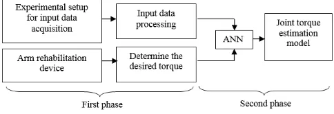
regardless of their percentile during the rehabilitation session specifically for flexion motion. To ensure the robustness of the model, the EMG data acquisition need to be conducted to several percentile subjects with several repetitions. This is crucially important to obtain the generalized model that applicable to a wide range of patients. Besides it can compensate the error that is cause due to the device’s measurement inaccuracy. Fig. 1 shows a block diagram of our research that consists of two major phases. First phase is input data processing and desired torque determination. Second phase is the ANN construction and testing. The collected data is used to develop and validate ANN model in the second phase [14].
Figure 1. Research methods
A. Experimental Setup
Implementation of arm rehabilitation device based on movement is recorded from the EMG signal of healthy subjects. From the human anatomy studies, different angle movements of upper limb with elbow as the reference is depends on relation of agonist and antagonist. In this study is focusing on the behaviour of biceps muscle as agonist and the triceps as the antagonist respectively. Muscle that involved in this movement is biceps and triceps, however in this study to understand the electrical activity during muscle contraction, the biceps is the only muscle that taking into account. The movements’ ranges in between position of arm flexion until arm fully extend.
flexion). Data was collected from two subjects by 5 repetitions of each flexion movements [8].
(a) (b)
Figure 2. (a) Subject is set-up with arm rehabilitation device for experiment , (b) Simulation of subject’s to lift up the dumbbell
Prior of data collection process, the skin needs a preparation. The preparation of skin is ruled by the Surface Electromyography for the Non-Invasive Assessment of Muscles (SENIAM) procedure for non-invasive methods. The subject’s skin has to be shaved by using small electrical shaver and cleaned with sterile alcohol swabs saturated with 70% Isopropyl Alcohol. This step is to be taken for minimizing the noise and to have a good contact with the electrodes of the skin by decreasing the impedance of the skin. The skin has to be clean from any contamination of body oil, body salt, hair and the dead cells. The preparation of skin can be done by wiping the alcohol swab into the area of skin that electrode placement to be applied. The placements of the electrode have to be at the belly of the muscles not in the tendon or motor unit. This ensured the detecting surface intersects most of the same muscle on subject as in Fig. 3(a) at the biceps brachii, and as a result, an improved superimposed signal is observed. Reference electrode has to be at the bone as the ground, for this experiment it placed at elbow joint as shown in Fig. 3(b). These electrodes are connected to the combination of hardware Olimex EKG-EMG-PA and Arduino Mega for data collection.
(a) (b)
Figure 3. (a) The biceps brachii muscles for electrode positions, (b)The electrode placements on subject skin
B. Input Data Processing
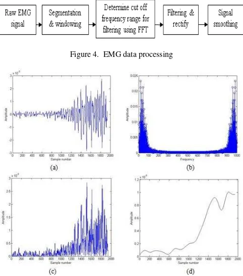
Fig. 4 shows the EMG data processing block diagram. After obtained satisfactory EMG signal as shown in Fig. 5(a), the signals are segmented with 0.969ms length. The time duration for one cycle of flexion movement need to be carefully defined because it will be one of the data of the input neuron. Hence the windowing method is conducted prior to Fast Fourier Transform (FFT) is performed to the signal to analyses the frequency content of the signal. The EMG signal is break into its frequency component and it is presented as function of probability of their occurrence. In order to observe the variation of signal in different frequency components, the FFT signal is represented by Power Spectral Density (PSD). From the PSD we can describes how the signal energy or power is distributed across frequency. Fig. 5(b) shows that most of the power is in the range of below 10Hz, therefore the EMG signal should be filtered in the range of above 10Hz as a cut off frequency for low pass filtered. After decide the cut off frequency for filtering, the DC offset of the EMG signal is removed and is rectified to obtain its absolute value as shown in Fig. 5(c). Finally, the signal was smoothed and normalized passing it through a 5th order Butterworth type low-pass filter with cut off frequency 10Hz and the smooth signal is illustrate in Fig. 5(d) [8][9].
Figure 4. EMG data processing
C. Desired Torque
Desired torque of the elbow joint is used as target data for our ANN techniques as well as act as output signal for muscle [14]. The data is collected throughout the angle from 00 to 1200 angle with increment of 0.06190 each step to align with the sample number of EMG signal. Fig. 6(a) shows the arm rehabilitation device position for torque calculation. The desired torque for elbow joint is determined by applying standard torque equation:-
τ = (rloadFload + rarmFarm) cos θ (1)
Where rload is distance from the elbow joint to the load, Fload is force due to load, Farm is the force due to the mass of the lever arm and rarm is evenly distributed distance of mass of arm distance which is half of rload. The angle θ between r and F is drawn from the same origin. A applied load is 2.268kg dumbbell and the distance from the elbow joint is 0.25m while the mass of the lever arm is 0.1kg . Fig. 6(b) shows the desired torque characteristic for the elbow joint.
(a) (b)
Figure 6. (a) Arm rehabilitation device position, (b) Desired torque characteristic
D. Artificial Neural Network (ANN) Techniques
ANN is a computing paradigm that is loosely modeled after cortical structures of the brain. It consists of interconnected processing elements called nodes or neuron that work together to produce an output function. It capable to map a data set of numeric inputs with a set of numeric outputs. It is also the most widely applied training network which has input layer, hidden layer and output layer. The neurons on each layer need to be considered carefully to produce high accuracy network. The number of hidden neurons could affect the performance of the network. The network performance not always been improved if the hidden layer and its neurons is increased [4]. Therefore the number of hidden neurons is tested to achieve the optimized network. However there is constraint in determining the number of neurons. If the numbers of hidden neurons is too large, the network requires more memory and the network become more complicated while if the number of hidden neurons is too small, the network would face difficulty to adjust the weigh properly and could cause over fitting which
is problem where the network cannot be generalized with slightly different inputs [10].
The input signal is propagated forward through network layer using back propagation algorithm. An array of predetermined input is compared with the desired output response to compute the value of error function. This error is propagated back through the network in opposite direction of synaptic connections. This will adjust the synaptic weight so that the actual response value of the network moved closer to the desired response [11]. BPNN has two-layer feed-forward network with hidden neurons and linear output neurons. The function used in the hidden layer of network is sigmoid function that generates values in range of -1 to 1 [8]. There are layers of hidden processing units in between the input and output neurons. For each epoch of data presented to the neural network, the weights (connections between the neurons) and biases are updated in the connections to the output, and the learned error between the predicted and expected output, the deltas, is propagated back through the network [12].
A Lavenberg-Marquardt training back propagation algorithm is implemented for this work to model the EMG to torque signal. Input layer consist of two nodes which is denoted as b that represent EMG signal from biceps and t that represent the movement time that act as a training data while output layer has only one node which is denoted as τ that represent the desired torque that act as a target data. The network is optimized for 20 hidden neurons as shown in Fig. 7[4]. The network was trained using 1839 sets of EMG data for arm flexion motion from 00 angles to 1200 angle. It also has output data which is torque of correspondent arm motion. The training process was iteratively adjusted to minimize the error and increased the rate of network performance [10]. MATLAB software is used to construct the BPNN network. The network requires data for learning and testing in order to determine the weights each node uses. The training has been done by dividing the input data of 70% for training, 15% for validation and 15% for testing [9]. The network will be trained until the following condition fulfilled before it stop [10]:-
i- reach maximum number of epochs
ii- gradient performance became less than the minimum gradient
iii- validation performance increased more than the maximum fail times since the last decreased one
still considered unsatisfactory because the network is not generalized. Therefore further tuning and training need to be conducted in order to improve the network performance [10].
Figure 7. BPNN model
IV. EXPERIMENTAL RESULTS
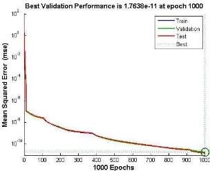
In order to optimize the network performance, different number of hidden neurons is simulated for several times until achieved the satisfactory results [1 0 ].The network is trained using Lavemberg-Marquardt algorithm and the performance of the network is measured using MSE and R. The best validation performance of the network is 1.7638e-11 at epoch 1000 as shown in Fig. 8. It is an acceptable result since the test set error and the validations set error have similar behavior. It shows that the MSE has decreased rapidly along the epochs during training. The regression also produces a very good curve fitness for training, test and validation result 1.000 which give an optimal value for our model as shown in Fig. 9 [9][13].
Figure 8. Best validation performance
Error sizes are well distributed since most error approaching zero values that make the trained model perform better as shown in Fig. 10(a). The network has maximum instance around 275 of MSE distributed around the zero line of the error histogram [13]. Fig. 10(b) shows a comparison between prediction output from the trained network and target torque. The prediction output has totally
had a good agreement with the characteristics of the target data.
Figure 9. Regression of the trained model
(a)
(b)
Figure 10. (a) Error histogram of the trained model, (b) Target vs trained network output
The results can be summarizing in Table I:-
TABLE I. SUMMARY OF THE EXPERIMENT RESULTS
No. Stop
Epochs
Regression MSE Time Elapsed
(Seconds)
V. CONCLUSIONS
Based on the result, it can be concluded that the ANN model with two nodes at the input layer produce MSE of 1.7638e-11 at epoch 1000 and average regression of 1.000. It is definitely a good performance result as it shows that this neural network model can well represent the relationship between EMG signals and elbow joint torque. Hence this model can be used for motor torque control of the arm rehabilitation devices. However even though the R and MSE produce almost perfect result, there are still further improvement can be done in term of stop epochs and training time elapsed. The model can be further improved by applying other artificial intelligence algorithm such as genetic algorithm and particle swarm optimization to shorten the training time and stop epochs.
ACKNOWLEDGMENT
The authors would like to thanks Universiti Teknikal Malaysia Melaka (UTeM) and Ministry of Education, Malaysia for the financial supports given through Research Grant.
REFERENCES
[1]S. D. C. G. C. Louise Ada.:Strengthening Interventions Increase Strength and Improve Activity After Stroke: A Systematic Review. Australian Journal of Physiotherapy, vol. 52, pp. 241-248, 2006.
[2]B. B.:Adult Hemiplegia: Evaluation and treatment..Oxford, Butterworth-Heinemann., 1990.
[3]J. Muthuswamy.:Biomedical Signal Analysis in Standard Handbook Of Biomedical Engineering And Design. Mc-Graw Hill, 2004, pp. 18.1 - 18.30.
[4]Li Dapeng , Zhang Yaxiong.: Artificial Neural Network Prediction of Angle Based on Surface Electromyography. International Conference on Control, Automation and Systems Engineering (CASE).pp. 1-3, 2011.
[5]M. B. I. Reaz, M. S. Hussain, and F. Mohd-Yasin.: Techniques of EMG Signal Analysis: Detection, Processing, Classification and Applications. Biological Procedures Online, vol. 8, pp. 11-35, 2006.
[6]Morita, S. Kondo, T. Ito, K. : Estimation of Forearm Movement from EMG Signal and Application to Prosthetic Hand Control, IEEE
International Conference on Robotics and
Automation(ICRA).vol.4.pp.3692-36972. 2001.
[7]Kent, L.M. Siegler, S. Guez, A. ; Freedman, W. .: Modelling Of Muscle EMG To Torque By The Neural Network Model Of Backpropagation . IEEE International Conference of the Engineering in Medicine and Biology Society, Proceedings of the Twelfth Annual. Pp 1477-1478.1990.
[8]Favieiro, G.W. Balbinot, A. Barreto, M.M.G. : Decoding Arm Movements by Myoeletric Signals and Artificial Neural Networks. Conference of Biosignals and Biorobotics(BRC). pp 1-6. 2011.
[9]Naeem, U.J. Abdullah, A.A. ; Caihua Xiong.:Estimating human arm's muscle force using Artificial Neural Network . IEEE International Symposium on Medical Measurements and Applications Proceedings (MeMeA),pp 1-6.2012
[10]Ahsan, M.R. ; Ibrahimy, M.I. ; Khalifa, O.O. EMG Motion Pattern Classification through Design and Optimization of Neural Network. International Conference on Biomedical Engineering (ICoBE), pp 175-179.2012
[11]B. Hudgins, P. Parker, and R. Scott.:A new strategy for multifunction myoelectric control. IEEE Trans. Biomed. Eng., vol. 40, no. 1, pp. 82– 94, Jan. 1993.
[12]Mars,P, Chen JR, Nambiar, R Learning Algorithms.: Theory and applications in signal processing, control and communications. CRC 1996.
[13]Supeni, E. E. and Epaarachchi, J. A. and Islam, M. M. and Lau, K. T. (2013) Development of artificial neural network model in predicting performance of the smart wind turbine blade. In: 3rd Malaysian Postgraduate Conference (MPC 2013), 4-5 Jul 2013, Sydney Australia.
![Fig. 7[4]. The network was trained using 1839 sets of EMG](https://thumb-ap.123doks.com/thumbv2/123dok/515218.58759/4.612.55.285.323.424/fig-network-trained-using-sets-emg.webp)