VoI I1, No 2, ApnI
-
June2002
Natural anticoagulant deficiencies in acute coronarysyndrome
87Anti-thrombin
IItr, Protein
C,
and Protein
S
defÏciency
in acute
coronary
syndfome
Dasnan
Ismailx,
SHarun*, Idrus
Alwi*,
Karmel
L Tambunan**,
ShufrieEffendy**
Abstrak
Akhir perjalanan yang sangat sering ditemukan pada sebagian besar penyakit iantung
koroner
adalah oklusi pembuluh darah koroner. Pada keadaan normnl, antithrombinIII
(ATIII),
protein C dan protein S sebagai l<nfaktor proteinC
aktif, merupakan antikoagulanalami
(pengontrol hemostasis) yang berperan menyeimbangl<an aktivitas prokoagulan (keseimbangan kompleksantithrombin-thrombin), untuk mencegah terjadinya trombosis. Dalam keadaan tak seimbang, antikoagulan alami dan prokoagulan dapat menyebabpan tejadinya trombosis. Dilakukan penelitian pada tigapuluh subyek dengan sindrom koroner akut (SKA) untuk melihat angka kejadian defisiensi anrithrombin
III
(ATIII),
protein C dan protein S, dan hasilnya dibandinglcan dengan kelompok kontrol. Pida pasien SKA, distribusifrekuensiAT-lll
dengan aktivitas<
75 Vo adalah 23,3 Vo (7 dari 30), di mana hanya ditemukan 6,7 7o (2dari
30) pada kelompok kontrol. Ti^dak ditemukan defisiensi protein C pada 30 subyek kelompok kontrol, sedangl<an padaSKA di mana aktiiitas
<
70 Vo ditemuknn 13,3 Vo (4dan
30). Limc puluh persen (15dan
30) subyek kelompok kontroL mengalami defisiensi alctivitas protein S, sedangkan pada SKA dengan aktivitas protein S<
70 Vo ditemukan 73,3 Vo (22 dan 30). Pada analisis regresi linier, lcoefisien determinan defsiensi aktivitasAT-lll
untuk terjadinya SKA adalah 13,25 Vo dan koefisien determinan defisiensi aktivitas protein C untuk terjadinya SKA adalah 9,06 %, cut-off point akrtvitas ATIII
tanpa defi.siensi protein S yang diperkiralcan untuk terjalinya SKA adalnh 45 Va. Pada analisis diskiminan, defsiensi aktivitas protein C mempunyai estirutsiisiko
unnk terjadinya SKA sebesar 4,5 kati dibandinglann nnpa defisiensi protein C, dan defisiensi aktivitas ATIII
mempunyai estimasi risilco untuk terjadinya SKA sebesar 3,5 kati dibandingkan tanpa defisensi AT III. pada regresi logistik binary, defsiensi aktivitas' protein S hanya merupalcan faktor penguat defisiensi alaivitas ATIII untuk terjadinya SKA. Defisierisi protein C dnn ATIII
dapat merupalcnnfaktor pemicu unruk terjadinyaSKA, di mana koefisien determinan untL.k terjadinya SKA adalah sebesar 9,06 dan I 3,25Vo. Aktivitas protein C rendah membeikan risiko Iebih
tinggi
tntuk terjadinya SKA dibandingkan alctivitas ATIII
rendah. Defisiensi protein S merupalan faktor penguat defisiensi faktor ATIII
untuk terjadinya SKA. Cut-offpoint aktivitas ATIII
tanpa defisiensi protein S unruk terjadinya penyakit saa pembuLuh adalah 45 7odan unruk penyakit tiga pembuluh adalah 9,05 Vo. (Med J Indones 2002; 11 : 87'92)
Abstract
The
final
most common pathwayfor
the mojortÇof
coronary artery diseaseis
occlusion ofa
coronary vessel.Under
normal conditions, antithrombinIII
(AT III), protein C, and protein S as an active protein C cofactor, are natural anticoagulants (hemostatics control) that balances procoagulant activity (thrombin antithrombin complex balance) to Prevent thrombosis.If
the conditionbecomes unbalanced,
nanral
anticoagtilants and the procoagulants canleaà
to thrombosis. Thirty subiects with acute coronary syndrome(
ACS ) were studiedfor the incidence of antithrombinIII
(AT III), protein C, and protein S deficiencies, and the result werecompareto thecontro!group.AmongpatientswithACS,the frequencyof distributionof
AT-lllwith
activity<75Vowere23,3Vo(7 of30), and
only 6,7Vo ( 2 of 30 ) in control subject. No oneof
the 30 control subject have protein C activity deficient,in
ACS withactivity<70Vowerel3,3Vo(4of 30).Fifteenoutofthe
30(50Vo)controlsubjects hadproteinSactivitydeficiency,whileproteinS deficiency activity<
70Vo wasfound
7j,jVo (22 outof
30). On linear regression, the deterministic coefficient ofAT-lll
activity deficiency to the development ACS was 13,25 Vo, and the deterministic cofficientof
protein C activity deficientto
the developmentof
ACS was 9,06 Vo. The cut-ofrpoint
for
AT-Lil without protein S defciency expected to contibute to the development of vessel disease was 45%. On discriminant analysis, protein C activity deficiency poseda
riskfor
ACSof 4,5
greater than non deficient subjects, andAT-|il
auiviry
defciency poseda
riskfor
ACSof
3,5 times greater than non deficient subjects. On binary Logistic regression, protein S activity acted only asa
reinforcingfactor
of
AT-lll activity defciency in the developmentof
ACS. Protein Cand AT
Itl
deficiency can trigger ACS, with determinant coefficientsof
9,06Vo and I3,25Vo respectively. Low leveLs of protein C posed a greater risk of ACS than low levels of ATIII.
Protein S deficiency was a reinforcingfacbr
on AT-ttl deJtcient to developmentof
ACS. The cut-off point ofAT-\il
without prorein S defciency expected to give single vessel disease was 45Vo, and 9,5V0for
the developmentof
triple vessel disease. (Med J Indones 2002;1l:
87-92)Keywords: acute coronary syndrome, Anti-thrombin II[, Protein C, Protein S
'
Division of Cardiology, Departrnent of Internal Medicine, Faculty of Medicine University of Indonesia / Dr. Cipto Mangunkusumo Flospital, Jakarta, Indonesn88
Ismail et alCoronary artery
diseaseis
the leading
causeof
deathin
western society, and
acutecoronary syndrome
is
avery common rnanifestation
of
this
disease.In
theUniled
States,
1,5
million
individuals suffer from
acutemyocardial
infarction
per
year;
50
7oof
which
are
falal.
In
Indonesia,
basedon
the
National Hold
Survey
on
Health
by
the
Department
of
Health,Indonesia
in
1992,
cardiovascular disease
was
theleading
cause
of
mortality.z
Of
these
coronarythrombotic
events,l
67
Vo
of
patients harbour
a coagulationblood
protein or platelet defect leading
tothrombosis.
Fifty
percentof
thesecoagulation
protein orplatelet
defects arehereditary,
thus emphasizing the importanceof
defining
the presence and typeof
defectin
survivors
of
acutecoronary
syndrome.Defining
thedefect also
allows
one
to
optimize
antithrombotic
therapy
for
secondary prevenlion. IThe final
most common pathway
for
the
majority of
coronary
artery
disease
is
atherosclerotic
plaqueinstability, ruplure,
and subsequent thrombus formation,which
lead
to
occlusion
of
a coronary vessel.l Acute
myocardial
infarction
(MI)
and unstable angina pectorisare
two
components
of
acute coronary
syndÏome.Atheiosclerosis
is
a
dynamic process where
rapidprogression may
occu-rin
young
plaques at the siteof
intenseinflammation.t
Th"
inflammation
disrupts
theendothelium, exposing thrombogenic material
thattriggers platelet
aggregation
and
intravascularcoagulation.t
Th"
int"raction
of
platelets, thrombin,
and leukocytes
in
unstable plaqueresult
in
the releaseof
various active products,including matrix
degradation molecules, procoagulantfactors
and protein, cytokines,and
growth factors (EDGF,
VEGD
that
can
lead
tothe
tbrmation
of
an
obstructive
endovascular thrombus, andto
accelerated atherosclerosis.lUnder
normal conditions, antithrombin
III
(AT
III),
protein C, andprotein
S as an active protein C cofactor,are natural
anticoagulants (hemosta[ics
control)
lhat
balances procoagulant
activity
(thrombin antithrombin
complex balance)
to
prevent thrombosis.
If
thecondition becomes unbalanced,
natural anticoagulantsand
procoagulants canlead
to thrombosis.3'aThe role
of
thrombus formation in
the pathogenesisof
acule coronary syndrome
hasbeen
well
established.However,
comprehensive
and
systema[ic studies
of
the nalural
coagulation
inhibitor
are
not
avaiiable
in
patientswith
theseconditions.
Med
I Indones
METIIODS
Sample size was calculated using correlation with
95Vo confidences
two
tailed
by
90% power
andconelation
estimate
0.41, which
is
30
subjects
for
cases andcontrols.
Caseswere
subjectswith 2
out
of
3 clinical criterion
for
acute
myocardial
infarction;)
typical
chestpain, characteristic
evolution
changes in12lead ECG,
andsignificant
cardiac enzyme elevation and unstable anginaaccording to Braunwald
criteria.6All
of
the
casesshowed
coronary artery
disease oncoronary
angiography.
Controls were
subjects
whowere
not
detected
with
ischemic pattern
on
thetreadmill
test/electrocardiography,
or did
not
havecoronary artery
diseaseon coronary angiography.
AT
lII,
protein C,
andprotein
Sactivity
were
determinedfrom
the
chromogenic substrate
system
usingchromotimer machine.
Data were
analyzed
usingSPSS 10.0.1
by
discriminant,
linier,
and
binary
logistic
regressionto
calculaterisk
estimateof AT Itr,
protein C,
and
protein S
and
their interaction
to
the developmentof
ACS.
RESULT
Case
Summary
AT-III,
protein C,
and
protein S
activity
assay wasperformed
on 30
subjects
with
acule
coronarysyndrome
(ACS) on
coronary
artery
studies, and
30subjects as control. Among
the
control group,
20subjects
werenot
found
with
ischemic paltern on
thetreadmill
test,two
of
thecontrol
subjectswere found
with
ischernic pattems on thetreadmill
testbut did
nothave coronary
artery
disease
on
coronary
angio-graphy, and the restof
lhem
(8 subjects)did
not
showischaemiciinfarction
pattern.
The
distribution
of
nurnber
of
vessel disease
in
the control and
casesgroup
is shownin Table
1.Table
L.
Distribution of number of vessel disease in the conrol and cases groupSex Non S ingte Double Triple
lvlale
20 (66,67Vo) 6 (66,51Eo)Female
l0 (33,33Vo) 3 (33,337o)5
(28,6Vo)
9 (64,3%)2 (1
l,4To)
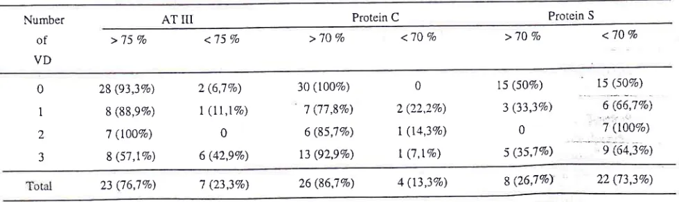
5 (35 ,7 7o)The minimum
activity
of
AT-Itr
was 60Voin
AÔS and 5'7Vo incontrol group,
where themedian
of
AT-III
activity
wzs
9'7Vo inthe
control group,
897o
in'-t
VoL I
l,
No 2, April-
June 2002with
double
vessel disease, and80lo in
subjectswith
triple
vessel disease(Figure
l).
The minimum protein C activity
was 1Voin
subjectswith ACS
andSIVo
in
thecontrol
groups. The medianprotein
C
activity was
146,57oin
the control
group, 1357oin
subjectswith
single
vessel disease, l24Voin
subjects
with
double
vessel
disease,and
ll},SVo
in
subjects
with triple
vessel disease(Figure
1).The minimum protein
Sactivity
was
20Voin
subjectswith ACS
and
ISVo
in
control
grouPs,
where
themedian
of protein
Sactivity
was 69,57oin
thecontrol
group,
567o in
subjecs
with
single
vessel
disease,Natural anticoagulant deficiencies in acute coronary
syndrome
89327o
in
subjectswith
double
vessel disease, and 617oin
subjectswith triple
vessel disease@igure
1).The
frequency
oi
distribution
of
AT-m,
protein
C,and protein
S
according
to
the
number
of
vesseldisease
are
shown in
Table
2.
The
frequency of
distribution of
AT-Itr
deficiency
(<75Vo)
were7
out o130 (23,3To)in
thosewith
ACS,
andonly
2out of
30 (6,7Vo)in
thecontrol
subjects.None
of
the 30 control
subjects had
protein
C
deficiency (levels
<707o),while 4
out
of
30 (l3,3To)
subjectswith ACS.
Fifteen
out
of
30
(50Vo)
control
subjects
had protein
Sdeficiency (levels
<70Vo),whi\e 22 out
of
30
(73,3Vo) subjectswith ACS
hadprotein
Sdeficiency.
90% Centrol ronge
70
50 140
r30
'I 30
I t0
90 r20
lt0
70
6
9oo
iro
120
6
-9
so 6É
.ô
30o
o-
t09oo
o
o40
c
'6
zo oo-O
Numbar of vsssel disaosê
Box-plot AT
m
Non Single Doublo lriPle
Number of vessel dlseose
Box-plot
Protein CNumber of vessôl dlseose
Box-plot
Protein
S [image:3.612.86.535.305.502.2]and protein S activity according to the number
Figure I . Box and whisker plot of 90vo central range frequency on AT III, protein c, of vessels disease.
Table 2. The frequency distribution oi AT
III,
protein C and protein S according to the number of vessel disease (VD).Number AT
III
Protein C Protein Sof
>75Vo
<'75Vo VD<70 Vo >'lO
Vo
<70 % >'10 vo0
I
2
3
28 (93,3Vo)
8 (88,97o)
7 (t00%)
8 (s7,1%)
2 (6,7Vo)
1
(ll,lVo)
06 (42,9%)
30 (l00Vo)
7 ('t7,8%)
6 (85,7Vo)
13 (92,9Vo)
0 ) (')) 1a7^\
|
(r4,3%)I
(7,lVo)rs (s0%) 3 (33,3%)
0
5 (3sJ%)
t5 (s0%\ 6 (65|lVo)
i
ooora
-
--
eie+,zqa
[image:3.612.55.545.592.738.2]90
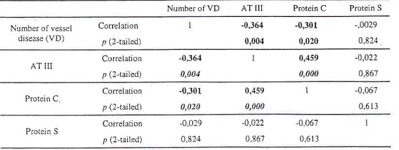
Ismail et alBoth
AT-III
andprotein C
hadsignificant conelations
with
the number
of
vessel disease, as shownin
Table3. There was no
correlalion
belweenprotein
Sactivity
and the
number of
vessel diseaseor level
of AT-Itr,
or
level of protein C
(Table 3).According to
the definition,
AT-III
deficiency
refersto
below 75Vo,
whlle
protein
C, or
protein
Sdeficiency refers
to
below
707o.The
discriminant
analysis
on the
frequency
of
distribution
of
AT-III,
protein C,
andprotein
Sdeficiency
demonstrated thatprotein C
deficiency
poseda
greaterrisk
ACS
thanAT-III
deficiency. Protein
S
deficiency
apparentlywas
a
reinforcing factor
on
AT-Itr
deficiency for
ACS
while
AT-III
deficiency
without protein
Sdeficiency did not
pose a significant risk.On linear regression analysis
with
the number
of
vessels disease
as
a
dependent
variable, and
theactivity
of AT-III
as
an
independent
variable,
theactivity of
AT
III
has
a
determinant
coefficiency of
13,25
Voin
contributing
to
ACS.
The
expectedactivity of
AT-III
that
posesa risk
of
single
vesseldisease
is
3
457o, and
to
triple
vessel
disease
if
activity
of AT-III
is
3
9,57o.
With
the activity of
protein C
as an independent variable, the
activity of
protein
C
has a determinantcoefficiency
of
9,06
Voin
conlributing to
the developmentof ACS.
Logistics regression analysis
by
backward
methodwere performed
using
vessel diseaseas'a
dependentvariable
andAT-III,
protein
C,
andAT-m
by
protein
Med J Indonei
C
interaction as
an
independent variables entered in
step one, aswell
as similar modelwith AT-III,
protein
S,
andAT-m by protein
Sinteraction
as independent variables enteredin
step one.On
both
analysisAT-III
posed
a
significant
risk
of
ACS.
These
analysisshowed that
protein S
had nospecific correlation
with
the development
of
ACS.
From the
sameanalysis by backward method with
vessel disease as a dependent
variable and
protein
C,protein S,
and
protein
C by protein S interaction
asindependent variables entered
in
step one,
protein
Cposed
a
significant
risk
of
ACS.
This
analysis
alsoshowed
that, protein
S
had
no
specific
correlation
with
thedevelopment
of
ACS.
With
double
andtriple
vessel disease as dependent
variables, and
protein
C,protein S,
and
protein C by protein S interaction
asindependent
variables
are enteredin
step one,protein
C
posed a significantrisk of
ACS. From this
analysis,protein
Salso
demonstratedno specific
correlation
to the development ACSFrom the
analysis
by
backward method
with
doubleand
triple
vessel diseaseas
dependentvariables,
andAT-III,
protein S,
andAT-III
by protein
S interaction
as independentvariables
enteredin
step
one, AT-III
by protein
S interaction
posed
a
significant
risk
of
ACS. This
analysis demonstrated
that protein S
hadno
specific correlation
with
the development
of
ACS,
insleadmight
have acted as areinforcing
factor
onAT
III
deficiency to
the developmentof
ACS.
Table 3. Pearson correlations matrix on the number of vessel disease, activity of AT-III, protein C, and protein S
Number of VD AT
III
ProteinC
Protein SNumber of vessel disease (VD)
Correlation p (2-tarlecJ)
-0,364 0,004
-0,301
-,00290,020
0,824AT
III
Correlationp (2-tailed)
-0,364 0,004
0,459
-0,0220,000
0,867Protein C Correlation p (2-r^iled)
-0,301 0,020
0,459 0,000
-0,067 0,6r3
Correlation p (2-tailed)
-0,0?9
0,824
-0,022
0,867
[image:4.612.67.478.535.690.2]Vol I
l,
No 2, April-
June 2002DISCUSSION
Pathophysiologic
mechanismsthat
can
trigger
onsetsof
acute coronary
syndromes
are:
(a)
Thrombosis,
occurring on previously coronary endothelial pertubations
or
"disrupted
or
intact
plaqLres",
when
systemicthrombotic tendency
is
high
because
of
platelet
activation,
ahypercoagulability
state, and/orimpaired
fibrinolysis
system;
(b)
Plaquedisruption; the
risk of
plaque disruption
depending
more
on
plaque
com-position
and
vulnerabiLity (piaque type)'
than
on degreeof
stenosis(plaque size).
Major
determinantsof
vulnerability
of
plaque
rupture
are
size
andconsistency
of
atheromatous
core,
and
ongoinginflammation within
the
cap.7'8(c)
Vasoconstriction,
generalized
or
occurring
local
around
a
coronaryplaque.
In
coronary
thrombosis, three aspect of
the haemostatic system leading tofibrin
formation associatedwith
the
onsetof
ischaemic
heart disease(IHD),
are:(1)
Hyperfunction of
platelets,
leading
to the
releaseof
Platelet Factor-2 (PF2) causingfibrinogen
activation,PF3 causing prothrombin complex activation,
andPF4
asan anti-heparin
factor; (2) High
levels
of:
(a)Factor
VIIc,
VIIIa,
and
vWF
leading
to thrombin
formation;
@)Fibrinogen;
(c)Inhibitor
to
plasminogenactivating factor; (3)
Functional
deficiency
of AT
III,
protein
S, andprotein
C.Hypercoagulability
slateis
acondition of high
serumlevels of
peptide materials,
anFl
and F2fragment
of
prothrombin,
where
endothelial perturbation
anddeficiency
in
one
or
both
hemostatics
control
andfibrinolytics system lead
to
thrombosis.
Thrombo-modulin
releasedby
endothelial
cells
activateAT III,
and protein
S
binding protein
C,
are the
mainmechanisms
of
hemostaticscontrol,
and plasminogenactivators are
the
main
mechanisms
of
fibrinolytic
sys[em.
Polential defense mechanisms against
thrombosisinclude
AT
III
found
agradient increasing level
from
those
at
low
risk
of
ischemic heart
disease. Meacleareported
AT-III
activity
were positively
conelated
with
VIIc
and fibrinogen.
In contrast,
low
activity
were
found in
those
with
ahistory
infarction or
othermanilestations
of
arterial
disease.Suchlow
activity
was
associated
with increased
incidence
of
major
thrombotic
episodes,
simrlar
to
that
found
in
this study,with
theexclusion
of protein
S.a'eOn
palients
of presenting
with
the
first
episode
of
thrombosis,
the
incidence
of
AT
III
deficiency
wasNatural anticoagulant deficiencies in acute coronary syndrome
9l
slightly higher (1,29-1,5 times) than
in
protein
Cdeficiency,a
which
was
no
different
in
this
shrdy,where
AT
III
andprotein C deficiency
contributed
as determinants to thedevelopment
of
ACS
l3,25Vo
and9,06Vo (1,45
times) respectively.
AT trI activity
below
50Vo are
prognostic
to
thrombosis2 near
the level of
AT
m
activify
expected to contibute to the developmentof
ACS in this
studiy.
Innerfietd
I
et
al.ereported
2out
of
23
(8,7Vo)
control
subject
with
AT-III
deficiency
whics
was
nearly equal
with our
studies,where
6,7Vocontrol
subjects had
AT-IItr
deficiency.
However,
in
subjectswith
ACS
22 ost
o124 (9l,7Vo)
suffered
from
AT-trI
deficiency
which
differ from
our
studies,
whereonly
23,3Vo areAT-III
deficient.
Protein
C
deficiency
is
usually classified
into
two
types:3
type
I
is
characterized.by reduced
antigeniclevels
and
functional
activity
in
degrading
activated factorsVc
andVIIIc,
while
type
II
ischaracterizedby
normal
antigenic
levels
with
reduced
functional
activity
in
degrading activated factors
Vc
and
VIIIc.
Both
typeS are associatedwith
athrombophilic
state.AT
trI
deficiency
is
commonly
divided
into
threetypes:3
type
I
is
quantitative deficiency, type
tI
isqualitative abnormal
ability
to
inactivate factor Xa
and
thrombin, type III
is
dueto
mutation that
reducesthe
ability
of
AT
III
to bind with
heparin.
In
the
lasttwo
typesof
AT
trI
deficiency,
AT
III
activity
mayin
normal limits.
That
explains
AT
III
deficiency
can not be ascertained solely
from
activity
levels. Inslead,what
it
is
themost important
to
assesAT
Itr
effectiveness
in
inactivating
factor
Xa
andthrombin.
Cases
with normal or high level
AT
III
activity
mayhave deficient function.
In
contrast,
in
control
subjects,
low-level activity
of
AT
III
maybe
effective
in
inactivating
factor
Xa
and thrombin.
This
alsoexplains
why
low
levelsof
protein C
poseda
greaterrisk
of ACS
thanlow
levels of
AT Itr.
Protein
Sfunction in
hemostaticsis
acofactor
for
theanticoagulant
effect
of
protein
C.
In
the
circulation
approximately
60Voof
protein
S
is
bound
to
Crtb-binding protein,while
the remainder are free (unbound)or
bound
to
protein
C3.
Low
level activity
of total
protein
Smay or
maynot involve protein
Sbinding
toprotein
C.
That
explains
why
low-levels
of
only
protein
S
did not
contribute much
to
development
of
ACS. Protein S binding protein
C
assaywere
morevalid
thantotal
activity
assayin determining'the
risk
protein S deficiency
alone
wasnot a significant risk
for ACS.
On the other
hand,AT
trI
deficiency,
all of
themdid
not
haveactivity
below 50% or below
457o.However
in
subjects
with protein S
deficiency,
there was asignificant
risk for
ACS. This fact
suggests thatprotein
Sdeficiency
is
areinforcing factor
on
AT
Itr
deficiency
to the developmentof ACS.
CONCLUSION
Protein
C
and
AT
Itr
deficiency could trigger
ACS
with
determinant
coefficients
of
9,06%
and.l3,25Vorespectively.
Low-level
activity of protein C
posed a greaterrisk for
ACS
thanlow-level activity of AT Itr.
Protein
Sdeficiency
was
areinforcing factor
onAT-Itr
deficiency
for
ACS. The cut-off point activity of
AT-Itr without
protein
S
deficiency
expected
tocontribute
single
vessel diseaseis
45Vo, and 9,5Vo totriple
vessel disease. [smail et alty
level
were thetotal
level, out
of
29
subjectsREFERENCES
Med J Indones
Bick RL,
Kaplan
H.
Syndromesof
thrombosis and hypercoag';lability. Congenital and acquired causes of thrombosis. Med Clin North Am 1998;82:409-58. National household
surveyon
health-Department of Health, lndonesia 1992.Ginsberg
J
at al, Eds. Critical Decisionsin
Thrombosisand Hemost4sis. London, BC Decker Inc. Hammilton. 1998
Meade TW, Cooper J,
Miller
GJ, Howartt DJ, Striling Y. AntithrombinIII and
arterial disease. Lancet 1991:338.The Joint
European Socielyof
Cardiology/American College of Cardiology Committee. Myocardial infarction redefined-A consensus documentof
The Joint European Societyof
Cardiology/American Collegeof
Cardiology Committee for the Redefinition of Myocardial Infar*ion. Eur Heart J 2000;21:1502-13.Braunwald E. Unstable angina : a classification. Circulalion 1989;80:410-4.
Falk E, Shah PK, Fuster
V.
Coronary plaque disruption. Circulation 199 5 ;92:651 -7l.
Shah
PK.
New insight
into the
pathogenesis andprevention
of
acute coronary syndrome.Am
J
Cardiol 1997 ;7 9(t2B):17 -23.Innerfield
I,
GoldfischerJD,
ReissHR,
Greenberg J.Serum antithrombin in coronary-artery disease. Am J Clin Pathol 1976;6:64-8
1.
2.
3.
4.
5.
6
7
8