ISSN: 1978-3019
Genetic Differentiations among the Populations of
Salvia japonica
(Lamiaceae) and Its Related Species
SUDARMONO1∗∗∗∗∗, HIROSHI OKADA2
1Center for Plant Conservation, Bogor Botanical Gardens, Indonesian Institute of Sciences (LIPI),
Jalan Ir. H. Juanda No. 13, Bogor 16003
2Botanical Gardens, Graduate School of Science, Osaka City University, 2000 Kisaichi, Katano, Osaka 576-0004, Japan
Received November 5, 2007/Accepted March 28, 2008
Morphological and genetic variations within Salvia japonica (Lamiaceae) and its related species in Japan were analyzed for clarifying their taxonomic significance. The genetic variations were explored through chloroplast and nuclear ribosomal DNA sequences and allozyme polymorphisms. Since chromosome numbers characterized the genus of Salvia, we also examined whether the karyotypes were different. We examined 58 populations of S. japonica and 14 populations of others species of Salvia. Among the populations of S. japonica represented four forms (f. japonica, f. longipes, f. lanuginosa and f.
albiflora). The size of chromosomes were various among Salvia spp. Based on the allozyme as well as the DNA sequence, the populations of S. japonica separated from the others Salvia species. The populations of S. japonica exhibited four combinations of the morphological characters. However, these combinations did not correlate to the four forms of S. japonica. In addition, the morphological variations did not correlate to the allozyme and DNA sequences. It is suggested that the four morphological variations as well as the four form of S. japonica should not considered to be a taxonomic unit; accordingly, S. japonica were considered to be still at the early stage of speciation process.
Key words: allozyme, DNA, morphological variations, Salvia japonica
___________________________________________________________________________
_________________
∗∗∗∗∗Corresponding author. Phone/Fax: +62-251-322187,
E-mail: s_darmono@yahoo.com
INTRODUCTION
Much debate still exists as to which mechanism is responsible for the majority of speciation events. The process of speciation in plants at minimum must involve: (i) divergence in some feature(s), usually morphological, such that plants are distinguishable; (ii) development of reproductive isolation sufficient to maintain these distinguishing features (Crawford 1985). The various ways by which divergence and reproductive isolation develop represent the modes of speciation. From these considerations, it follows that any study of speciation must consider the factors isolating species and the amount of genetic divergence between species. Various classifications of the modes of speciation have been proposed recently (Grant 1981; Gottlieb 2003); all have merit and they differ primarily in emphasis on different aspects of the process. The outline of Grant (1981) referred modes of plant speciation to two basic categories; i.e. evolutionary divergence (or primary speciation) and hybrid speciation. Primary speciation may involve either quantum or geographical speciation. The former is a rapid process whereas the latter involves gradual divergence. Hybrid speciation may occur at the diploid or polyploidy levels (Takano & Okada 2002).
Salvia japonica is a widespread species in Japan and
vicinity areas, such as Korea, China, Taiwan and so on, while the other 8 species of Salvia are endemic species to Japan. Murata and Yamazaki (1993) treated that S. japonica Thunb., S. isensis Nakai ex Hara, S. lutescens (Koidz.) Koidz., S. ranzaniana belongs to the series Japonicae C. Y.
Wu, where as S. pygmaea Matsum. belongs to series
Appendiculatae. Salvia japonica contains morphological
variations, and is divided into two varieties, namely, var. japonica Thunb. and var. formosana Murata. Further
S. japonica var. japonica is composed of four forms, namely,
f. albiflora Hiyama, f. japonica, f. lanuginosa (Franch)
Stib., and f. longipes (Nakai) Sugimoto (Murata 1952). T h e i n f o r m a t i o n a b o u t w i d e - range morphological variations in S. japonica suggests that the taxonomic treatment within the species remains questionable. Murata and Yamazaki (1993) regarded S. japonica as the most variable in form of leaves. The question is whether they belong to the same species, or they can be divided into several taxa that are distinguishable based on genetic characteristics.
We analyzed variations of morphological characters in
S. japonica and compared those variations with genetic
variations detected from chloroplast DNA, nuclear ribosomal DNA and allozymes within S. japonica and its related species in Japan.
MATERIALS AND METHODS
Sample Collection.A total sum of 2,138 individuals of
Salvia were used. They were include in S.japonica (58
populations), S. lutescens (two populations), S. nipponica (three populations), S. glabrescens (three populations),
S. pygmaea (one population), S. isensis (one population),
S. plebeia (one population), S. hayatana (one population),
S. arisanensis (one population), and S. ranzaniana (one
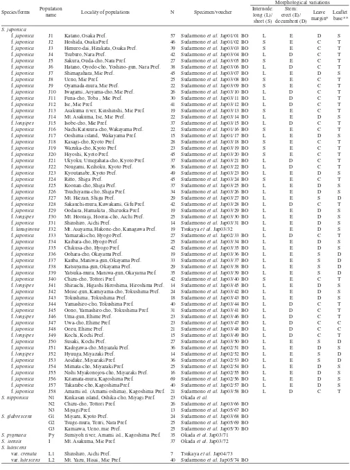
Table 1. Species, form, and name of populations, localities, numbers of individuals for allozyme analysis and chromosomes observation of
Salvia spp. and morphological characters of S. japonica populations
Morphological variations
Species/forms Locality of populations N Specimen/voucher
S. japonica
f. japonica J10
f. japonica J11
f. japonica J12
f. japonica J13
f. japonica J14
f. longipes J15
f. japonica J16
f. japonica J17
f. japonica J18
f. japonica J19
f. japonica J20
f. japonica J21
f. japonica J22
f. japonica J23
f. japonica J24
f. japonica J25
f. japonica J26
f. japonica J27
f. japonica J28
f. japonica J29
f. longipes J30
f. japonica J31
f. lanuginosa J32
f. japonica J33
f. japonica J34
f. japonica J35
f. japonica J36
f. japonica J37
f. japonica J38
f. japonica J39
f. japonica J40
f. longipes J41
f. japonica J42
f. japonica J43
f. japonica J44
f. japonica J45
f. longipes J46
f. japonica J47
f. japonica J48
f. longipes J49
f. japonica J50
f. japonica J51
f. longipes J52
f. japonica J53
f. japonica J54
f. japonica J55
f. japonica J56
f. japonica J57
f. japonica J58
S. nipponica N1
N2 N3
S. glabrescens G1
G2 G3
S. pygmaea Py
S. isensis I
S. lutescens
var. crenata L1
var. lutescens L2
Katano, Osaka Pref. Hoshida, Osaka Pref.
Himuro-dai, Hirakata, Osaka Pref. Tsuburo, Nara Pref.
Sakura, Ouda-cho, Nara Pref.
Hatano, Oyodo-cho, Yoshino-gun, Nara Pref. Shimagahara, Mie Pref.
Ueno, Mie Pref. Oyamada-mura, Mie Pref. Iwagami, Aoyama-cho, Mie Pref. Funa-cho, Toba , Mie Pref. Ise, Mie Pref.
Asakuma river, Kinshinshi, Mie Pref. Mt. Asakuma, Ise, Mie Pref. Isobe-cho, Mie Pref.
Nachi Katsuura-cho, Wakayama Pref. Ooshima island, Wakayama Pref. Kasagi-cho, Kyoto Pref. Wazuka-cho, Kyoto Pref. Hiyoshi, Kyoto Pref.
Ukyoku, Umegahata-cho, Kyoto Pref. Nougami, Keihoku, Kyoto Pref. Kyoutanabe, Kyoto Pref. Ritto, Shiga Pref. Koonan-cho, Shiga Pref. Tsuchiyama-cho, Shiga Pref. Mt. Hiezan, Shiga Pref.
Sakauchi-mura, Kawakami, Gifu Pref. Oodaira, Hamakita , Shizuoka Pref. Mt. Hooraiji, Hoorai-cho, Aichi Pref. Shinshiro, Aichi Pref.
Mt. Asayama, Hakone-cho, Kanagawa Pref. Yamazaki-cho, Hyogo Pref.
Kaibara-cho, Hyogo Pref. Chikusa-cho, Hyogo Pref. Oohara-cho, Okayama Pref. Kanba, Maniwa-gun, Okayama Pref. Katsuyama-gun, Okayama Pref.
Yatsuka-mura, Maniwa-gun, Okayama Pref. Chizu-cho, Tottori Pref.
Shiraichi, Higashi Hiroshima, Hiroshima Pref. Meise-gun, Kamiyama-cho, Tokushima Pref. Tokushima, Tokushima Pref.
Yamashiro-cho, Tokushima Pref. Oono, Yamashiro-cho, Tokushima Pref. Uma-gun, Ehime Pref.
Uwa-cho, Ehime Pref. Oozu, Ehime Pref. Kochi, Kochi Pref. Susaki, Kochi Pref.
Kadogawa-cho, Miyazaki Pref. Hyuuga, Miyazaki Pref. Aoidake, Miyazaki Pref. Mimata-cho, Miyazaki Pref.
Nishi Myakonojou-cho, Miyazaki Pref. Kitamata-mura, Kagoshima Pref. Takarabe-cho, Kagoshima Pref.
Amami isl. (Amami oshima), Kagoshima Pref. Kinkasan island, Oshika-cho, Miyagi Pref. Chizu-cho, Tottori Pref.
Miyagi Pref. Miyazu, Kyoto Pref. Tsuge-mura, Tenri, Nara Pref. Kamiawa, Ueno, mie Pref.
Sumiyoh river, Amami isl., Kagoshima Pref. Mt. Asakuma, Mie Pref.
Shinshiro, Aichi Pref. Mt. Yazu, Hisai, Mie Pref.
57
Sudarmono et al. Jap01/01 BO
Sudarmono et al. Jap01/02 BO
Sudarmono et al. Jap03/03 BO
Sudarmono et al. Jap03/04 BO
Sudarmono et al. Jap03/05 BO
Sudarmono et al. Jap03/06 BO
Sudarmono et al. Jap03/07 BO
Sudarmono et al. Jap03/08 BO
Sudarmono et al. Jap03/09 BO
Sudarmono et al. Jap03/10 BO
Sudarmono et al. Jap03/11 BO
Sudarmono et al. Jap03/12 BO
Sudarmono et al. Jap03/13 BO
Sudarmono et al. Jap03/14 BO
Sudarmono et al. Jap03/15 BO
Sudarmono et al. Jap01/16 BO
Sudarmono et al. Jap01/17 BO
Sudarmono et al. Jap03/18 BO
Sudarmono et al. Jap03/19 BO
Sudarmono et al. Jap03/20 BO
Sudarmono et al. Jap03/21 BO
Sudarmono et al. Jap03/22 BO
Sudarmono et al. Jap03/23 BO
Sudarmono et al. Jap03/24 BO
Sudarmono et al. Jap03/25 BO
Sudarmono et al. Jap03/26 BO
Sudarmono et al. Jap03/27 BO
Sudarmono et al. Jap03/28 BO
Sudarmono et al. Jap03/29 BO
Sudarmono et al. Jap03/30 BO
Sudarmono et al. Jap03/31 BO
Tsukaya et al. Jap03/32
Sudarmono et al. Jap02/33 BO
Sudarmono et al. Jap03/34 BO
Sudarmono et al. Jap03/35 BO
Sudarmono et al. Jap03/36 BO
Sudarmono et al. Jap03/37 BO
Sudarmono et al. Jap03/38 BO
Sudarmono et al. Jap03/39 BO
Sudarmono et al. Jap03/40 BO
Sudarmono et al. Jap03/45 BO
Sudarmono et al. Jap03/42 BO
Sudarmono et al. Jap03/43 BO
Sudarmono et al. Jap03/44 BO
Sudarmono et al. Jap03/41 BO
Sudarmono et al. Jap03/46 BO
Sudarmono et al. Jap03/47 BO
Sudarmono et al. Jap03/48 BO
Sudarmono et al. Jap03/49 BO
Sudarmono et al. Jap03/50 BO
Sudarmono et al. Jap02/51 BO
Sudarmono et al. Jap02/52 BO
Sudarmono et al. Jap02/53 BO
Sudarmono et al. Jap02/54 BO
Sudarmono et al. Jap02/55 BO
Sudarmono et al. Jap02/56 BO
Sudarmono et al. Jap02/57 BO
Sudarmono et al. Jap03/58 BO
Okada et al.
Sudarmono et al. Jap03/66 BO
Sudarmono et al. Jap05/67 BO
Sudarmono et al. Jap03/68 BO
Sudarmono et al. Jap05/69 BO
Sudarmono et al. Jap05/70 BO
Okada et al. Jap03/71
Okada et al. Jap03/72
Tsukaya et al. Jap04/73
Sudarmono et al. Jap05/74 BO
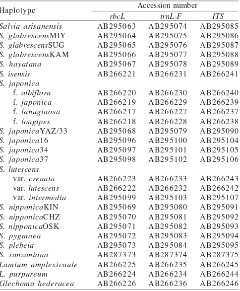
from four different populations that have peculiar karyotype and morphological characters, i.e. population Yamazaki, Hyogo Pref.(J33), population Nachi-katsuura, Wakayama Pref. (J16), population Kanba, Okayama Pref. (J37), and population Kaibara, Okayama (J34) were used for DNA analysis. In addition, individuals of S. nipponica as well as S. glabrescens from three different localities were also used for DNA analyses. Template DNA of twenty-three taxa of Salvia and three taxa of outgroup (Table 2) were analyzed for estimating the relationships among species. These taxa included S. japonica, which was analyzed from four individuals of different populations, S. nipponica and S. glabrescens, which were analyzed from three individuals of different localities, and others Salvia species, which their DNA sequences were obtained from DNA Data Bank of Japan. Living materials transplanted in the screen house of Botanical Gardens, Faculty of Science, Osaka City University. Voucher specimens are kept in BO (Herbarium Bogoriense) and Botanical Gardens, Osaka City University.
Variations of Morphological Characters. In order to examine the morphological variations of leaves, all individuals of 58 populations of S. japonica were observed (Table 1). Four morphological characters were examined, those are, (i) the internode length, i.e., Long (L): more than 5 cm or Short (S): less than 1.5 cm; (ii) the stem habit, i.e., erect (E) or decumbent (D); (iii) the leaf margin, i.e., crenate (C): tooth rounded, dentate (D): tooth obtuse angled, or serrate (S): tooth acute angled; (iv) the leaflets base, i.e., truncate (T), shallowly cuneate (S), or deeply cuneate (D) (Table 1).
Chromosome Observation.Chromosome number and the photomicrographs obtained during our examinations revealed that chromosome lengths of the best cell of all taxa referred Table 1. Growing root tips were incubated in 0.05% colchicine aqueous solution for 2 hours at 18 oC. They were fixed with
the fixative fluid (ethanol: chloroform:glacial acetic acid = 2:1:1) for more than 45 minutes at 5 oC. The root tips were then
macerated with 1N HCl at 60 oC for 18 seconds. The
meristematic tissues were stained with 2% aceto-orcein for 5-10 minutes on a slide glass. After then, one drop of 45% acetic acid was added and the slide glass was covered by cover slip and gently squashed.
DNA Extraction and Amplification. Total DNA was isolated from 0.7 to 1.5 grams of fresh or silica gel-dried leaves, using a modification of the 2x cetyltrimethylammonium bromide (CTAB) extraction protocol of Doyle and Doyle (1987). The chloroplast DNA (hereafter cpDNA) sequences were amplified with primer pairs rbcL 1-1 as the forward primer and
rbcL NN3-2 as the reverse primer (Hasebe et al. 1994), and for the intergenic spacer region of trnL-F FRF as the forward primer and 5FR as the reverse primer (Sudarmono & Okada 2007). The highest yields of polymerase chain reaction (PCR) products of rbcL and trnL-F were achieved using the following conditions. The PCR reaction mixture consisted of 5 µl of 5% rTaq-polymerase (TAKARA, Japan) reaction buffer, 4 µl of 0.2 mM each dNTP, 2.5 µl of 20 pM each primer, 0.25 µl of 0.5 units Taq DNA polymerase, 10-50 ng of 5 µl template total DNA and mess up by sterilized water in a total volume of 50 µl. The PCR samples were heated to 94 oC for 3 min, followed
by 37 cycles of 94 oC for 1 min, 54 oC for 1 min, 72 oC for 2 min
30 sec, and a final extension at 72 oC for 5 min.
Additional pairs of forward and reverse sequence primers used for the amplification of nuclear ribosomal DNA (hereafter nrDNA) in this study were ITS A and ITS B (Blattner 1999). The PCR reaction with the ITS primers included 5 µl dimethyl sulfoxide (DMSO) with a reduced amount of sterilized water to compensate. The PCR conditions were denaturation at 94 oC for
Salvia arisanensis S. glabrescensMIY
S. glabrescensSUG
S. glabrescensKAM
S. hayatana S. isensis S. japonica
f. albiflora
f. japonica
f. lanuginosa
f. longipes S. japonicaYAZ/33
S. japonica16
S. japonica34
S. japonica37
S. lutescens
var. crenata
var. lutescens
var. intermedia S. nipponicaKIN
S. nipponicaCHZ
S. nipponicaOSK
S. pygmaea
Table 2. Accession number (DNA Data Bank of Japan) of DNA sequences of the taxa studied
Accession number Table 1. Continued
Morphological variations
Species/forms Locality of populations N Specimen/voucher Leave
margin*
S. plebeia P1
S. hayatana H
S. arisanensis A
S. renzaniana R
Total individuals
*Leaf margin: crenate (C); dentate (D); serrate (S); **Leaflet base: truncate (T); shallowly cuneate (S); deeply cuneate (D); N = number of individuals
Sudarmono et al. Jap04/75 BO
Okada et al.
Okada et al.
Okada et al. Okada5698
Kizu River, Kyotanabe, Kyoto Pref. Wu shinpi, Wu-yen Chiao, Taiwan Mt. Ho-huan Shan, Taiwan
Shinogo, Kitayama vill., Wakayama Pref.
5 min, followed by 35 cycles of 94 oC for 1 min 30 sec, 60 oC for
30 sec, 72 oC for 40 sec, and a final extension at 72 oC for 5 min.
Amplified fragments were subjected to electrophoresis in a 1.5 % agarose gel and purified using Microspin S-300 HR Columns following the manufacturer’s protocol (GE Healthcare Biosciences, USA). DNA cycle sequencing with BigDye Terminators v1.1 (Applied Biosystems, USA) and PCR primers were performed in 10 µl volumes on the cleaned PCR products (25 cycles, 10 sec denaturation at 96 oC, 5 sec annealing at
50 oC, and 4 min extension at 60 oC for rbcL and trnL-F, or
25 cycles, 10 sec denaturation at 96 oC, and 4 min annealing/
extension at 60 oC for ITS). Cycle sequencing reactions were
purified by ethanol precipitation, and then denatured in HiDi Formamide 25 µl, 95 oC for 2 min. The denatured samples were
cooled on ice and run on an ABI PRISM 310 genetic analyzer (Perkin Elmers Co., Applied Biosystems, USA).
Sequence Alignment and Phylogenetic Analysis. Alignments were obtained using the program BioEdit 5.0.9 (Hall 2005), and adjusted visually. Alignments of rbcL and the intergenic spacer region of trnL-F of cpDNA were combined. The alignment of the nrDNA region included the ITS1-5.8S rDNA-ITS2 region. Gaps were treated as missing data. Phylogenetic relationships were analyzed using maximum parsimony (MP) approaches with a strict consensus, implemented with the computer program, PAUP*, Phylogenetic Analysis Using Parsimony, version 4.0 Beta10 (Swofford 2002). Heuristic searches were conducted with SIMPLE addition, tree-bisection-reconnection (TBR) branch swapping, and MULPARS options. Bootstrap analysis (Felsenstein 1985) was performed with PAUP* v4.0 using 1,000 bootstrap replications to assess the amount of support for monophyletic groups. Branch lengths were used in preference to cladogram, in which nucleotide substitutions occurring between taxa and character-state changes were detected by distance-based methods.
Allozymic Analysis.Young, fresh leaves of about 0.5 cm2
from every individual were used for allozymic analyses. They were homogenized with 0.1 M TRIS-HCl grinding buffer, pH 7.5. The extract was absorbed by filter paper (Whatmann No. 3) and run on a 12% starch gel (horizontal electrophoresis system) and on a 7.5-10% polyacrylamide gel (vertical electrophoresis system) (Sudarmono & Okada 2007). A total of eight enzyme systems were analyzed. Six of the eight enzyme systems, Phosphoglucoisomerase (PGI), Phosphoglucomutase (PGM), Menadione reductase (MNR), Isocitrate dehydrogenase (IDH), 6-phosphogluconate dehydrogenase (6-PGD), and Malate dehydrogenase (MDH), were analyzed using the horizontal system. The remaining two systems, Aspartate aminotransferase (AAT), and Shikimate dehydrogenase (SKDH), were resolved using vertical gel electrophoresis as described by Shiraishi (1988). Staining was followed with the procedure of Soltis et al. (1983), with some modification in buffer pH from pH 8.0-8.5 to pH 7.5 for PGM. Genetic interpretation of the present isozyme gel banding pattern was based on the evaluation of allozymic polymorphisms in other well documented investigations (Shield et al. 1983; Kephart 1990; Syamsuardi & Okada 2002).
The genetic identities and genetic distances for each pair-wise combination of populations were also estimated following Nei (1978). In this study the unbiased genetic identity was used to accommodate bias, because of small sample size (< 50 individuals). Allele frequencies were analyzed using POPGENE ver. 1.31 (Yeh et al. 1999).
For the analysis of genetic relationships between 72 populations of Salvia based on allozymic polymorphisms, We used the Unweighted Pair Group Method using Arithmetic averages (UPGMA) phylogram and employed NTSYS-pc 2.0 (Rohlf 2000).
RESULTS
Morphological Variations.Among the morphological characters of S. japonica, i.e., the internode length: long (L) or short (S), the stem habit: erect (E) or decumbent (D), the leaf margin: crenate (C), dentate (D) or serrate (S), and the leaflet base: truncate (T), shallowly cuneate (S) or deeply cuneate (D), could be combined into 36 combinations. However, only four combinations were found in this study (Table 1). Those are; 12 populations showed SECT: the combination with short internode (S), erect stem (E), crenate leaf margin type (C) and truncate leaflet shape (T), 17 populations had LDCT: long internode (L), decumbent stem (D), crenate leaf margin (C) and truncate leaflet (T), eight populations displayed LESD: long internode (L), erect stem (E), serrate leaf margin (S) and deeply cuneate leaflet (D), and 21 populations exhibited LEDS: long internode (L), erect stem (E), dentate leaf margin (D), shallowly cuneate leaflet (S). These four combinations might be considered as taxonomic units. However, the geographic distribution of the majority populations which have LEDS combination of morphological characters tend to be separated each other (Table 1).
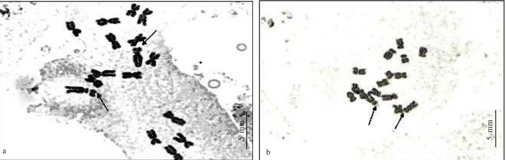
Chromosome Analysis.Chromosome numbers of the S.
japonica (Figure 1), S. lutescens, S. isensis, S. pygmaea,
S. hayatana, S. arisanensis, S. nipponica, S. glabrescens
and S. plebeia were 2n = 16. This is the same result as the
report by Funamoto et al. (2000), but different from that of Wu and Huang (1975) who reported 2n = 16-18 for species of S.
japonica. Mitotic metaphase chromosomes varied ranging
from 0.8 to 3.8 µm in length (Figure 1). The smallest chromosome of each species karyotipe ranged from 0.8 µm (S.
japonica f. longipes) to 2.0 µm (S. arisanensis). The longest
chromosome of those karyotype was 2.0 up to 3.8 µm. However, the longest chromosome of the karyotypes of S.
japonica Yamazaki populations (Hyogo Pref.) was various
(2.4 – 3.8 µm). Chromosome complements of S. nipponica, S.
glabrescens, S. pygmaea, and S. isensis were similar, they
consisted of two subtelocentrics, eight submetacentrics and six metacentrics. Satellite chromosomes were observed only
in S. japonica, S. glabrescens, and S. pygmaea. Chromosomes
of individuals in Yamazaki population had one set chromosome having longer satellite than the short arm. This finding was the first time so far.
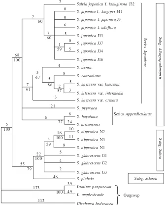
The Phylogeny Constructed from DNA. Monophyly of
a b
5 mm
Figure 1. Mitotic metaphase chromosomes of S. japonica from Ichinomiya, Kaibara-cho, Hyogo Pref (J34)2n = 16(a), and S. japonica from Yamazaki, Hyogo Pref (J33)2n = 16 (b). Arrows indicate satellite chromosome.
5 mm
value (100%) in the phylogenetic tree, which constructed based on combined data of the cpDNA (rbcL and intergenic spacer trnL-trnF) and ITS region of nrDNA (Figure 2). There were two clades in Salvia in this study, one of them was the clade of Salvia of subg. Allagospadonopsis with high bootstrap values (100%), while the other consisted the clade of subg. Salvia and subg. Sclarea with moderate bootstrap values (79%). In this clade, S. glabrescens and S. nipponica formed a subclade supported high bootstrap values (100%). Clade of subg. Allagospadonopsis consisted of all taxa of S.
japonica (60% bootstrap value), clade of S. isensis, S.
lutescens, and S. ranzaniana (69% bootstrap value), and
clade of S. hayatana, S. arisanensis (77% bootstrap value) and S. pygmaea. Branch lengths of Salvia species from the node of the Salvia clade varied from 77 changes per site in f. longipes, f. japonica to 101 changes per site in S.
plebeia (Figure 2).
Allozyme Variation. Seven enzyme systems (AAT, PGI, PGM, MNR, IDH, 6-PGD, and MDH) showed consistent banding patterns, and all of them were polymorphic. These enzyme systems contained 9 allozyme loci; AAT was composed of 2 loci (Aat-1 and Aat-2), PGI 1 locus, PGM 1 locus (Pgm-2), MNR 1 locus, IDH 1 locus, 6-PGD 1 locus
(6-Pgd-2) and MDH 2 loci (Mdh-1 and Mdh-2). Three loci
(Pgm-2, Mdh-1, and Mdh-2) were interpreted as a monomer,
five loci (Aat-1, Aat-2, Pgi, Idh, and 6-Pgd-2) as a dimer, and one locus (MNR) as tetramer according to the previous studies (Weeden & Wendel 1989; Kephart 1990).
Genetic Differentiation at the Species Level.Mean of total genetic diversity (HT) of S. japonica was calculated as 0.408 (41%), most of which was partitioned as 26% within populations (HS = 0.261) and 15% among populations (DST = 0.147) (Table 3). Almost all of loci showed high genetic diversity within populations except Aat-1 and Pgm-2. Mean of genetic differentiation coefficient (GST) of S.
japonica was 0.372, in the other words, 37% occurred among
populations and 63% within populations. Genetic differentiation in S. japonica was similar to other conspecific species of Salvia, which their genetic diversity was majority occured among populations (Table 3). Although gene flow of S. japonica (Nm = 0.486) was lower
than endemic species of S. lutescens (Nm = 2.38), it was comparable to the so-called “widespread species” (Nm = 0.149) or “out-crossing species by animal” (Nm = 0.634) (cf. Hamrick & Godt 1990).
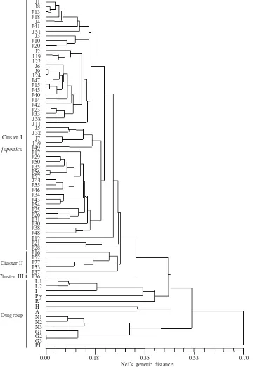
Genetic Distance.Three main clusters were constructed within populations of S. japonica. The base cluster (cluster III) was composed of only population of J36, the second cluster (cluster II) consisted of populations J16, J52, J27, J53, J37, where as the remaining populations formed cluster I (Figure 3 & 4). Other Salvia species were separated from the cluster of
S. japonica. However, S. japonica were closely related to
the cluster of S. lutescens, S. isensis, S. pycmaya, and S.
ranzaniyana (Figure 3).
Comparison Morphological Variations with Genetic Variations. Combinations of variations of morphological character of S. japonica did not correlate to allozymic variations (Figure 4). Cluster III consisted of only one population of f. japonica (Figure 4). Cluster II was composed of heterogeneous morphological variations, i.e. f. japonica with similar combination of morphological characters (LESD), population J16 of f. japonica with morphological combination SECT, and f. longipes (population J52). Cluster I contained highly heterogeneous morphological variations, i.e., populations with all of the combinations available (SECT, LDCT, LESD, and LEDS), f. japonica, f. lanuginosa, and f.
longipes (see Table 1, Figure 4). Further, some subclusters
seemingly existed, but no correlation was detected between subclusters and any morphological variations.
DISCUSSION
Salvia japonica f. lanuginosa J32
S. japonica f. longipes J41
S. japonica f. japonica J3
S. japonica f. albiflora S. japonica J33
S. japonica J37
S. japonica J34
S. japonica J16
S. isensis
S. ranzaniana
S. lutescens var. lutescens S. lutescens var. intermedia S. lutescens var. crenata S. pygmaea
S. hayatana
S. arisanensis
S. nipponica N2
S. nipponica N3
S. nipponica N1
S. glabrescens G1
S. glabrescens G2
S. glabrescens G3
S. plebeia
Lamium purpureum
L. amplexicaule
Glechoma hederacea
Series Appendiculatae
Subg. Sclarea
Outgroup
Series
Japonicae
Subg.
Allagospadonopsis
Subg.
Salvia
132
100 4 9 3 9 173
4 6 7 9
5 5
100 2 2
5 9 4
2 4 5 9 100
1 6 1 1 1 0 2 4 7 7
8 6
100 5
2 1 3 6 1
7
5 7 3 2
5 8
8 6 5 6 7
3 100
6 8
4 5 9 5
1 0 6 0
7 6 0 2
5 6 0 0 7
Figure 2. A strict consensus tree of 48 most parsimonious trees from combined analysis of rbcL, trnL-F and ITS DNA data set of 23 taxa of Salvia. Length 719, CI = 0.847, RI = 0.830. Bootstrap values are below nodes and branch lengths are above branches. Population numbers after the species names refer to Table 1.
Table 3. Genetic diversity indices and estimation gene flow for S. japonica, S. lutescens, and comparing with outcrossing and widespread species (Hamrick & Godt 1990). Standarderror are in parentheses
Genetic diversity indices
HS DST HT GST
Species Gene flow Nm References
S. japonica S. lutescens
Outcrossing by animal Widespread
0.261 (0.038) 0.190 (0.059) 0.243 (0.010) 0.267 (0.014)
0.147 (0.024) 0.034 (0.017)
0.408 (0.050) 0.224 (0.062) 0.310 (0.010) 0.347 (0.013)
0.372 (0.043) 0.095 (0.036) 0.197 (0.017) 0.210 (0.025)
0.486 (0.076) 2.380 (1.890) 0.634 0.149
Present study Present study
Hamrick and Godt (1990) Hamrick and Godt (1990)
HS:the genetic diversity within populations; DST: the genetic diversity among populations; HT: the total genetic diversity; GST: the coefficient of gene differentiation among populations; Nm: the gene flow estimate.
however, no one has attempted to examine its taxonomic significance from viewpoints of genetic relationships.
At the first, we tried to find the correlation between chromosomal variations and morphological ones. As the results, the peculiar satellite chromosome was found only in population Yamazaki, Hyogo Pref., it was not from any other populations. However, it had no relationships to any morphological variations. Then, we looked for the other points of view.
In general, it is accepted that ITS of nrDNA evidence is frequently useful for analysis of low taxonomic level (Mort & Crawford 2004), because the nuclear ribosomal DNA has topology of both parental species (e.g. Choi & Pak 1999). Therefore, we employed the ITS as well as cpDNA for analysing the relationships among Salvia species in Japan and understanding the situation of the variations observed
J1 J8 J13 J18 J4 J41 J51 J3 J10 J20 J2 J19 J22 J6 J9 J24 J47 J15 J45 J40 J14 J42 J23 J33 J58 J11 J5 J32 J7 J39 J49 J17 J29 J50 J35 J56 J57 J44 J55 J46 J34 J43 J54 J25 J26 J31 J30 J38 J48 J12 J21 J28 J16 J52 J27 J53 J37 J36 L 1 L 2 I P y R H A N1 N2 N3 G1 G2 G3 P I
0.00 0.18 0.35 0.53 0.70
S. japonica
Outgroup
Nei’s genetic distance
Figure 3. UPGMA dendrogram of allozyme divergence based on Nei’s genetic distance among 72 populations of ten species of Salvia. Name of populations refer to Table 1. Cluster I of S. japonica are JI to J28 by column; cluster II are J16 to J37 by column, and cluster III is J36.
Cluster I
Cluster II Cluster III
The results of cpDNA analysis as well as nrDNA suggested that at present, all of the Salvia species in Japan showed monophyly, although Walker et al. (2004) reported nonmonophyly of American species of Salvia. Further, the species belong to subg. Allagospadonopsis, namely, S.
japonica,S. isensis, S. lutescens, S. ranzaniana, S. pygmaea,
S. hayatana, and S. arisanensis were closely related to each
other and formed one clade, and the other two subgenera, subg. Salvia and subg. Sclarea, formed the other clade. Relatively low bootstrap supports of DNA analyses in S.
japonica were detected. S. japonica showed wide-range
morphological variations in vegetative organs (Table 1) as well as characters of flower, which were recognized as intraspecific taxa, i.e., four forms (Murata 1952). We compared four combinations of morphological variations and four forms
of S. japonica (Table 1) with genetic variations. As the results,
variations and genetic variations detected from DNA and allozymic analyses, observed in S. japonica were not evaluated as criteria to identified intraspecific taxonomic units.
S. japonica populations might be still at the early stage of
speciation process.
ACKNOWLEDGEMENT
We deeply grateful to Minoru Nomura Tamura (Botanical Gardens of Osaka City University) and Jun Yamashita (Okayama University) for their critical comment and thanks are also given to Hirokazu Tsukaya (University of Tokyo), Goto Seto (Osaka Museum of Natural History), Shigeru Ohba Inoue (Kashiwara city), and Mamoru Horiuchi (Katano city) for their help in obtaining materials, to Moritoshi Kato (University of Tokyo) for permission to use facilities, to Yoko Kita (University of Tokyo) for technical help, and to Hiroshi
J1 J8 J13 J18 J4 J41 J51 J3 J10 J20 J2 J19 J22 J6 J9 J24 J47 J15 J45 J40 J14 J42 J23 J33 J58 J11 J5 J32 J7 J39 J49 J17 J29 J50 J35 J56 J57 J44 J55 J46 J34 J43 J54 J25 J26 J31 J30 J38 J48 J12 J21 J28 J16 J52 J27 J53 J37 J36 Cluster III Cluster II Cluster I
Figure 4. UPGMA dendrogram of allozyme divergence based on Nei’s genetic distance between all investigated populations of S. japonica. Name of populations refer to Table 1. Populations of long internode, erect stem, dentate margin leaf, shallowly cuneate leaflet (LEDS) morphological characters are represented by filled box; populations of long internode, decumbent, crenate leaf, truncate leaflet (LDCT) morphological characters are represented by open box; populations of short internode, erect stem, crenate margin leaf, truncate leaflet (SECT) morphological characters are represented by filled circles; populations of long internode, erect stem, serrate margin leaf, deeply cuneate leaflet (LESD) morphological characters are represented by filled triangle; as well as S. japonica form
japonica are represented by open triangle, form longipes are represented by O, and form lanuginosa are represented by U.
Yamada for his technical support using PAUP*. We also acknowledged anonymous reviewers for valuable comments to improve this manuscript.
REFERENCES
Blattner FR. 1999. Direct amplification of the entire ITS region from pure preserved plant material using recombinant PCR.
Biotechniques 29:1180-1186.
Brunell MS, Whitkus R. 1999. Assesment of morphological variation in Eriastrum densifolium (Polemoniaceae): Implications for subspecific delimitation and conservation. Syst Bot 23:351-368. Choi K, Pak JH. 1999. A natural hybrid between Pseudostellaria
heterophylla and P. palibiniana (Caryophyllaceae). Acta Phytotaxonomica et Geobotanica 50:161-171.
Crawford DJ. 1985. Electrophoretic data and plant speciation. Syst Bot 10:405-416.
Felsenstein J. 1985. Confidence limits on phylogenies: an approach using the bootstrap. Evolution 39:783-791.
Funamoto T, Zushi M, Harana T, Nakamura T. 2000. Comparative karyomorphology of the Japanese species of Salvia L. (Lamiaceae).
J Phytogeo Tax 48:11-18.
Gottlieb LD. 2003. Rethinking classic examples of recent speciation in plants. Phytologist 161:71-82.
Grant V. 1981. Plant speciation, 2nd ed. New York, USA: Colombia
Univ Pr.
Hall BG. 2005. Phylogenetic Trees Made Easy, How-to Manual for Molecular Biologists. Massachusetts: Sinauer Assoc.
Hamrick JL, Godt JW. 1990. Allozyme diversity in plant species. In: Brown AHD, Clegg MT, Kahler AL, Weir BS (eds). Plant Population Genetics, Breeding and Genetic Resources. Sunderland: Sinauer. p 43-63.
Hasebe M, Omori T, Nakazawa M. 1994. RbcL gene sequences provide evidence for the evolutionary lineages of leptosprangiate ferns.
Proc Nat Acad Sci USA 91:5730-5734.
Kephart SR. 1990. Starch gel electrophoresis of plant isozymes: a comparative analysis of techniques. Am J Bot 77:693-712. Mort ME, Crawford DJ. 2004. The continuing search: low-copy nuclear
sequences for low level plant molecular phylogenetic studies. Taxon
53:257-261.
Murata G. 1952. Salvia subgen. Allagospadonopsis of Japan and Formosa. Acta Phytotax Geobot (In Japanese) 14:184-190. Murata G, Yamazaki T. 1993. Salvia L. In: Iwatsuki K, Yamazaki T,
Boufford DE, Ohba H (eds). Flora of Japan IIIa, Kodansha, Tokyo. p 302-307.
Nei M. 1978. Estimation of average heterozygosity and genetic distance from a small number of individuals. Genetics 89:583-590. Rohlf FJ. 2000. NTSYS, Numerical Taxonomy and Multivariate
Analysis System, Version 2.0.2j. New York: Applied Biostatistics Inc. Shield CR, Orton TJ, Stuber CW. 1983. An outline general resource needs an procedures for the electrophoretic separation of active enzymes from plant tissue. In: Tanksley SD, Orton TJ (eds).
Isozyme in Plant Genetics and Breeding, Part A, B. Amsterdam: Elsevier. p 443-468.
Shiraishi S. 1988. Inheritance of isozyme variations in Japanese black pine, Pinus thunbergii Parl. Silvae Genet 37:93-100.
Soltis DE, Hauffler CH, Darrow DC, Gastony DC. 1983. Starch gel electrophoresis of ferns: a compilation of grinding buffers, gel and electrode buffers, and staining schedules. Am Fern J 73:9-27. Sudarmono 2007. Genetic differentiations among populations of Salvia
spp. (Lamiaceae) in Japan and its biosystematic significance [Thesis]. Osaka Pref: Osaka City University.
Sudarmono, Okada H. 2007. Speciation process of Salvia isensis
(Lamiaceae), an endemic species of serpentine areas in the Ise-Tokai district, Japan, from the viewpoint of the contradictory phylogenetik trees between chloroplast and nuclear DNA. J Plant Res 120:483-490.
Swofford D. 2002. PAUP 3.1.1: Phylogenetic Analysis Using Parsimony.-Champaign, III.: Illinois Natural History Survey. Syamsuardi, Okada H. 2002. Genetic diversity and genetic structure of
populations of Ranunculus japonicus Thunb (Ranunculaceae).
Plant Sp Biol 17:59-69.
Takano A, Okada H. 2002. Multiple occurrences of triploid formation in Globba (Zingiberaceae) from molecular evidence. Plant Syst Evol 230:143-159.
Walker JB, Sytsma KJ, Treutlein J, Wink M. 2004. Salvia (Lamiaceae) is not monophyletic: implications for the systematics, radiation, and ecological specializations of Salvia and tribe Mentheae. Am J Bot 91:1115-1125.
Weeden NF, Wendel JF. 1989 Visualization and interpretation of plant isozymes. In: Soltis DE, Soltis PS (eds). Isozyme in Plant Biology.
Portland: Dioscorides Pr. 5-45.
Wu JT, Huang TC. 1975. Biosystematic studies of Formosan Salvia.
Taiwania 20:78-98.