DETEKSI CENDAWAN ENDOFIT PADA TEMBAKAU (Nicotiana tabacum L.) DAN PENGARUHNYA
TERHADAP KESEHATAN TANAMAN
THE DETECTION OF ENDOPHYTIC FUNGI ON TOBACCO (Nicotiana tabacum L.) AND THEIR EFFECT
ON PLANT HEALTH
SKRIPSI
Oleh
AMIRUDIN AKHMAD FAUZI NIM. 111510501141
PROGRAM STUDI AGROTEKNOLOGI FAKULTAS PERTANIAN
DETEKSI CENDAWAN ENDOFIT PADA TEMBAKAU (Nicotiana tabacum L.) DAN PENGARUHNYA
TERHADAP KESEHATAN TANAMAN
THE DETECTION OF ENDOPHYTIC FUNGI ON TOBACCO (Nicotiana tabacum L.) AND THEIR EFFECT
ON PLANT HEALTH
SKRIPSI
Diajukan guna memenuhi salah satu persyaratan untuk menyelesaikan program sarjana pada program studi Agroteknologi
Fakultas Pertanian Universitas jember
Oleh
AMIRUDIN AKHMAD FAUZI NIM. 111510501141
PROGRAM STUDI AGROTEKNOLOGI FAKULTAS PERTANIAN
UNIVERSITAS JEMBER 2015
PERSEMBAHAN
Dengan memanjatkan puji syukur kehadirat Allah SWT Yang Maha Pengasih lagi Maha Penyayang, Saya persembahkan Skripsi ini kepada :
1. Keluarga tercinta, ibunda Bawon Damayanti, kakakku Zulfikar Vanni Hanjaya dan istrinya Ratih Ekamaysla, Adikku Figo Muhaimin Yahya, Nenekku, dan Alm. Kakekku yang senantiasa memberikan dukungan dan do’a yang tiada henti kepada saya.
2. Semua Guru dan Dosen yang telah memberi bimbingan yang besar sepanjang hidup saya.
3. Almamater yang saya banggakan Program Studi Agroteknologi Fakultas Pertanian Universitas Jember.
MOTTO
“Barangsiapa sungguh-sungguh, sesungguhnya kesungguhannya itu adalah untuk
dirinya sendiri” (QS Al-Ankabut : 6)
“Musuh yang paling berbahaya di atas dunia ini adalah penakut dan bimbang.
Teman yang paling setia, hanyalah keberanian dan keyakinan yang teguh.”
(Andrew Jackson)
“Kita melihat kebahagiaan itu seperti pelangi, tidak pernah berada di atas kepala
kita sendiri, tetapi selalu berada di atas kepala orang lain.”
(Thomas Hardy)
PERNYATAAN
Saya yang bertanda tangan di bawah ini: Nama : Amirudin Akhmad Fauzi NIM : 111510501041
Menyatakan dengan sesungguhnya bahwa karya ilmiah yang berjudul Deteksi Cendawan Endofit pada Tembakau (Nicotiana tabacum L.) dan Pengaruhnya terhadap Kesehatan Tanaman, adalah benar-benar hasil karya saya sendiri, kecuali jika dalam pengutipan substansi disebutkan sumbernya, dan belum pernah diajukan pada institusi manapun, serta bukan karya jiplakan. Saya bertanggung jawab atas keabsahan dan kebenaran isinya sesuai dengan sikap dan etika ilmiah yang harus dijunjung tinggi.
Demikian pernyataan ini saya buat dengan sebenarnya, tanpa adanya tekanan dan paksaan dari pihak manapun serta bersedia mendapat sanksi akademik jika ternyata dikemudian hari pernyataan ini tidak benar.
Jember, 25 Juni 2015 Yang menyatakan,
Amirudin Akhmad Fauzi NIM. 111510501141
SKRIPSI
DETEKSI CENDAWAN ENDOFIT PADA TEMBAKAU (Nicotiana tabacum L.) DAN PENGARUHNYA
TERHADAP KESEHATAN TANAMAN
THE DETECTION OF ENDOPHYTIC FUNGI ON TOBACCO (Nicotiana tabacum L.) AND THEIR EFFECT
ON PLANT HEALTH
Oleh:
AMIRUDIN AKHMAD FAUZI NIM. 111510501141
Pembimbing:
Pembimbing Utama : Ir. Hari Purnomo, M.Si., Ph.D., DIC.
NIP : 19660630 199003 1 002
Pembimbing Anggota : Ir. Sigit Prastowo, M.P.
NIP : 19650801 199002 1 001
PENGESAHAN
Skripsi berjudul “Deteksi Cendawan Endofit pada Tembakau (Nicotiana tabacum L.) dan Pengaruhnya terhadap Kesehatan Tanaman” telah diuji dan disahkan pada :
Hari : Kamis
Tanggal : 01 Juni 2015
Tempat : Fakultas Pertanian Universitas Jember
DPU,
Ir. Hari Purnomo, M.Si., Ph.D., DIC. NIP. 19660630 199003 1 002
DPA,
Ir. Sigit Prastowo, M.P. NIP. 19650801 199002 1 001
Penguji 1,
Ir. Kacung Haryono, M.S., Ph.D. NIP. 19640814 199512 1 001
Penguji 2,
Ir. Sundahri, M.P. NIP. 19670412 199303 1 007
Mengesahkan Dekan,
Dr. Ir. Jani Januar, MT NIP. 19590102 198803 1 002
RINGKASAN
Deteksi Cendawan Endofit pada Tembakau (Nicotiana tabacum L.) dan Pengaruhnya Terhadap Kesehatan Tanaman; Amirudin Akhmad Fauzi; 111510501141; 2015; 51 halaman; Program Studi Agroteknologi Fakultas Pertanian, Universitas Jember.
Tembakau merupakan salah satu komoditas penting di Indonesia. Kualitas tembakau seringkali tidak tercapai akibat adanya serangan OPT sebagai faktornya. Kesehatan tanaman tembakau on farm banyak dipengaruhi oleh teknik budidaya yang masih konvensional dan rendahnya pengetahuan petani. Penyakit tanaman tembakau antara lain mozaik, lanas, dan busuk batang dapat menurunkan kualitas dan produksi tembakau. Cendawan endofit merupakan cendawan yang hidup didalam jaringan tanaman dan tidak bersifat parasit terhadap tanaman. Cendawan endofit dapat berperan sebagai bio-proteksi yang dapat melindungi tanaman dari serangan OPT terutama patogen penyebab penyakit. Penelitian ini bertujuan untuk mendeteksi cendawan endofit yang ada pada tanaman tembakau yang berkaitan langsung dengan kesehatan tanaman tembakau.
Penelitian ini memiliki 2 eksperimen yang berbeda. Eksperimen pertama menggunakan rancangan acak lengkap sederhana dengan 3 taraf dan 5 replikasi. 3 taraf tersebut antara lain adalah kontrol, media+Chaetomium dan media+Trichoderma. Sementara itu, eksperimen kedua menggunakan rancangan acak lengkap sederhana dengan 2 taraf ddan 5 replikasi. 2 taraf tersebut antara lain adalah media+Chaetomium dan media+Trichoderma yang masing-masing diinokulasikan cairan zoospora dari Phytophtora spp.
Hasil penelitian menunjukkan bahwa Trichoderma spp. dan Chaetomium sp. merupakan cendawan endofit yang berada di jaringan tanaman tembakau. Aplikasi kedua cendawan endofit ini tidak memberikan pengaruh nyata terhadap parameter pertumbuhan, akan tetapi memberikan perlindungan terhadap serangan
Phytophtora spp. Cendawan Trichoderma spp. yang ditemukan merupakan cendawan Trichoderma spp. yang diaplikasikan pada media sesuai dengan hasil PCR yang dilakukan pada yang terdeteksi pada 1.5 kB.
SUMMARY
The Detection of Endophytic Fungi on Tobacco (Nicotiana Tabacum L.) and Their Effect on Plant Health; Amirudin Akhmad Fauzi; Study Program of Agrotechnology, Faculty of Agriculture, University of Jember.
Tobacco is an important commodity in Indonesia. Tobacco quality is oftenly unfulfilled because of biotic interference as the factor. On-farm plant health is much affected by conventional practice and unskilled farmer. Tobacco disease such as mosaic, black shank, and rotten stem are able to decrease tobacco quantity and quality. Endophytic fungi is a microorganism which stays or colonizes in the plants without incurring negative effect to the host. Endophytic fungi play role as bio-protection that protect the plant from biotic interference especially from plant pathogen. This research objective is to detect and determine the present of endophytic fungi on tobacco that relate with plant health.
This research contained 2 different experiments. The first experiment used complete randomized design with 3 levels and 5 replications. The levels include control, Chaetomium+media, and Trichoderma+media. Meanwhile, the second experiment used complete randomized design with 2 levels and 5 replications. The levels include Chaetomium+media, and Trichoderma+media which both of them were inoculated with zoospore of Phytophtora spp.
The result shown that Trichoderma spp. and Chetomium spp. were found as endophytes in the tobacco tissue. Application of these endophytes was not show significant different on tobacco plant growth, but both of them protected the plants from Phytophtora spp. infection. Trichoderma spp. which found in the tissue was declared as the same type with the inoculum that applied to the media by the result from PCR technique in 1.5 kB.
PRAKATA
Puji syukur kepada Allah SWT karena hanya dengan limpahan rahmat dan hidayah-Nya penulis dapat menyelesaikan skripsi yang berjudul “Deteksi Cendawan Endofit pada Tembakau (Nicotiana tabacum L.) dan Pengaruhnya terhadap Kesahatan Tanaman”. Skripsi ini disusun sebagai salah satu syarat untuk menyelesaikan Pendidikan Program Sarjana (S1) pada Program Studi Agroteknologi Fakultas Pertanian Universitas Jember. Penyusunan tugas akhir ini tidak terlepas dari bantuan berbagai pihak, oleh karena itu penulis ingin menyampaikan ucapan terima kasih kepada:
1. Dr. Ir. Jani Januar, M.T. selaku Dekan Fakultas PertanianUniversitas Jember. 2. Ir. Hari Purnomo, M. Si., Ph. D, DIC, selaku Dosen Pembimbing Utama dan
Ir. Sigit Prastowo, M.P., selaku Dosen Pembimbing Anggota, yang memberikan perhatian, bimbingan, meluangkan waktu, dan pikiran sehingga skripsi ini dapat diselesaikan.
3. Ir. Kacung Haryono, M.S., Ph.D., selaku Dosen Penguji satu dan Ir. Sundahri, M.P., selaku Dosen Penguji dua yang memberikan arahan dan pikiran sehingga skripsi ini dapat diselesaikan.
4. Nanang Tri Haryadi, S.P., M.Sc., selaku Dosen Pembimbing Akademik yang telah membimbing selama menjadi mahasiswa.
5. Ibunda Bawon Damayanti, Kakak Zulfikar Vanni Hanjaya beserta istrinya Ratih Ekamayslah, Adik Figo Muhaimin Yahya, Nenek dan Alm. Kakek yang senantiasa menjadi cermin semangat penulis dalam menjalani kehidupan.
6. Segenap Dosen dan Tim Student Exchange di Fakultas Pertanian yang telah memberikan dukungan dan kerja kerasnya dalam membimbing penulis selama menjalani pertukaran pelajar di Kasetsart University, Thailand.
7. Dr. Chainarong Rattanakreetakul selaku Supervisor dan segenap civitas akademika di Kasetsart University selama penulis menjalani pertukaran pelajar di Kasetsart University, Thailand.
8. Kerabat-kerabatku Mbak Nungky, Dina Ajeng, Mas Ian, Mas Rahmat, Pisut Keawmanee, Ratchanok Piwtamai, Dewi Masitoh, dan teman-teman lainnya yang tidak dapat disebutkan satu per satu, terima kasih atas dukungan, motivasi, pengalaman, dan saran yang bermanfaat bagi kehidupan penulis. 9. Teman-teman IMAGRO (Ikatan Mahasiswa Agroteknologi), UKM CR
(Chorus Rusticarum), dan UKM Plantarum yang telah bersedia membagi ilmu dan pengalaman berorganisasi selama menjadi mahasiswa.
10. Teman-teman Program Studi Agroteknologi angkatan 2011 dan semua pihak yang tidak dapat disebutkan satu persatu, terima kasih telah meluangkan pikiran dan memberikan semangat serta pengalaman hidup yang tidak terlupakan.
Penulis menyadari bahwa skripsi ini sangat jauh dari kesempurnaan. Oleh karena itu, dengan segala kerendahan hati penulis senantiasa mengharapkan saran dan kritik yang membangun demi kesempurnaan skripsi ini. Akhirnya penulis berharap, semoga skripsi ini dapat bermanfaat bagi semua pihak, terutama bagi perkembangan ilmu di bidang pertanian.
Jember, Juni 2015 Penulis
DAFTAR ISI
Halaman
HALAMAN JUDUL ... i
HALAMAN PERSEMBAHAN ... ii
HALAMAN MOTTO ... iii
HALAMAN PERNYATAAN ... iv
HALAMAN PEMBIMBING ... v
1.3 Problem Identification ... 2
1.4 Benefit ... 3
CHAPTER 2. LITERATURE REVIEW ... 4
2.1 Tobacco in Indonesia ... 4
2.2 Diseases on Tobacco ... 4
2.3 Endophytic Fungi as Biological Control ... 6
1. Preparation of Endophytic Fungi ... 10
2. Preparation of Phytophtora spp. zoospore Suspension ... 10
3. Preparation of Tobacco Seedling ... 11
4. Preparation of Media ... 11
5. Preparation of Detection of Endophytic Fungi ... 11
3.2.2. Experimental Design ... 12
3.3.3. Research procedure ... 12
1. Efficacy of Endophytic Fungi on Tobacco Growth ... 12
2. Effect of Endophytic Fungi to Control Phytophtora spp ... 13
3. Detection of Endophytic Fungi Using Morphological Characteristic ... 13
4. Detection of Endophytic Fungi with PCR Method ... 14
a. Trichoderma DNA Extraction ... 14
b. Tobacco Parts Extraction ... 14
4.1.1 Effect of Endophytic Fungi on Tobacco Growth ... 17
4.1.2 Effect of Endophytic Fungi Against Phytophtora spp. Infection ... 29
4.2 Discussion ... 34
4.2.1 Effect of Endophytic Fungi on Tobacco Growth ... 34
DAFTAR TABEL
Halaman 3.1. Analysis of varians (ANOVA) on effect of endophytic fungi
on tobacco growth variable ... 17 3.2. Population of endophytic fungi on media ... 21 3.3. Number of identified potential endophytic fungi ... 21 3.4. Population of applied endophytic fungiin peatmoss media after
2 months ... 31
DAFTAR GAMBAR
Halaman
3.1 Experimental design ... 12
4.1 Tobacco plant height ... 18
4.2 Tobacco leaf number ... 18
4.3 Tobacco stem size ... 19
4.4 Growth comparison between treatments after 5 months ... 19
4.5 Tobacco root length ... 20
4.6 Root length comparison between treatment after 5 months plantation ... 20
4.7 Fungi 1 growth on PDA from top and bottom view ... 22
4.8 Microscopic view of fungi 1. ... 22
4.9 Fungi 2 growth on PDA from top and bottom view ... 23
4.10 Microscopic view of fungi 2. ... 23
4.11 Fungi 3 growth on PDA from top and bottom view ... 24
4.12 Microscopic view of fungi 3. ... 24
4.13 Fungi 4 growth on PDA from top and bottom view ... 25
4.14 Microscopic view of fungi 4. ... 25
4.15 Fungi 5 growth on PDA from top and bottom view ... 26
4.16 Microscopic view of fungi 5. ... 26
4.17 Fungi 6 growth on PDA from top and bottom view ... 27
4.18 Microscopic view of fungi 6. ... 27
4.19 Fungi 7 growth on PDA from top and bottom view ... 28
4.20 Microscopic view of fungi 7. ... 29
4.21 Tobacco seedling height ... 30
4.22 Tobacco seedling leaf number ... 30
4.23 Dual culture of Trichoderma spp. against Phytophtora spp. for pathogenicity test. ... 31
4.24 Population of applied endophytic fungi on RBA media ... 32
4.25 Detection of applied endophytes on tobacco plant parts
by tissue transplanting method. ... 33 4.26 Agarose gel showing PCR product of tobacco plant parts ... 34
DAFTAR LAMPIRAN
Halaman
1. Potato Dextrose Agar (PDA) Media ...50
2. Rose Bengal Agar (RBA) Media ...50
3. V8 Juice Broth Media ...50
4. Surface Sterilization of Plant Parts Material ...50
5. Raw data of tobacco height (cm) and ANOVA table in the effect of endophytic fungi on tobacco growth at 1st observation. ...51
6. Raw data of tobacco height (cm) and ANOVA table in the effect of endophytic fungi on tobacco growth at 2nd observation. ...51
7. Raw data of tobacco height (cm) and ANOVA table in the effect of endophytic fungi on tobacco growth at 3rd observation. ...52
8. Raw data of tobacco height (cm) and ANOVA table in the effect of endophytic fungi on tobacco growth at 4th observation. ...52
9. Raw data of tobacco height (cm) and ANOVA table in the effect of endophytic fungi on tobacco growth at 5th observation. ...53
10. Raw data of leaf number and ANOVA table in the effect of endophytic fungi on tobacco growth at 1st observation. ...53
11. Raw data of tobacco leaf number and ANOVA table in the effect of endophytic fungi on tobacco growth at 2nd observation. ...54
12. Raw data of tobacco leaf number and ANOVA table in the effect of endophytic fungi on tobacco growth at 3rd observation. ...54
13. Raw data of tobacco leaf number and ANOVA table in the effect of endophytic fungi on tobacco growth at 4th observation. ...55
14. Raw data of tobacco leaf number and ANOVA table in the effect of endophytic fungi on tobacco growth at 5th observation. ...55
15. Raw data of tobacco stem size (cm) and ANOVA table in the effect of endophytic fungi on tobacco growth at 1st observation. ...56
16. Raw data of tobacco stem size (cm) and ANOVA table in the effect of endophytic fungi on tobacco growth at 2nd observation. ...56
17. Raw data of tobacco stem size (cm) and ANOVA table in the effect
of endophytic fungi on tobacco growth at 3rd observation. ...57 18. Raw data of tobacco stem size (cm) and ANOVA table in the effect
of endophytic fungi on tobacco growth at 4th observation. ...57 19. Raw data of tobacco stem size (cm) and ANOVA table in the effect
of endophytic fungi on tobacco growth at 5th observation. ...58 20. Raw data of tobacco seedling height (cm) and ANOVA table in the
effect of endophytic fungi against Phytophtora spp. at 1st observation ...58 22. Raw data of tobacco seedling height (cm) and ANOVA table in the
effect of endophytic fungi against Phytophtora spp. at 2nd observation ....58 23. Raw data of tobacco seedling height (cm) and ANOVA table in the
effect of endophytic fungi against Phytophtora spp. at 3rd observation ...59 24. Raw data of tobacco seedling height (cm) and ANOVA table in the
effect of endophytic fungi against Phytophtora spp. at 4th observation ...59 25. Raw data of tobacco leaf number and ANOVA table in the
effect of endophytic fungi against Phytophtora spp. at 1st observation ...60 26. Raw data of tobacco leaf number and ANOVA table in the
effect of endophytic fungi against Phytophtora spp. at 2nd observation ....60 27. Raw data of tobacco leaf number and ANOVA table in the
effect of endophytic fungi against Phytophtora spp. at 3rd observation ...60 28. Raw data of tobacco leaf number and ANOVA table in the
effect of endophytic fungi against Phytophtora spp. at 4th observation ...61
CHAPTER I. PREFACE
1.1 Background
Tobacco (Nicotiana tabacum L.) is one of the most important commodities in Indonesia. Main product of tobacco is the leaves which become the main material of cigarettes. The demand of tobacco in the world is rising, for that reason farmers should increase the productivity of tobacco yield. Farmers have to manage their tobacco production technique till postharvest stage. Indonesia is one of the biggest tobacco exporters in the world. Since 1970 until 2007 Indonesian tobacco yield was under China (38.87%), Brazil (14.73%), India (8.43), USA (4.73%), and Argentina (2.86%) (FAO, 2009). Therefore, the productivity of tobacco yield is very important for Indonesia. Because it’s yield give huge contribution for Indonesian economic.
Tobacco is an annual plant which grows on tropical country such as Indonesia with elevation between 0 until 900 ASL (Deptan, 2012). Indonesian tobacco is categorized on Na-Oogst and Voor-Oogst (Rachman, 2013). Voor-Oogst (VO) tobacco is the main material for the filler on the cigarettes, but Na-Oogst (NO) tobacco is the material for covering the cigar (dekblad). VO tobacco is usually planted on dry season. It is waterless, but requirement of water must be provided. NO tobacco is usually planted on rainy season, because it needs more water than VO tobacco. Perfect yield of NO tobacco have to be far from deformedless which commonly caused by the pests.
Farmers in Indonesia basically used their local wisdom to manage their field. Moreover, regeneration of the new farmers is very low. In 2013, the growth rate decreased about 6% from 31.17 million to 26.13 million farmers (Tempo, 2013). This condition was severed by the unstable climate. Climate became one of nature aspect that can determine the plant yield in which pest and disease emergence was related with. On farm management of tobacco pay so much attention from other sector. This condition relate with how the farmers overcome major pest problem. Uncontrolled application of pesticide causes many environmental issues. According to USEPA (United State Environment Protection Agency), more than
2,600 pesticide chemical ingredients are published in the market and more than 3,500 formulation of pesticide are marketed around the world. This enhancement happened especially on developed country (Yuliani et al., 2011). Most of Indonesian farmers still depend on chemical pesticide to overcome pest invation which against the concept of sustainable agricultural.
Tobacco diseases is caused by various microorganism (include fungi and bacteria). They have their specific symptom which allowed us to diagnose and find the best way to control them. Plant protection is the most reliable activity to safe the product from disease disorder. Preventive and curative methods are the best way to protect the plants from biotic disturbance. The decreasing of chemical prevention is the purpose of sustainable agriculture and mostly supported by using endophytic fungi. Endophytic fungi are located on plant tissue such as leaves, branch, or on plant roots (Purwanto, 2008). Endophytic fungi produced some antibiotic compound to against the pathogenic bacteria or fungi (Worang, 2003).
In modern agriculture, the endophytic fungi can be used as a biological control which is the best way to protect plants from disease disorder. In this study, the detection of endophytic fungi on tobacco is very useful method to realize the sustainable agriculture.
1.2 Problem identification
There are several problems which identified based on the background described before.
1. What kind(s) of the endophytic fungi that can be used in tobacco (Nicotiana tabacum L.)?
2. What is the role of endophytic fungi to tobacco?
1.3 Objective
3
1.4 Benefit
1. To promote the used of biological control to reduce the disease disorder. 2. Implementation of the research will reduce the application of chemical
CHAPTER II. LITERATURE REVIEW
2.1 Tobacco in Indonesia
Tobacco (Nicotiana tabacum L.) is an annual plant which it is one of
solanaceae family from genus Nicotiana. In this genus, N. tabacum is the famous one than another Nicotiana’s plants such as N. rustica, N. silvestris, N. glutinosa,
and N. petunoides which they are less valueable (Santoso, 2001 ; Deptan, 2012). They can adapt and develop well in a few region of Indonesia and it makes various common name of tobacco in Indonesia, such as Selopuro Tobacco, Temanggung Tobacco, Kendal Tobacco, Madurese Tobacco, and Paiton Tobacco. Tobacco is divided according to the harvested season as, Na-Oogst Tobacco (Harvested on the end of rainy season) and Voor-Oogst tobacco (Harvested on the end of dry season). But, according to the use of tobacco, it is grouped in to filler tobacco, cover of cigarillos (dek-omblad), filler of filter cigerettes (Rachman, 2013).
Indonesian tobacco is characterized as ascociated tobacco, as they have much cultivated by small farmer in Indonesia. Ascociate tobacco is produced to fulfil the market cigarettes needs (Santoso, 2001). Production of tobacco, especially for NO tobacco in Indonesia is focused on 3 region, respectively in Deli (Sumatra Island), Klaten (Center Java), and Jember (East Java). The region is divided based on the ecological system that very suitable to get the best production of NO tobacco. But, historically that area was managed to product VO tobacco which used as the filler of cigarettes (Djajadi, 2008).
2.2 Diseases on Tobacco
Tobacco production is affected by several factors and one of them is diseases disorder that can reduce up to 80% of tobacco production. Here is several diseases which commonly found in Tobacco cultivation.
a) Mosaic (Tobacco Mosaic Virus)
The infection of this disease is very easy, and commonly infected another host plant by the material of tobacco cultivation or by the friction between the
5
infected host plants to the healthy one. TMV is a soil borne virus which able to penetrate from injured roots. TMV live well on the debris in the soil and will infect the host plant whenever the injury happened on roots (Akin and Nurdin, 2003). TMV is considered as the most heat resistant plant pathogen, stable in vitro, and able to survive over 10 years in dried leaves and cigarettes or cigars (Lucas 1975). Therefore, the infected dried leaves, cigar or cigarettes can be the source of inoculums of TMV. In the field, the most common source of virus inoculums is the debris of infected plants which play role as reservoirs for virus transmission to the halthy plants. Inproper disposal of these plant residues contribute as the recycle of virus as pathogens (Conway, 1996).
b) Black Shank (Phytophtora nicotianae)
Black shank which it is caused by P. nicotianae is a dangerous tobacco disease especially for NO tobacco. The infection occurs from seeding till mature tobacco which ready to be harvested (Csinos and Bertrand, 1994). This disease can be detected by the symptom such as withered plant, chlorosis, and brown rotten stem and if it’s splitted, heartwood sectional will appear. P. nicotianae is a soil borne fungi which live well in the soil and become the vector of Black Shank disease. Zoospore is flagellate and able to move well on water. The irrigation system and rainflow are the best way to spread the infection to the whole field. (Jaarsveld et al., 2002). Wet and moist condition is the suitable condition for P. nicotianae to infect the host plant. Black shank disease reported able to decrease about 25% from total yield product (Suripno and Yulianti, 2009).
c) Black Stalk (Erwinia carotovora)
2.3 Endophytic Fungi as Biological Control
Endophytic is a microorganism which stays or colonizes in the plants without incurring negative effect to the host. It can help to protecting the host from biotic or abiotic interference. Every plant reported as the host of various endophytic fungi. Endophytic fungi have an important role to increasing the tolerance of drought and hold the expansion of herbivores insect, pathogenic fungi, virus, and roots nematode. As a biological control, endophytic fungi have a mechanism as parasitism, antibiotic, nutrient competition, and resistance induction. Endophytic fungi produce fungtional metabolism which include on terpenoids, steroids, xanthones, chinones, phenol, iso-coumarins, benzopyranones, tetralones, cytochalacins and enniatines which play role as antibacterial, antirival, and antifungal (Suryanarayanan et al, 2009).
Several endophytic fungi are able to decrease the infection of some pathogen treat. Endophytic fungi as Chaetomium sp. and Phoma sp. species have been successful to reduced number of pustuls and leaves widespreak attacks on grain caused by Puccinia recondita f.sp. tritici. Moreover, leaching media Chaetomium
sp.and Phoma sp. isolates have been activated active defence reaction from the plant, that limit the multiplication of the pathogen (Dingle and Mcgee, 2003). Emergence of endophytic fungi has good chance as a biological agents, because fungi included on host plant system and suitable as promotor to introducing foreign gene on the plants tissue. Host of endophytic fungi can be manipulated genetically to produce the active compound which prospected for the host plants via genetic engineering, such as biopesticide (Petrini, 1992).
2.3.1 Trichoderma spp. as Endophytic Fungi
7
species have been extensively documented (Harman et al., 2004). Some species of Trichoderma that also are found as endophytes in stems of woody plants include
T. aggressivum Samuels & W. Gams, T. caribbaeum Samuels & Schroers, T. erinaceus Bissett, C. P. Kubicek & Szakacs, T. evansii Samuels, T. hamatum
(Bonord.) Bainier, T. harzianum Rifai, T. koningiopsis Samuels, C. Sua´rez & H. C. Evans, T. martiale Samuels, T. ovalisporum Samuels & Schroers, T. pubescens
Bissett, T. spirale Bissett, T. stilbohypoxyli Samuels & Schroers, T. strigosum
Bissett, T. stromaticum Samuels & Pardo-Schulth, T. theobromicola Samuels & H. C. Evans, T. viridescens (A. S. Horne & H. S. Will.) Jaklitsch & Samuels and
T. virens J. H. Mill., Giddens & A. A. Foster) Arx (Evans et al., 2003, Crozier et al., 2006, Samuels et al., 2006, Hanada et al., 2008, Samuels and Ismaiel 2009).
Fungal antagonistic Trichoderma species are considered as promising biological control agents against numerous phytopathogenic fungi include
F.oxysporum (Sarhan et al., 1999). These filamentous fungi are very common in nature, with high population densities in soil and plant litters (Samuels, 1996). They are saprophytic, promptly growing and easy to culture, in addition to producing huge quantities of conidia of long lifetime (Mohamed and Haggag, 2006). Trichoderma species have shown efficiency on biocontrol of plant pathogens. According to Dolatabadi et al., (2012), Trichoderma harzianum can effectively avoid Lentil (Lens culinaris) from Fusarium oxysporum infection. In addition, Trichoderma has more report on the plant induced resistant to Fusarium
species.
2.3.2 Chaetomium spp. as Endophytic Fungi
Chaetomium species are normally found in soil and organic compost.
Chaetomium is one of the largest genera of saprobic ascomycetes with more than 300 species worldwide. Chaetomium species are potential degraders of cellulose and other organic material and can be antagonistic against various soil microorganisms. Chaetomium globosum and C. cochlioides are antagonistic to species of Fusarium and Helminthosporium (Soytong et al., 2001). The mechanism of disease control is competition, antibiosis/lysis, antagonism, induced immunity in plants and hyphal interference. C. cupreum found to produce rotiorinol (Kanokmedhakul et al., 2006) and C. globosum produces chaetoglobosin-c (Sibounnavong et al., 2011).
It has been found that by using specific strains of C. globosum, it is possible to obtain promising control over many plant pathogens. By coating seeds of corn with spores of C. globosum it was possible to prevent seedling blight caused by
Fusarium roseum f sp. cerealis 'graminearum'. Such seed coating treatments were also found to reduce disease incidence of apple scab caused by Venturia inequalis
(Soytong et al., 2001). It has also been reported that some isolates of C. globosum
produce antibiotics that can suppress damping-off of sugar beet caused by
Pythium ultimum (Di Pietro et al., 1991). A further isolate of C. globosum was found to be antagonist against Rhizoctonia solani and Alternaria brassicicola
(Soytong et al., 2001) and also reduced the quantity of sporulation of Botrytis cinerea on dead lily leaves exposed in the field (Kohl et al., 1995).
Zhang et al., (2013) reported that Chaetomium have antifungal substance as
chaetoglobosin and it can inhibit the growth of Rizopus stolonifer. The mycelia growths of 24 h cultivation of R. stolonifer and 72 h cultivation of C. diplodiella
were nearly completely inhibited as Chaetoglobosin A concentration 40 and 20 g/mL, respectively. Chaetoglobosin A had acute antifungal activity against phytopathogenic fungi, which indicated that the studied propolis had the potential to be a natural preservative that can be applied to control plant diseases.
9
CHAPTER III. METHOD OF RESEARCH
3.1 Time and Place
This research was implemented during December 2013 till June 2014 in the Greenhouse and Laboratory of Physiology of Plant Disease, Department of Plant Pathology, Faculty of agriculture at Kamphaeng Saen Campus, Kasetsart University.
3.2 Research Method 3.2.1. Preparation
1. Preparation of Endophytic Fungi
Trichoderma culture was taken from Biological Control Laboratory, Department of Plant Pathology, Faculty of Agriculture at Kamphaeng Saen campus, Kasetsart University. The culture was multiplied via steamed rice as carrier. Two hundred grams of fresh steamed rice was put into the plastic bag (1 kg size) with aseptic technique, sealed the plastic bag after the steamed rice filled in. After the temperature reduced, Trichoderma culture was poured to the rice about 0,5 g respectively. The plastic bag was stabbed with the needle to make aeration in to the plastic bag ecactly on the upside part. Number of the holes was about 15 holes to each bag and the bag was placed in the incubation room with room temperature and with lying position to get equal surface on Trichoderma
growth. The incubation took about 3-4 days to be harvested. After Trichoderma
completely growth in the steamed rice, then it was stored in the refrigerator.
Chaetomium spp. was collected from laboratory culture and the whole method for make Chaetomium inoculum was the same as explained in
Trichoderma inoculum, except steamed corn subtituted steamed rice as the carrier.
2. Preparation of Phytophtora spp. Zoospore Suspension.
Phyophthora spp. was grown on V8 agar plates at 25 °C under dark condition. For zoospore production, mycelial plugs were transferred to a flask containing 25 mL of V8 vegetable juice and incubated in the dark at 25 ± 2 °C.
11
After 1-2 weeks, mycelial plugs were removed from the medium and macerated with sterile distilled water in a sterile blender for 30 s. Drops of mycelial suspension were placed onto the surface of water-agar plates using a sterile syringe. Sporangial formation in Phytophthora spp. was induced by removing uncolonized agar from around the mycelium and incubating the culture for an additional 3 days at 25 °C; plates were placed under fluorescent lights (40 W, daylight) at a distance of 12 cm. Zoospores were induced to release by incubating the culture plates in sterile water at 4°C at room temperature for 1 hour. The zoospores were collected and filtered through Whatman No. 54 to remove sporangial cases and mycelial. The concentration was then adjusted to 102, 103, and 104 zoospores per milliliter using a hemocytometer (Ward and Stoessl, 1974).
3. Tobacco Seedling Preparation
Tobacco was seedled using tobacco seed and organic matter as the media. Media was moisted with water addition. Tobacco seed was poured in to the media and then covered with double-layer filter paper to keep the moisture. After 20-25 days filter paper was removed from the media and then tobacco seedling grew untill reach the vigorous stage (about 40-45 days) and ready to be transplanted to the new media.
4. Media Preparation
Media was composed as sterilized soil and sterilized peatmoss. Soil was sterilized in mass media sterilization. Peatmoss was sterilized manually in autoclave with 121oC and 72 psi/1atm for 15 minutes. Media was mixed with proportion 3:1 of soil:peatmoss (v/v). Media was added with 20 g of inoculum of endophytic fungi (Trichoderma spp. and Chaetomium sp.) as treated media, the rest which was not added with inoculum of endophytic fungi as the control media.
5. Detection of Endophytic Fungi Preparation
Dextrose Agar) and RBA (Rose Bengal Agar) as the culture media. PDA contain of potato as carbon source, dextrose as sugar source and agar. RBA was a selective media contain of KH2PO4, MgSO4, Rose Bengal, Agar, Peptone, and Glucose. PCR method was prefaced by DNA extraction using extraction kit (Thermo.ltd) for pure Trichoderma culture extraction. Trichoderma in tobacco plant parts were extracted by modified extraction which used lysis buffer (250 mM Tris-HCl (pH 8.0), 50 mM EDTA (pH 8.0), 100 mM NaCl and 2% SDS), PCI, 100% ethanol, and TE buffer.
3.2.2. Experimental Design
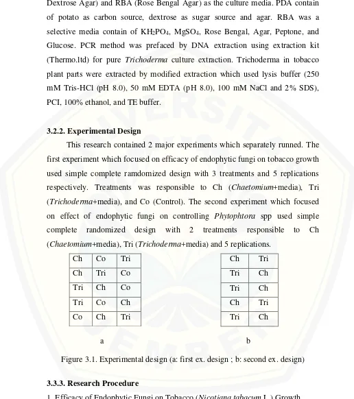
This research contained 2 major experiments which separately runned. The first experiment which focused on efficacy of endophytic fungi on tobacco growth used simple complete ramdomized design with 3 treatments and 5 replications respectively. Treatments was responsible to Ch (Chaetomium+media), Tri (Trichoderma+media), and Co (Control). The second experiment which focused on effect of endophytic fungi on controlling Phytophtora spp used simple complete randomized design with 2 treatments responsible to Ch (Chaetomium+media), Tri (Trichoderma+media) and 5 replications.
Ch Co Tri
Figure 3.1. Experimental design (a: first ex. design ; b: second ex. design) 3.3.3. Research Procedure
1. Efficacy of Endophytic Fungi on Tobacco (Nicotiana tabacum L.) Growth. The efficacy of endophytic fungi on plant growth was tested on tobacco seedling.The endophytic fungi were mixed with mixed sterile media. 41 days age
13
tobacco seedling which have already prepared in the nursery was planted to each treatments. The growth variable measurements included height, leaf number, stem size, and root length was concerned.
Three and five months after tobacco was transplanted, five grams of media in each treatment were taken for checking the availability of the both endophytic fungus. Five grams of the treated media was diluted upto 10-5 using sterilized deionizing water. Dilution 10-3 and 10-5 were used as the sample of colony availibility measurement. One hundred microliter (100µl) of the designed dilution was poured into RBA media with 3 replication respectively, so that each fungi will be responsible to 6 RBA plates. After 5 and 7 days of incubation, colony was counted manually according to the characteristic of each endophytic fungus.
In the end of plantation, some parts of tobacco, especially for the root zone and the lower stem was isolated using tissue transplanting method to detect the presence of the endophytic fungi.
2. Effect of Endophytic Fungi to Control Phytophtora spp.
Vigorous tobacco seedling was transplanted to peatmoss that have been inoculated with twenty gram of tested endophytic fungi. After seven days transplanted in the media, plants root were injured by pinning knife vertically surrounding the plants. Then, media were soaked with fifty milliliter of each fungus zoospore suspension. To accomplish the best distribution of the inoculum within the root system, each pot was watered immediately after inoculation with no water outflow (Mitchell and Rayside, 1986). Favorable ecology for disease development was created by watered the plants in 2-3 days intervals to keep the soil moisture stay high.
3. Detection of Endophytic Fungi Using Morphological Characteristic
fungal mycelium will be identified morphologically (Legiastuti and Aminingsih.,
2012).
4. Detection of Endophytic Fungi with PCR Method a). Trichoderma DNA Extraction
Endophytic fungi which isolated from tobacco plant part were cultured on PDA for 4 days. After the fungus fully covered the PDA, the culture was chopped into small agar plug, about 0,5 cm. The agar plug was soaked into flasks contained 100 mL of Czapek Dox Broth and incubated the cultures at 25oC for 3-5 days with an orbital shaker (120 rpm). Mycelia were harvested, washed with sterilized distilled water, frozen, and lyophilized. Total DNA was extracted from 50 mg of freeze-dried mycelia. DNA extracted was obtained using the DNA extraction kit according to manufacturer’s instructions. The DNA extracted from each sample was resuspended in 100 ml of TE buffer (Rubio et al., 2005).
b). Tobacco Parts Extraction
Tobacco parts were separated into small piece and grinded with lysis buffer. Plant solution was incubated at 55-65oC for 1 hr followed by centrifugation at 12,000 rpm for 15 min. The supernatant was then extracted with equal volume of water saturated phenol and further centrifuged at 12,000 rpm for 10 min. The supernatant was further extracted with equal volume of phenol: chloroform: isoamyl alcohol (25:24:1) and centrifuged at 12,000 rpm for 15 min; the supernatant was then transferred in a new tube and the DNA was precipitated with chilled ethanol (100%). DNA was pelleted by centrifuged at 12000 rpm for 15 min and washed with 70% ethanol by centrifugation. The pellets were air dried and suspended with TE buffer (pH 8.0) (Chakraborty et al., 2010).
c). DNA Amplification
15
reaction contained 5.0 µl of fungal DNA, 1.0 µl of Taq polymerase, 2,5 µl of DNA polymerase buffer, 2,5 µl of each deoxynucleoside triphosphate, 2,5 µl of MgCl2, 10 µl of H2O, and 2,0 of soluted primer in a total volume of 25,5 ��. PCR conditions included an initial denaturation of 5 min at 94oC, followed by 35 cycles of 1 min at 94oC, 1 min 30 s at 65oC, and 2 min at 72oC, with a final extension step of 72oC for 7 min, with a thermal cycler. PCR products were analysed by electrophoresis in a 0.8% agarose gel in 1 × TAE buffer (40 mM Tris, 20 mM
acetic acid, 1 mM EDTA [pH 8]), stained with Gel Star. Lambda pstl 24 (Fermentas, Inc) is used to be the marker (Rubio et al., 2005).
3.2.4. Research Variable
Some parameters were measured on this research include: 1. Plant Growth Development
a. Plant height (cm): Measured from the surface of the media till the last growing point. Measurement was implemented once in a month.
b. Leaf number (unit): Measured by counting the vigourous leaves manually. Measurement was implemented once in a month.
c. Stem diameter (cm): Measured using vernier caliper in the 2 cm above the surface. Measurement was implemented once in a month.
d. Root length: Measured from the root stock to the end of the root parts below. Measurement was implemented in the end of plantation.
2. Endophytic fungi colony (cfu/g): measured by counting the colony manually and converted the number into cfu/g.
3. Mortality
Plant mortality was responsible to the respon of tobacco plant on
Phytophtora spp. inoculation. Data was recorded by counting the dead plant and presentaged the number of missed plant using formula:
� = � − �
� × 100%
Note :
3.2.5. Data Analysis
CHAPTER IV. RESULT AND DISCUSSION
4.1 Result
4.1.1 Effect of Endophytic Fungi on Tobacco Growth
The effect of Trichoderma and Chaetomium treated media was evaluated on tobacco growth. Tobacco was grown in the growing media (3:1 soil:peatmoss v/v) added with Trichoderma and Chaetomium inoculum separately. The variable of plant height, leaf number, stem size, and root length was determined. Growth comparasion of each treatments based on ANOVA result was described below. Table 4.1. Analysis of varians (ANOVA) on effect of endophytic fungi on tobacco
growth variable.
Source of Variation df F-value Pr>F
Height 2 2,545ns 0,12
Leaf number 2 2,668ns 0,11
Stem size 2 6,536* 0,012
Root length 2 938,9** 0,000000655
*) significant in α=0.05 ; **) significant in α=0.01
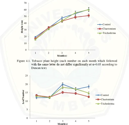
Endophytic fungi were observed on tobacco plant growth for 5 months observation. Analysis of varians was tested using Duncan Multiple Range Test (DMRT) with alpha (α) = 0,05. Result on tobacco plant height varian as the effect of endophytic fungi addition on the media was shown on figure 4.1. Tobacco plant height average was not significantly different between control treatment and endophytic added media treatments both on Chaetomium+media and
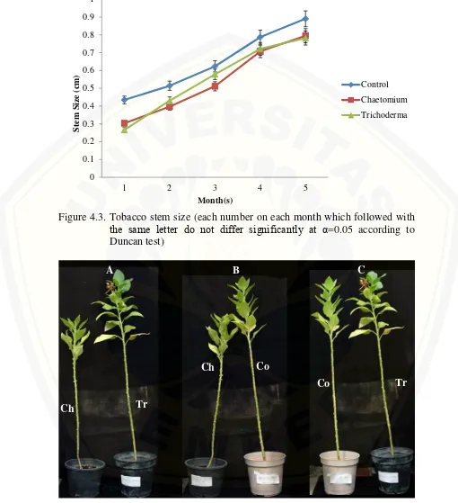
Trichoderma+media on the whole months. This result claimed that endophytic fungi addition to the media didn’t play role on tobacco plant height variable. Based on the tren of the figure 1, shown that Trichoderma+media have potential effect compared than both control and Chaetomium+media treatment. Result on leaf number varian shown that there was no significant differ between leaf number on control treatment with Trichoderma+media treatment in the last month (Figure 4.2). The difference was shown at control treatment versus Chaetomium+media treatment. Stem size varian result shown that both treatments didn’t give any
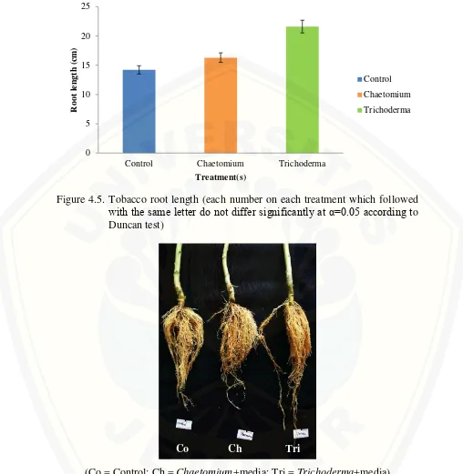
contribution on tobacco stem development untill the last month (Figure 4.3). The last growth variable, root length, shown significant difference between treatments (Figure 4.5). Trichoderma+media treatment gave the best result with 21,6 cm average and significantly different compared with Chaetomium+media treatment with 16,3 cm average and control treatment with 14,2 cm average.
Figure 4.1. Tobacco plant height (each number on each month which followed with the same letter do not differ significantly at α=0.05 according to Duncan test)
19
Figure 4.3. Tobacco stem size (each number on each month which followed with the same letter do not differ significantly at α=0.05 according to Duncan test)
(Co: Control ; Ch: Peatmoss+Chaetomium ; Tr: Peatmoss+Trichoderma) Figure 4.4. Growth comparasion between treatments after 5 months plantation.
Figure 4.5. Tobacco root length (each number on each treatment which followed with the same letter do not differ significantly at α=0.05 according to Duncan test)
(Co = Control; Ch = Chaetomium+media; Tri = Trichoderma+media) Figure 4.6. Root length comparision between treatments after 5 months plantation
Persistancy of applied endophytic fungi to the media was determined on 3 month and 5 month after planting [MAP] (Table 2). The result shown that applied
21
MAP. Otherwise, Trichoderma spp. persistancy was increase from 1,3 x 10³ in the 3 MAP to 6,6 x 10⁵ in the 5 MAP.
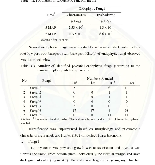
Table 4.2. Population of Endophytic fungi on media
Time1
Several endophytic fungi were isolated from tobacco plant parts include root-low part, root-basepart, stem-base part. Kind(s) of endophytic fungi observed was described below.
Table 4.3. Number of identified potential endophytic fungi (according to the number of plant parts transplanted).
No Fungi Numbers founded
Co1 Cha2 Tri3 Total
Control; 2Chaetomium treated media; 3Trichoderma treated media. Total of tissue transplanted was 144.
Identification was implemented based on morphology and microscopic character using Barnett and Hunter (1972) imperfecti fungi taxonomy.
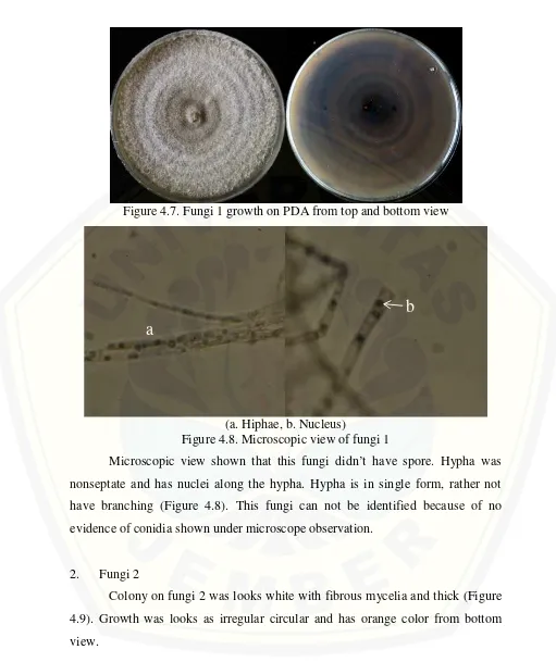
1. Fungi 1
Figure 4.7. Fungi 1 growth on PDA from top and bottom view
(a. Hiphae, b. Nucleus)
Figure 4.8. Microscopic view of fungi 1
Microscopic view shown that this fungi didn’t have spore. Hypha was nonseptate and has nuclei along the hypha. Hypha is in single form, rather not have branching (Figure 4.8). This fungi can not be identified because of no evidence of conidia shown under microscope observation.
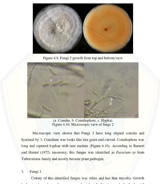
2. Fungi 2
Colony on fungi 2 was looks white with fibrous mycelia and thick (Figure 4.9). Growth was looks as irregular circular and has orange color from bottom view.
a
23
Figure 4.9. Fungi 2 growth from top and bottom view
(a. Conidia, b. Conidiophore, c. Hypha) Figure 4.10. Microscopic view of fungi 2
Microscopic view shown that Fungi 2 have long elipsed conidia and hyalined by 3. Conidium was looks like rice grain and curved. Conidiophore was long and septated hyphae with rare nucleus (Figure 4.10). According to Barnett and Hunter (1972) taxonomy, this fungus was identified as Fusarium sp from Tubercularia family and mostly became plant pathogen.
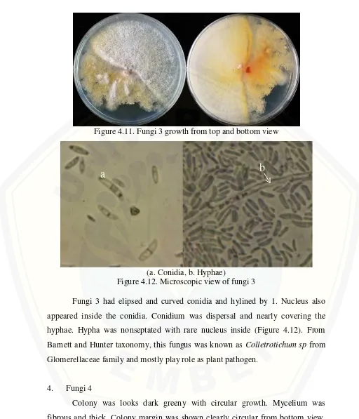
3. Fungi 3
Colony of this identified fungus was white and has thin mycelia. Growth looks circular and mycelium was spread out fast. In this picture, looks the fungi 3 was inhibted by chaetomium which being the contaminant in the culture (Figure 4.11).
a
Figure 4.11. Fungi 3 growth from top and bottom view
(a. Conidia, b. Hyphae)
Figure 4.12. Microscopic view of fungi 3
Fungi 3 had elipsed and curved conidia and hylined by 1. Nucleus also appeared inside the conidia. Conidium was dispersal and nearly covering the hyphae. Hypha was nonseptated with rare nucleus inside (Figure 4.12). From Barnett and Hunter taxonomy, this fungus was known as Colletrotichum sp from Glomerellaceae family and mostly play role as plant pathogen.
4. Fungi 4
Colony was looks dark greeny with circular growth. Mycelium was fibrous and thick. Colony margin was shown clearly circular from bottom view. Darker color was shown on the center of culture (Figure 4.13).
25
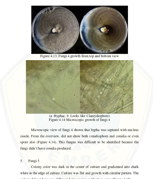
Figure 4.13. Fungi 4 growth from top and bottom view
(a. Hyphae, b. Looks like Clamydiophore) Figure 4.14 Microscopic growth of fungi 4
Microscopic view of fungi 4 shown that hypha was septated with nucleus inside. From the overview, did not show both conidiophore and conidia or even spore also (Figure 4.14). This fungus was difficult to be identified because the fungi didn’t have conidia produced.
5. Fungi 5
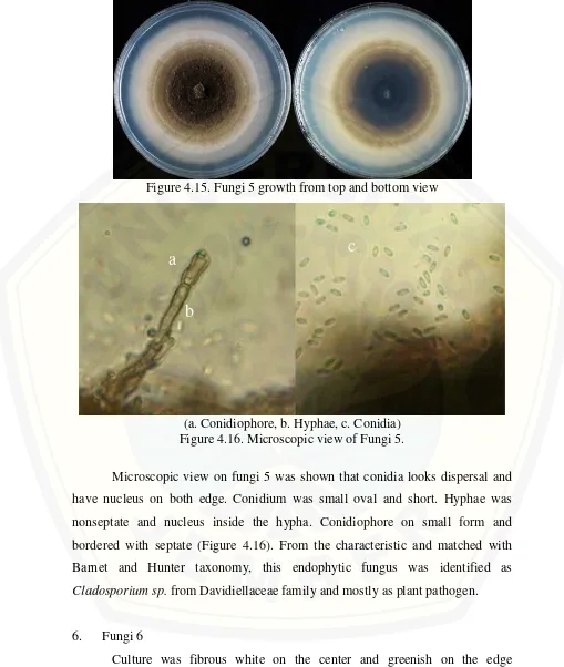
Colony color was dark in the center of culture and gradiented into chalk white in the edge of culture. Culture was flat and growth with circular pattern. The colony did not have any different between top and bottom view (Figure 4.15).
a
Figure 4.15. Fungi 5 growth from top and bottom view
(a. Conidiophore, b. Hyphae, c. Conidia) Figure 4.16. Microscopic view of Fungi 5.
Microscopic view on fungi 5 was shown that conidia looks dispersal and have nucleus on both edge. Conidium was small oval and short. Hyphae was nonseptate and nucleus inside the hypha. Conidiophore on small form and bordered with septate (Figure 4.16). From the characteristic and matched with Barnet and Hunter taxonomy, this endophytic fungus was identified as
Cladosporium sp. from Davidiellaceae family and mostly as plant pathogen.
6. Fungi 6
Culture was fibrous white on the center and greenish on the edge irregularly surrounding the center. From bottom view, the center of culture shown
a
c
27
have yellow colour. The culture was identified as Chaetomium which applied on the Chaetomium treatment (Figure 4.17).
Figure 4.17. Fungi 6 growth from top and bottom view
(a. Hyphae, b. Ascospores)
Figure 4.18. Microscopic view of fungi 6
This fungi was known as Chaetomium spp according to the characteristic which shown both in the microscopic view and macroscopic view (Figure 4.18.). Septate hyphae, perithecia, asci and ascospores are visualized. Perithecia are large, dark brown to black in color, fragile, globose to flask shaped and have filamentous, hair-like, brown to black appendages (setae) on their surface. Perithecia have ostioles (small rounded openings) and contain asci and ascospores inside. Asci are clavate to cylindrical in shape and rapidly dissolve to release their ascospores (4 to 8 in number). Ascospores are one-celled, olive brown in color,
a
and lemon shaped (de Hoog et al., 2000). This fungus was identified as potential endophytic fungi according to the role based on the literature reviewed.
7. Fungi 7
Colony was greenish and white in the surface of colony. Colony was fibrous with thick mycelia. Colony was glaucous on the bottom view with grey majority on the center of culture (Figure 4.19). This fungus was identified as Trichoderma
which founded on Trichoderma treatment media. The last fungi which isolated from tobacco plant was identified as Trichoderma spp. (Figure 4.20). Septate hyaline hyphae, conidiophores, phialides, and conidia are observed. Conidiophores are hyaline, branched, and may occasionally display a pyramidal arrangement. Phialides are hyaline, flask-shaped, and inflated at the base. They are attached to the conidiophores at right angles. The phialides may be solitary or arranged in clusters. Conidia are one-celled and round or ellipsoidal in shape. They are smooth or rough-walled and grouped in sticky heads at the tips of the phialides. These clusters frequently get disrupted during routine slide preparation procedure for microscopic examination. The color of the conidia is mostly green (de Hoog et al., 2000,). This fungus was identified as potential endophytic fungi according to the role based on the literature reviewed.
29
(a. Hyphae, b. Spores, c. Phialide) Figure 4.20. Microscopic view of fungi 7
4.1.2 Effect of Endophytic Fungi Aginst Phytophtora spp. Infection
Two endophytes, Chaetomium sp. and Trichoderma spp., were studied further against plant pathogen infection. Phytophtora spp. was known as the major disease which commonly appeared at seedling period even also in mature tobacco. Several variables include plant growth variable such as plant height and leaf number was measured and with addition of mortality variable as plant health defence variable. Significant difference was shown on tobacco plant height between Trichoderma+media and Chaetomium+media treatments. The difference was shown from week 4th untill the last data gained. On the last record,
Trichoderma+media induced tobacco plant height untill 49.15 cm average and
Chaetomium+media induced tobacco plant height reached 26,62 cm average.
According to DMRT with α=0.05 shown that Trichoderma+media treatment was
differ signicanly compared than Chaetomium+media treatment (F-value = 8.513, Pr>F = 0.0194). This result claimed that Trichoderma+media treatment induced tobacco seedling better than Chaetomium+media treatment.
a
b
Figure 4.21. Tobacco seedling height (each number on each period which followed with the same letter do not differ significantly at α=0.05 according to Duncan test)
Leaf number variable didn’t give significant difference between both treatments (Figure 4.22). On the last observation, leaf number on
Trichoderma+media treatment gained 9.8 leaves average and Chaetomium+media treatment with 7.8 leaves average (F-table = 1,923 ; Pr>F = 0.203).
Figure 4.22. Tobacco seedling leaf number (each number on each period which followed with the same letter do not differ significantly at α=0.05
1 week 4 weeks 7 weeks 10 weeks
H
1 week 4 weeks 7 weeks 10 weeks
31
Mortality variable shown to be concerned as the plant health parameters on this research. Both treatments successfully contributed to the plant defence induced system against Phytophtora spp. infection. No mortality recorded or even plant suffered as the effect of applied pathogen infection on both treatments. It means that Phytophtora did not work as literature marked. Dual-culture was implemented to proof the effectifity of endophytes (Trichoderma spp) against
Phytophtora spp (Figure 4.23).
Figure 4.23. Dual-culture of Trichoderma spp. against Phytophtora spp. for pathogencity test of Trichoderma spp. (left: Pure Phytophtora spp. culture ; middle: Pure Trichoderma spp. culture ; right: Trichoderma
spp. versus Phytophtora spp. culture)
Persistancy of applied endophytic fungi to the media was determined on the last observation (Table 4.5). The result shown that applied Chaetomium sp. population reached up to 3.2 x 10⁴ cfu/gand Trichoderma spp. population was higher with 3,3 x 10⁵ cfu/g.
Table 4.5. Population of applied endophytic fungi in peatmoss media after 2 months.
Figure 4.24. Population of applied endophytic fungi on RBA media. (T1= Trichoderma on 10-3 dilution ; T2= Trichoderma on 10-5 dilution ; C1= Chaetomium on 10-3 dilution ; C2= Chaetomium on 10-5 dilution)
Trichoderma spp. and Chaetomium sp. persistancy was observed in the tobacco plant parts include rootzone parts and stem-base part, morphologically by tissue transplanting method (Figure 4.25) and mollecular method (Figure 4.26) used PCR technique. The result on tissue transplanting method described that both applied endophytes were present inside the tobacco plant parts with the same character as the pure culture. It means that both applied endophytes penetrated well to plant tissue. The truth of this detection was supported by PCR technique result which implemented on Trichoderma spp. isolate.
33
parts band was shown that Trichoderma detected on each plants parts. About 1.5 kb band was detected in related to the previous study by Rubio et al., 2005 which detecting Trichoderma harzianum on the same size with SCAR a1 and a1c with 68oC annealing temperature. It was meant that applied Trichoderma applied and identified as T. harzianum.
(CS= +Chaetomium Stem ; CRB = +Chaetomium Root Base ; CRL= +Chaetomium Root Low ; TS= +Trichoderma Stem ; TRB= +Trichoderma Root Base ; TRL= +Trichoderma Root Low) Figure 4.25. Detection of applied endophytes on tobacco plant parts.
Figure 4.26. Agarose gel showing PCR product of tobacco tobacco plant parts. Root (R1, R2, R3, R4) and tobacco stem (S1, S2, S3, S4) inoculated with Trichoderma (T). Amplified using SCAR a1 and a1c at annealing temperature 62oC for 1.5 minutes. Marker used lambda pstl 24 (Fermentas.ltd)
CS TS
CRB TRB
4.2 Discussion
4.2.1 Effect of Endophytic Fungi on Plant Growth
Endophytic fungi have been widely studied have role as plant growth promotion. Especially for Trichoderma spp. that has been known as plant growth regulator and biocontrol agents. Chaetomium spp. also have secondary metabolism that induce plant growth such as high chloropyl content, higher biomass, and increase shoot growth (Schulz and Boyle, 2005). Tobacco growth on after 5 month shown that there were no differences between each treatments on plant height in the end of observation. There were no differences on leaf number and stem size in all treatments. Looking back from each month growth, there were no difference growths between treated tobaccos with control. Growth rate between treated tobacco and control also did not have any different at all.
Role of endophytic fungi on plant growth variables were shown on tobacco seedling together with plant pathogen infection. Trichoderma+media treatment induced plant height and the result was significantly different compared than
Chaetomium+media treatment. Both treatments were not significantly different on plant leaf number. This result shown that applied Trichoderma spp. gave better result in case of plant growth induced system. Better result on Trichoderma treatments shown that Trichoderma application is more effective than
Chaetomium treatments. Peatmoss, as the growing media of tobacco plant seem to be the factor affecting of Trichoderma success result. Trichoderma can survive well on peatmoss media because of high organic matter inside the peatmoss. Zaidi and Singh (2004) and Gangadharan (1989) reported that Trichoderma was on high population in several organic substrate like cow dung, FYM (farm yard manure), and paddy husk. The existence of Trichoderma inside organic matter will highly multiplicate, but they will reduced slightly under longer incubation.
35
that Trichoderma were going inside and contributed as plant growth regulator. Another reason was based on the result of persistancy of Trichoderma spp. inside the media was increased. Both endophytic fungi was added only once in the beginning of the plantation. Trichoderma had their ability to grow in wide range substrates like pigeonpea, farmyard manure, wheat bran, neem cake, mustard cake, saw dust, coffee husk, vermicompost, sorghum grains etc but their use in mass multiplication and formulation remained little explored (Kumar et al., 2013). In this experiment, there is no replacement of endophytic fungi to media after tobacco planted. Long period of plantation and also different weather between the plantation period also affecting the shelf life of both endophytic fungi in the media. According to the data, the existence of Trichoderma was very low on the third month and increased up to 2 times amount. The result overtook the population of Chaetomium which otherwisely decreased. According to Agosin et al (1997) Trichoderma shelf life was affected by pH of media, C/N ratio media, harvesting time of Trichoderma spp., and also duration spores were left in the culture. This result shown that Trichoderma spp. was slowly adapt to the media and become more convenience for Trichoderma spp. multiplication after then. It can be proven by the growth tren of Trichoderma+media treatment was better than other treatments.
Trichoderma was well known as great biocontrol agents that widely control many pathogens, especially soil borne pathogens like Pythium sp., Phytophtora
Chaetomium sp. population also recorded according to 3 MAP and 5 MAP. This amount was indicated that existence of Chaetomium sp. and it was not affected by the environment or any factors. Chaetomium sp. as biocontrol agents are known have long shelf life in the favorable environments such as soil pH, content of organic matter, soil aeration, moisture and chemical residue in the soil. As a biological control agent, ecology and climate might be affected on poor result of biological control. Chaetomium sp. can survive at biopellets formulation about 77% on 1 year storage and can survival about 59% at biopowder formulation on 1 year storage (Soytong et al., 2001). According to the result, this amount of Chaetomium sp. seems not to be enough to give effect on tobacco plant growth.
The role of endophytic fungi was also detected by tissue transplanting method. Both applied endophytic fungi were observed as the main endophytes inside tobacco tissue. Several fungi include the inoculated endophytic fungi were identified in 3 parts from the 5 months old tobacco atroot low (RL), root base (RB), and stem base (SB). Tobacco plant parts were transplanted into PDA media (Radji, 2005) and incubated for 7 days. Several fungi were successfully identified according to the macroscopic and microscopic morphology using Barnett and Hunter taxonomy (1972). Most of identified fungi were identified as Ascomycota (Chaetomium spp., Trichoderma spp., Fusarium spp., and Colletrotichum sp.) and Deutromycota division (Petrini, 1992). Only Trichoderma spp. and Chaetomium
sp. which described as endophytic fungi, and the rest (Fusarium spp., and
Colletrotichum sp. and Cladosporium sp.) were identified as plant pathogen. The result also determined the effectivity of Chaetomium and Trichoderma
37
give any effect on plant growth, even Chaetomium treatment was similar to be the worst treatment between Control and Trichoderma treatment.
4.2.2 Effect of Endophytic Fungi against Phytophtora spp. Infection.
Tobacco seedling was transplanted into single pot and was inoculated by
Phytophtora spp zoospores suspension which already prepared as previously. Inoculation was implemented after the seedling vigourous. The inoculation of
Phytophtora spp to tobacco seedling did not affect on the tobacco growth and development. Tobacco plant lived well without any interference from Phytophtora
spp infection. Phytophtora spp. has been known well as plant pathogen especially on tobacco seedling. More than 43 species of Phytophthora have now been described. Members of this genus cause a wide variety of diseases on major food crops, forest, fruit, and nut trees, and many ornamental plants. The species
Phytophthora parasitica causes root, stem, and fruit rot on more than 90 plant species, including tobacco (Ribeiro, 1978). Phytophtora is one of pathogen which causing of dumping-off on tobacco and any economic plants seedling together with Pythium spp., Rhizoctonia solani, Sclerotium rolfsii, and Fusarium oxysporum (Loliam et al., 2012). Phytophtora which infected the plants will lived and produced mainly diploid hyphae, oospores and chlamydospores within plant tissue. Zoospores are biflagellate and able to swim in the water. Oospores of this pathogen also have ability to be long lasting in the organic part of media (Lilja et al., 2006).
Inoculation of Phytophtora spp to tobacco seedling did not successfuly infect to tobacco. Percentage of tobacco seedling which infected by Phytophtora
spp. were nothing. It means that Phytophtora did not work as literature marked. From the result above, applied endophytic fungi seem to be the reason why was the Phytophtora did not give any effect on tobacco plant growth. Chaetomium and
Trichoderma was applied to the media which composed by 100% peatmost. Both of endophytic fungi play role as biocontrol agents. The ability of these both endophytic fungi was known well especially for Trichoderma. Trichoderma spp.
various test. In this experiment, dual culture also implemented to measure the inhibition ability of Trichoderma spp. over Phytophtora spp.
After 7 days culture, the effect of Trichoderma spp. was shown with inhibition over Phytophtora spp. culture. Culture of Trichoderma spp. looks overdominant inhibit the growth of Phytophtora spp. by growing over the mycelia of Phytophtora spp. culture. It means that when Trichoderma spp. mycelia started to contact with Phytophtora spp., the inhibition by Trichoderma spp. was begun. Inhibitions of Trichoderma spp. make the hyphae lysed, parasitizides, and disorganited over the Phytophtora spp. cell host (Moayedi and Mustowfizadeh-ghalamfarsa., 2010). The lysed mycelium was caused by enzyme activity of
Trichoderma spp. isolates at the contact points (Elad et al., 1983).
Antagonism role of Trichoderma causing the significant inhibition to
Phytophtora spp. even it also destroy the hyphae by lysis effect. Application of endophytic fungi, especially for Trichoderma has given impact to Phytophtora
spp. shelf life. Phytophtora spp. was failed to penetrate too far because of the inhibition of endophytic fungi which protected the plants tissue from the infection. Uneffective inoculation of Phytophtora spp. also gave chance the plants to grown without any disturbance. Endophytic fungi also play role as plant growth regulator Shown that Trichoderma application was looks better than Chaetomium
Population was detected by using Rose Bengal Agar and continued with spread plate method. The result has shown that existence of Trichoderma in the peatmoss as major media shown greater than Chaetomium existence. Population of
Trichoderma reached up to 3,3 x 10⁵ cfu/g. The colony counting result shown on
Chaetomium population just only 3,2 x 10⁴cfu/g, and it meant that Chaetomium
needed to adapt longer in peatmoss media. Otherwise, Trichoderma was able to survive and multiplicate well in peatmoss media. Smaller amounts of Chaetomium
found shown that pure peatmoss give unfavorable condition to Chaetomium
compared with to previous experiment. This result also affected tobacco growth which unconstantly promoted by Chaetomium as occured on Trichoderma
39
The effectifity of both applied endophytic fungi on Phytophtora spp. infection were supported by detecting both endophytic fungi inside plant tissue used tissue transplanting method and PCR technique. Morphological identification has been used widely as endophytes isolation. Some researcher may need to use specific media to gain more success on endophytes isolation. Selective medium like Rose Bengal also used by Frohlich et al., 2000 was used to inhibit the fast growing endophytes. Specific technique may also needed reflecting on the objectives of each researcher want to do with. The result of tissue transplanting method detection shown, that both Chetomium and Trichoderma lived inside of the plant tissue. Chaetomium looks penetrated better than Trichoderma in all tobacco tissue parts. Trichoderma did not penetrate well on stem compared with
Chaetomium which have more penetration to this area. Otherwise, Trichoderma
similar to be suitable to stay at root base (RB) and much detected in this plant part. From the result can be determined that both of applied endophytic fungi penetrated well to the plant tissue to gain and were symbiosis with the host plants. Interaction between endophytic fungi and host plants was widely study.
Trichoderma which found in each part of treated tobacco claimed that
Trichoderma did not just only stay and play role in the rhizosphere, but they are also penetrated into the plant tissue and give contribution by promoting the growth and also protect from the pathogens. Endophytes also play role as environment tolerance like drought (Frohlich et al., 2000 ; Sieber, 2007).