Role of
lactamase in the susceptibility of clinical isolates to
lactam
antibiotics
A. Soebandrio*, N.N. Sri-Budayanti, A. Widayati, V. Wiwing, M.A.C.M. Nusatya
Abstrak
Kombinasi antibiotik -laktam dengan penghambat -laktamasa terbukti telah dapat mengatasi resistensi yang disebabkan oleh produksi -laktamasa. Konsentrasi Hambatan Minimal (KHM) beberapa antibiotik -laktam terhadap isolat penghasil -laktamasa akan dievaluasi. A.anitratus, E.koli, K.pneumoniae, Proteus sp, Pseudomonas sp, S.aureus, S.epidermidis, S.pneumoniae, S.viridans, dan -hemolitik Streptokokkus, dipaparkan terhadap Ampisilin/Sulbaktam (AMS), Seftriaksone (CRO), dan Sefotaksime (CTX) menggunakan teknik Etest. Produksi -laktamasa diidentifikasi menggunakan cakram Cefinase. Enampuluh empat persen isolat memiliki kemampuan menghasilkan -laktamasa. Semua E.koli dan K.pneumoniae yang diuji merupakan penghasil -laktamasa, namun tidak satupun Proteus sp, Pseudomonas sp, dan S.epidermidis yang diuji menghasilkan -laktamasa. Dalam kelompok penghasil -laktamasa, sulbaktam mampu menurunkan resistensi terhadap CFP dari 25% menjadi 5%. Sekitar 20% dari isolat penghasil -laktamasa yang resisten terhadap CFP, ternyata peka terhadap CSL. Kepekaan S.viridans terhadap AMS, AMC, CFP, dan CSL ternyata lebih dari 80%, tetapi kurang dari 50% terhadap CRO dan CTX. S.pneumoniae ternyata kurang peka terhadap antibiotik yang diuji. Kepekaan S.aureus terhadap antibiotik uji adalah 60 sampai 70%, sedangkan Streptokokus -haemolitikus memperlihatkan respons yang baik. Hanya 30% atau kurang K.pneumoniae dan E.koli yang peka terhadap AMS dan AMC. A.anitratus memperlihatkan kepekaan yang baik hanya terhadap AMS (78%) dan CSL (89%). Enampuluh empat persen isolat yang diamati ternyata menghasilkan -laktamasa. Penghambat -laktamasa dapat menurunkan resistensi organisma penghasil -laktamasa terhadap antibiotik -laktam dari 25 menjadi 5 persen. (Med J Indones 2004; 13: 140-5)
Abstract
Combination of lactam antibiotic with lactamase inhibitor has been proven to overcome resistance caused by lactamase production. An evaluation to the MIC of some lactam antibiotics to b-lactamase producing isolates will be reported. A.anitratus, E.coli, K.pneumoniae, Proteus sp, Pseudomonas sp, S.aureus, S.epidermidis, S.pneumoniae, S.viridans, and hemolytic Streptococcus, were challenged to Ampicillin/Sulbactam (AMS), Amoxicillin/Clavulanic acid (AMC), Cefoperazone (CFP), Cefoperazone/ Sulbactam (CSL), Ceftriaxone (CRO), dan Cefotaxime (CTX) using ETest techniques. -lactamase production was identified using Cefinase disk. Sixtyfour percent of isolates were capable of producing lactamase. All E.coli and K.pneumoniae tested were lactamase producer, none of Proteus sp, Pseudomonas sp, and S.epidermidis tested produced lactamase. In lactamase producing group, Sulbactam was able to reduce resistance to CFP from 25% to 5%. About 20% of lactamase producing isolates which were resistant to CFP, were susceptible to CSL. Susceptibility of S.viridans to AMS, AMC, CFP, and CSL was higher than 80%, but less than 50% to CRO and CTX. S.pneumoniae was less susceptible to tested antibiotics, 50 to 60% susceptibility was shown to AMC, CFP, and CSL. S.aureus was 60 to 70% susceptible, while haemolytic Streptococcus showed good response to the tested antibiotics. Only 30% or less of K.pneumoniae and E.coli was susceptible to AMS and AMC. A.anitratus showed good susceptibility only to AMS (78%) and CSL (89%). Sixtyfour percent of isolate studied produced -lactamase. -lactamase inhibitor could reduce resistance of -lactamase producing organism to -lactam antibiotic from 25 to 5 percent. (Med J Indones 2004; 13: 140-5) Keywords: Antibiotic Susceptibility, MIC, -lactam antibiotic, -lactamase inhibitor
Resistance of clinical isolates to antibiotic has been increasing from year to year. One major cause of the emergence of resistance is the production of lactamase by resistant bacteria. A statistically significant increase in extended spectrum lactamase (ESBL) producing bacteria occurred over the 2-year period from 9 (0.6%) of 1414 isolates in 2000 to 22 (1.8%) of 1218 isolates in 2001.1 Organisms that produce ESBL have important therapeutic implications as they exhibit resistance to a variety of antimicrobial agents, including third-generation cephalosporins, extended-spectrum penicillins, and monobactams.2lactamases are classified into four classes based on substrate affinity and amino acid sequence. Class A includes various plasmid mediated lactamases (TEM-1, SHV-1), the plasmid mediated extended spectrum
lactamases derived from TEM or SHV, and some chromosomal encoded lactamases, such as that produced by Klebsiella pneumoniae.3 Genes encoding
lactamase enzyme are located on transferable plasmid that often carry other resistance factors.4 In the last decades, combination of lactam antibiotic with lactamase inhibitor has been proven to overcome resistance caused by lactamase production. In this paper, an evaluation to the Minimal Inhibitory Concentration (MIC) of some lactam antibiotics, either alone or in combination with lactamase inhibitor, to a number of clinical isolates will be reported.
METHODS
Isolates. Ten species of aerobic bacteria, comprise of ten each of Acinetobacter anitratus, Escherichia coli,
Klebsiella pneumoniae, Proteus sp, Pseudomonas sp,
Staphylococcus aureus, Staphylococcus epidermidis,
Streptococcus pneumoniae, Streptococcus viridans, and hemolytic Streptococcus, were randomly selected from clinical isolates obtained in the Clinical Microbiology Laboratory of FKUI. Isolation and identification of target organisms were performed using procedure described elsewhere.5,6 In brief, specimens (except blood) were directly inoculated onto appropriate agar plate. Blood specimens were inoculated into BacT/Alert aerobic liquid media (Organon Teknika, Netherland); positive cultures were then inoculated onto blood agar plates. Suspected colonies were picked up for further processes. Beta-lactamase enzyme production was identified using Cefinase disk (Becton-Dickinson, USA).
Susceptibility test. Susceptibility of isolates to antibiotics was performed using ETest strip (AB Biodisk, Sweden) techniques, where the results were expressed as Minimum Inhibitory Concentration/MIC (g/mL). Antibiotics tested were Ampicillin / Sulbactam, Amoxicillin / Clavulanic acid, Cefoperazone, Cefoperazone / Sulbactam, Ceftriaxone, dan Cefotaxime. For practical reason, only six antibiotics were included, as on media plate could maximally accommodate six ETest strips. Selection of these set of antibiotics was based on the observation that they belong to the most prescribed parenteral antibiotics in hospital setting (unpublished observation). Two antibiotics representing Penicillin derivatives, two representing the third generation cephalosporins, and the other two are comparison between a third generation cephalosporin and its combination with a
lactamase inhibitor. The susceptibility test was performed referring to technical and interpretation guidelines as recommended by the NCCLS.7 In brief, maximally six antibiotic strips were placed on Mueller-Hinton agar plate pre-inoculated with tested bacteria at the density equal to 0.5 McFarland standards. WHONET 5.1 software.8 was used to process data on isolates and its susceptibility to tested antibiotics, including the calculation of Geometric Means and interpretation of antibiotic susceptibility.
RESULT
Out of 100 selected isolates, only 95 were evaluable, since one Proteus sp and 4 S.pneumoniae isolates were not evaluable (Table 1). This table also shows that there were 61 isolates (64%) with capability of producing lactamase. All E.coli and K.pneumoniae
tested were lactamase producer, none of Proteus sp,
Pseudomonas sp, and S.epidermidis tested produced
lactamase.
Following the NCCLS standard for dilution method of antibiotic susceptibility testing (7), MIC testing and the determination of resistance should refer to breakpoint value as describe in the Table 2.
Using the above breakpoint, it revealed that the highest susceptibility was shown by all isolates to Cefoperazon / Sulbactam (81,1%) followed by Cefoperazon (63,8%). Susceptibility to other tested antibiotics ranged from 45 to 53%, as shown in Table 3.
This study showed that Geometric Mean of MIC of
Table 1. List and number of isolates tested and producing lactamase (BL)
Organism No. of
Isolates
BL (+)
Acinetobacter anitratus 10 9
Escherichia coli 10 10
Klebsiella pneumoniae 10 10
Proteus sp. 9 0
Pseudomonas sp. 10 0
Staphylococcus aureus 10 9
Staphylococcus epidermidis 10 0
Streptococcus pneumoniae 6 6
Streptococcus viridans,. 10 8
Streptococcus, beta-haemolytic 10 9
Total: 95 61 (64%)
Table 2. Antibiotics, antibiotic codes, and MIC breakpoint value
Antibiotic name Breakpoint (g/mL)
Ampicillin/Sulbactam S<=8 R>=32 Amoxicillin/Clavulanic acid S<=8 R>=32 Cefoperazone S<=16 R>=64 Cefoperazone/Sulbactam S<=16 R>=64 Ceftriaxone S<=8 R>=64 Cefotaxime S<=8 R>=64
Using the above breakpoint, it revealed that the highest susceptibility was shown by all isolates to Cefoperazon / Sulbactam (81,1%) followed by Cefoperazon (63,8%). Susceptibility to other tested antibiotics ranged from 45 to 53%, as shown in Table 3.
This study showed that Geometric Mean of MIC of
lactamase producing bacteria (Table 4) is lower than those of non producing bacteria (Table 5).
Table 3. MIC of tested antibiotics to the whole isolates and their respective susceptibility interpretation
Antibiotics MIC range
(g/mL)
Geometric Mean (g/mL)
%R %I %S
Ampicillin/Sulbactam 0,016 – 256 5,949 37,4 9,9 52,7 Amoxicillin/Clavulanic acid 0,016 – 256 5,834 44,7 8,5 46,8 Cefoperazone 0,064 - 256 11,554 26,6 9,6 63,8 Cefoperazone/Sulbactam 0,125 - 256 5,058 11,6 7,4 81,1 Ceftriaxone 0,016 - 256 4,975 33 19,1 47,9 Cefotaxime 0,032 - 256 4,328 33,7 15,8 50,5
R=Resistance; I=Intermediate; S=Susceptible
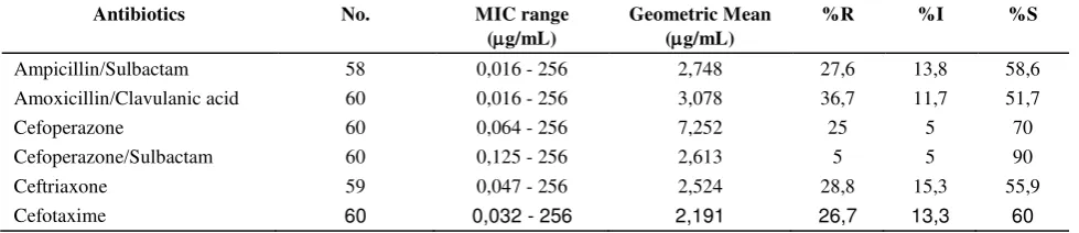
Table 4. MIC of tested antibiotics to lactamase producing isolates (61 isolates) and their respective susceptibility interpretation
Antibiotics No. MIC range
(g/mL)
Geometric Mean (g/mL)
%R %I %S
Ampicillin/Sulbactam 58 0,016 - 256 2,748 27,6 13,8 58,6 Amoxicillin/Clavulanic acid 60 0,016 - 256 3,078 36,7 11,7 51,7
Cefoperazone 60 0,064 - 256 7,252 25 5 70
Cefoperazone/Sulbactam 60 0,125 - 256 2,613 5 5 90 Ceftriaxone 59 0,047 - 256 2,524 28,8 15,3 55,9 Cefotaxime 60 0,032 - 256 2,191 26,7 13,3 60
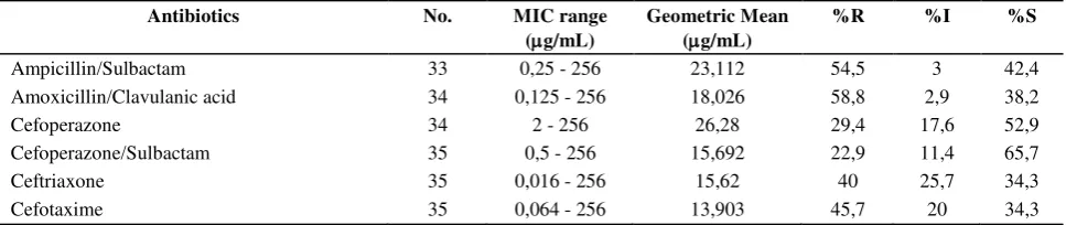
Table 5. MIC of tested antibiotics to non-lactamase producing isolates (34 isolates) and their respective susceptibility interpretation
Antibiotics No. MIC range
(g/mL)
Geometric Mean (g/mL)
%R %I %S
Ampicillin/Sulbactam 33 0,25 - 256 23,112 54,5 3 42,4 Amoxicillin/Clavulanic acid 34 0,125 - 256 18,026 58,8 2,9 38,2
Cefoperazone 34 2 - 256 26,28 29,4 17,6 52,9
Cefoperazone/Sulbactam 35 0,5 - 256 15,692 22,9 11,4 65,7 Ceftriaxone 35 0,016 - 256 15,62 40 25,7 34,3 Cefotaxime 35 0,064 - 256 13,903 45,7 20 34,3
R=Resistance; I=Intermediate; S=Susceptible
DISCUSSION
Although the number of isolates included in this study was relatively small, it was surprising that the percentage of lactamase producing isolates was quite high (61 out of 95 isolates). In 1988 the author found only about 23 percent of E.coli and about 30 percent of K.penumoniae had lactamase activity (data not shown). Calculation of the MIC Geometric Means showed that it has no correlation with the degree of susceptibility to each antibiotic tested, since it was merely influenced by the MIC range (Table 3). However, it could be used to compare susceptibility of different types of organism to a particular antibiotic, as will be discussed later. As shown in this table, susceptibility of all isolates was 81.1% to Cefoperazon/Sulbactam and 63,8% to Cefoperazon only. This brought to a suggestion that the
lactamase played an important role in reducing susceptibility of bacteria to antibiotic. It was interesting to observe that either Geometric Means of MIC of tested antibiotics and percent of susceptibility to tested antibiotics was higher in lactamase non-producing isolates compared to the lactamase producing isolates, as seen in Table 4 and Table 5. However, significant test was not performed to show its significance due to relatively low number of isolate tested. It was possible that mechanism of resistance other than lactamase played a role in this setting.
In lactamase producing group, highest susceptibility was shown to Cefoperazon/Sulbactam. The addition of sulbactam was able to increase susceptibility to Cefoperazon from 70% to 90%, or reduce resistance from 25% to 5%. This phenomenon was not clearly observed on non-lactamase producing group, where the addition of Sulbactam was only increase susceptibility to Cefoperazon from 52.9% to 65.6%, or reduces resistance from 29, 4% to 22, 9%. In both groups, it was clearly observed that resistance to Cefoperazon, either alone or in combination with
lactamase inhibitor, was the lowest among the tested antibiotics. To further observe the ability of
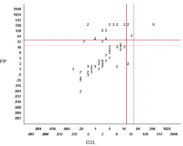
lactamase inhibitor in improving susceptibility to
lactam antibiotics, a comparison of individual isolate’s susceptibility to Cefoperazon alone and Cefoperazon/Sulbactam was apllied to scatter-plot diagram (Figure 1). As seen in the upper-left quadrant of the diagram, about 20% of isolates which were resistant to Cefoperazon (MIC =/> 64 g/mL), were susceptible to Cefoperazon/Sulbactam (MIC < 16
g/mL). On the other hand, isolates resistant to Cefoperazon/Sulbactam was also resistant to Cefoperazon alone, suggesting that the resistance was not due to production of lactamase. However, hyper-production of some groups of lactamase could also cause resistance to lactams and
Figure 1. Scatter-plot diagram of MIC (g/mL) of Cefoperazon (CFP, Y axis)) vs. MIC (g/mL) of Cefoperazon/Sulbactam (CSL, X axis) to lactamase producing isolates. Numbers on axes represent MIC values, while plotted numbers represent percent of isolates.
It is worth to observe that in both lactamase producing and non-producing groups, susceptibility, resistance to Amicillin/Sulbactam and Amoxycillin / Clavulanic acid, both are regarded as “old lactam derivatives”, are comparable to those of Ceftriaxone and Cefotaxime (Table 4 and 5). Ampicillin and Amoxicillin are usually more susceptible to hydrolysis by lactamase compared to third generation cephalosporins. The presence of lactamase inhibitor (Sulbactam or Clavulanic acid) could improve resistance of Ampicillin and Amoxicillin to the enzyme and therefore improve susceptibility of organism to these antibiotics. However, if one look at the susceptibility of individual lactamase producing isolate to tested antibiotics, as showed in Table 6, a different picture could be observed. Susceptibility of
Streptococcus viridans to Ampicillin/Sulbactam, Amoxicillin/Clavulanic acid, Cefoperazon, and Cefoperazon/Sulbactam was higher than 80%, but less than 50% to Ceftriaxone and Cefotaxime. Streptococcus pneumoniae was generally less susceptible to tested antibiotics, although 50 to 60% susceptibility was
shown to Amoxicillin/Clavulanic acid, Cefoperazon, and Cefoperazon/Sulbactam. Staphylococcus aureus was 60 to 70% susceptible to the antibiotics, while
haemolytic Streptococcus showed good response to the tested antibiotics. Only 30% or less of Klebsiella pneumoniae and Escherichia coli were not susceptible to Ampicillin/Sulbactam amd Amoxicillin / Clavulanic acid. Hyper-production of lactamase or possibly production of ESBL could be behind this finding.9,10 Further study involving larger number of isolates is needed to obtain clear explanation.
Interesting picture was shown by Acinetobacter anitratus, where it was susceptible only to Ampicillin / Sulbactam (78%) and Cefoperazon/Sulbactam (89%). This phenomenon could be best explained by the fact that Sulbactam, present in both Ampicillin/ Sulbactam and Cefoperazon/Sulbactam, possesses a specific antibacterial effect against Bacterioides fragillis and
Gene encoding this phenotype could be disseminated to other microbes previously susceptible to antibiotic by means of plasmid or other mechanism.4 A powerful means for the investigation of nosocomial outbreaks implicating lactamase producing bacteria, including combination of antibiotic susceptibility testing, enzyme characterization, and epidemiological data is required.9 The presence of lactamase producing species was significantly associated with the exposure to antibiotic. Moreover, more than one
lactamase-producing strain could be simultaneously found in patient organ.12 During anti-pseudomonal treatment in cystic fibrosis patient, high level of
lactamase activity was found in the sputum sample. This could be one of several explanations for the failure of treatment using lactam antibiotics.13 It should be noted that some antibiotics and some
lactamase inhibitor as well, are good inducer for
lactamase production. Our study showed that more than half of clinical isolate in fact had lactamase activity. An earlier study showed that the presence of
lactamase activity could increase post antibiotic effect of amoxicillin/clavulanic acid combination given twice or four times of MIC. Implication of this finding is that there is a room for reducing the dosage of the drug.14 It is therefore recommended for microbiology laboratories to include in their routine identification process detection of lactamase activity using relatively simple method as we showed. Further characterization of ESBL activity, particularly in suspected isolates such as
Klebsiella pneumonae etc. using more complicated procedure15 would give more informative data for either epidemiological purpose or selection of antimicrobial agents used in the treatment of infection.
Acknowledgment
The authors wish to thank the Head of the Department of Microbiology, Medical Faculty of the University of Indonesia (FKUI), for facilitating this study. Clinical isolates and data collection and processing have been made available by the Clinical Microbiology Laboratory, FKUI.
REFERENCES
1. Burgess DS, Hall II RG, Lewis II JS, Jorgensen JH, Patterson JE. Clinical and Microbiologic Analysis of a Hospital's Extended-Spectrum Beta-Lactamase-Producing Isolates over a 2-Year Period. Pharmacotherapy 2003; 23(10):1232-7.
2. Einhorn AE, Neuhauser MM, Bearden DT, Quinn JP, Pendland SL. Extended-Spectrum Beta-Lactamases: Frequency, Risk Factors, and Outcomes. Pharmacotherapy 2002; 22(1):14-20.
3. Fournier B, Roy PH, Lagrange PH, Philippon A. Chromosomal lactamase Genes of Klebsiella oxytoca
Are Devided into Two Main Groups, blaOXY-1 and blaOXY-2.
Antimicrob Agents Chemother 1996; 40(2):454-9. 4. French GL, Shannon KP, Simmons N. Hospital Outbreaks
of Klebsiella pneumoniae Resistant to Broad Spectrum Cephalosporins and Lactam-Lactamase Inhibitor Combinations by Hyperproduction of SHV-5 lactamase. J Clin Microbiol 1996; 34(2):358-63.
5. Koneman EW, Allen SD, Janda WM, Schreckenberger PC, Winn WC Jr, editors. Color atlas and textbook of Diagnostic Microbiology. 4th ed.Philadelphia: JB Lippincott; 1992. 6. Mahon CR, Manuselis G Jr, editors. Textbook of Diagnostic
Microbiology. Philadelphia: WB Saunders; 1995. 7. NCCLS (National Committee for Clinical Laboratory
Standards). Methods for dilution antimicrobial susceptibility tests for bacteria that grow aerobically. Wayne, Pennsylvania: National Committee for Clinical Laboratory Standards. NCCLS Document M7-A5, 2000. 8. WHO. WHONET 5 Microbiology Laboratory Database
Software. WHO/CDS/CSR/DRS/99.1, 1999
9. Nouvellon M, Pons JL, Sirot D, Combe ML, Lemeland JF. Clonal Outbreaks of Extended-Spectrum lactamase-producing strains of Klebsiella pneumoniae Demonstrated by Antibiotic Susceptibility Testing, lactamase Typing, and Multilocus Enzyme Electrophoresis. J Clin Microbiol 1994; 32(10):2625-7.
10. Kim JM, Kwon YM, Pai HJ, Kim JW, Cho DT. Survey of
Klebsiella pneumoniae Strains Producing Extended-Spectrum lactamases: Prevalence of 12 and SHV-2a in Korea. J Clin Microbiol 1998; 36(5):1446-9. 11. Williams JD. lactamase inhibition and in-vitro activity
of sulbactam and sulbactam/cefoperazone. Clin Infect Dis 1997; 24:494-7.
12. Nyfors S, Könönen E, Takala A, Jousimies-somer H. lactamase Production by Oral Anaerobic Gram-Negative Species in Infants in Relation to Previous Antimicrobial Therapy. Antimicrob Agents Chemother. 1999; 43(7):1591-4.
13. Giwercman B, Meyer C, Lambert PA, Reinert C, Høiby N. High-level lactamase Activity in Sputum samples from Cystic Fibrosis Patients during Antipseudomonal Treatment. Antimicrob Agents Chemother 1992; 36(1):71-6.
14. Thorburn CE, Molesworth SJ, Sutherland R, Rittenhouse S. Postantibiotic and Post-lactamase Inhibitor Effects of Amoxicillin plus Clavulanate. Antimicrob Agents Chemother 1996; 40(12):2796-801.