Laparoscopic resection versus myolysis in the management of symptomatic
uterine adenomyosis: alternatives to conventional treatment
Wachyu Hadisaputra, T. Dewi Anggraeni
Abstrak
Dalam kurun waktu Juni 2003 sampai dengan Juni 2004, pasien-pasien yang menderita adenomiosis berdasarkan ultrasonografi transvaginal dan memiliki keluhan menorhagia, dismenore, maupun nyeri pelvis diikutsertakan dalam penelitian. Randomisasi dilaku-kan untuk mengalokasidilaku-kan subjek ke dalam kelompok reseksi dan kelompok miolisis. Semua pasien dari kedua kelompok mendapat GnRH analog 3 siklus pasca-laparoskopi operatif. Penilaian dilakukan dalam jangka waktu 6 bulan, baik secara subjektif melalui kuesioner maupun secara objektif melalui evaluasi volume adenomiosis per ultrasonografi transvaginal di akhir semester. Terdapat 20 pasien yang menjalani pembedahan, 10 dalam kelompok reseksi dan 10 dalam kelompok miolisis. Komplikasi bermakna tidak ditemukan pada kedua kelompok. Evaluasi subyektif dapat dilakukan pada semua pasien sedangkan evaluasi objektif hanya dapat dilakukan pada 17 pasien. Tidak didapatkan perbedaan bermakna antar-kelompok dalam penurunan skor keluhan menorhagia (p = 0.399) dan dismenorea (p=0.213). Tidak ditemukan perbedaan bermakna dalam median penambahan volume adenomiosis (p = 0.630)antara kelompok reseksi (median=+15,35% (-100 – 159)) dengan kelompok miolisis (median=+48,43% (-100 – 553)). Lima pasien hamil, 3 dari kelompok reseksi, 2 dari kelompok miolisis, dengan satu kasus ruptur uteri pada usia kehamilan 8 bulan pada kelompok miolisis. Efektifitas reseksi adenomiosis per laparoskopi tidak berbeda bermakna dengan miolisis adenomiosis per laparoskopi dalam penatak-sanaan adenomiosis bergejala. Miolisis tidak disarankan bagi wanita yang masih ingin hamil. (Med J Indones 2006; 15:9-17)
Abstract
Effective therapy preserving reproductive function in adenomyosis is warranted. From June 2003 to June 2004, patients diagnosed as having adenomyosis by transvaginal ultrasound and had symptoms of menorrhagia, dysmenorrhea, and pelvic pain were randomly allocated to either receive laparoscopic resection or myolysis. GnRH analog was given for 3 cycles after surgery. Within 6 months, symptoms were evaluated using questionnaires and at the end of follow up, adenomyosis volume was assessed by transvaginal ultra-sound. There were 20 patients included, 10 patients had resection and the rest underwent myolysis. Both procedures did not yield sig-nificant complications. Subjective evaluation by questionnaires was done in all patients. Three patients could not be evaluated objec-tively by transvaginal ultrasound, 2 patients resigned and 1 was pregnant. There was no significant difference in menorrhagia and dysmenorrhea reduction score between the 2 groups (p=0.399 and 0.213, respectively). In both groups, dysmenorrhea was reduced significantly after treatment. No significant statistical difference was found in median adenomyosis volume increment (p=0.630) be-tween the resection (median=+15.35% (-100-159)} and myolysis groups (median=+48.43% (-100-553)). Five patients were pregnant, 3 from the resection group and 2 from the myolysis group. Uterine rupture was found in 1 patient (from the myolysis group) at the age of 8 months of pregnancy. The effectiveness of laparoscopic adenomyosis resection was not significantly different compared with lapa-rascopic myolysis as an alternative conservative surgery in treating symptomatic adenomyosis. Myolysis was not recommended for women who wish to be pregnant. (Med J Indones 2006; 15:9-17)
Keywords: laparascopy, resection, myolysis, conservative surgery, symptomatic adenomyosis
Adenomyosis is a benign invasion of the endome-trium into the myomeendome-trium through lymphatic or vas-cular duct resulting in diffuse enlargement of the ute-rus. Microscopically, it is seen as the presence of
ec-topic and non-neoplastic endometrial gland and stro-ma surrounded by hypertrophic and hyperplastic myometrium.1-3 To date, the etiology of this disease has not been clearly elucidated. However, several pa-thophysiological mechanisms have been proposed, such as damage of endometrial-myometrial border due to trauma and high estrogen biosynthesis asso-ciated with increased activities of aromatase en-zyme.1,4 The incidence rate of the disease varies be-Department of Obstetrics and Gynecology, Faculty of Medicine,
tween 5 and 70%. Generally it occurs in women aged between 40 and 50 years with prevalence rate of 70-80%.5-7 Adenomyosis was found in 23% of uterus which has been removed due to fibroma uteri.8
Adenomyosis constitutes the primary cause of infer-tility.4 Classical symptoms include menorrhagia (40-50%), dysmenorrhea 30%), and pelvic pain (15-25%). Other common symptoms are enlarged uterus, dyspareunia, suprapubic pain, and lower abdominal pain. The severity of the disease is generally associated with the severity of symptoms.1,5,9 However, in one-third of women the condition was asymptomatic.1,5
The conventional and definitive management of symp-tomatic adenomyosis is hysterectomy.2 It is estimated that in the United States approximately 650,000 hyste-rectomies were performed each year,6 with copious menstrual blood as the most frequent indications (20%). Nevertheless, problems arise with young women who still want to retain their reproductive functions. Drug therapy can be effective in controlling symptoms, but coexistence of endometriosis is frequently encoun-tered and the limited number of controlled trials make the evaluation of drug effectiveness is difficult.
Wood’s study showed that conservative surgeries, such as endometrial ablation, myometrial electrocoa-gulation, or laparoscopic excision were effective in >50% of patients, although the follow-up was limited to three years. The pre-operative administration of GnRH resulted in the average reduction in uterus vo-lume by 50.8%, while post-operative administration resulted in average reduction of 14.9%.8 However, except for clearly-defined adenomyosis, such as ade-nomyoma, it is difficult to ensure recovery after exci-sion or electrocoagulation. When uterus is preserved, there are uncertainties concerning the size of myome-trium removed, the rate of recurrence, disappearance of symptoms, and whether pregnancy and normal birth are still possible.8 To our knowledge, studies comparing the effectiveness of the aforementioned techniques are still limited. Therefore, we conducted a study to compare the effectiveness of laparasopic re-section versus myolysis, both were supplemented with post-operative GnRH administration, in the treatment of symptomatic adenomyosis.
METHODS
This study was a randomized trial comparing the ef-fectiveness of myolysis versus adenomyosis resection
by laparoscopy in eliminating subjective symptoms, such as menorrhagia, dysmenorrhea, and pelvic pain as well as objective signs such as reduced size of ade-nomyosis.
During the period of June 2003 to June 2004, adeno-myosis patients aged between 25-45 years who had symptoms of menorrhagia, dysmenorrhea, and pelvic pain were consecutively recruited at The Reproduc-tive Clinic of Raden Saleh, YPK Maternity Hospital, and Bunda Hospital. Diagnosis was established by transvaginal ultrasonography. Randomization was done to allocate subjects to myolysis or resection group.
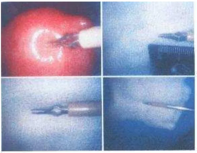
Surgical techniques
Myolysis of adenomyosis by laparoscopy
We used the electrodes of bipolar sharp forceps for myolysis. The posterior surface of adenomyosis was visualized continuously to protect the rectum, bladder, colon, and uterine blood vessels. The electrodes of bipolar forceps was put directly opposite to adeno-myosis so the the depth of penetration could be visua-lized and damages of colon and bladder could be pre-vented. After the depth for safe penetration had been determined, the electrodes were applied to adenomyo-sis at continuous power of 40-50 watts and then moved slowly to reach maximum coagulation. Fiber tip was cooled down by flowing fluid continuously. Excessive blood was suctioned and the tissue was cleaned up thoroughly. Adhesive barrier of interced Tc7 was administered to prevent adhesion.
Laparoscopic resection of adenomyosis
conti-nuous suturing. Excessive blood was suctioned and the tissue was cleaned up thoroughly. To prevent adhe-sion, we administered adhesive barrier of Intercede Tc7.
Post-operative follow-up and evaluation
After the surgical procedure, we observed the subjects for 24 hours. If their conditions were stable, they were discharged in the next day. We gave antibiotics and NSAID for 3 to 5 days and informed that stomach cramp or post-operative bleeding might occur. They were asked to come back to the hospitals if the cramp or bleeding became severe. If the symptoms could be overcome by medications, subjects could carry out normal activities in the next day. All subjects received GnRH analog therapy for 3 cycles. During the six-month follow up, they were requested to write down their subjective symptoms. At the end of follow up transvaginal ultrasonography was done by two feto-maternal counselors to assess the reduction of adeno-myosis volume and the subjective symptoms were
evaluated. The reduction of menorrhagia was eva-luated by measuring the number of pads used or by the length of its use i.e., < or > 6 days. Dysmenorrhea was assessed on the basis of loss of efficiency and the need for bed rest, while pelvic pain was evaluated based on analgesic drugs needed.
RESULTS
During the study period, 20 subjects underwent con-servative surgeries for symptomatic adenomyosis, 10 patients in the resection group and 10 patients in the myolysis group. Significant complications did not occur in both groups. Subjective symptoms could be evaluated in all patients while ultrasonographic reex-amination could only be done in 17 patients because two patients resigned from the study and one became pregnant before the time of the evaluation. Baseline characteristics of the subjects are depicted in Table 1, which were comparable between the groups.
Table 1. Baseline characteristics of study subjects
Baseline characteristics Group N
Resection Myolysis
Symptom scores Menorhagia
0
Adenomyosis volume Median (range)
Mean (SD)
76.85 (15-799) 153.42 (238.02)
In this study, mean age in the resection group was 37.7 (SD 5.7), ranged from 32 to 48 years. In the myolysis group, the mean age was 34.3 (4.6) years, ranged from 28 to years. Most patients were in the age group of 31-35 years (5/10 and 4/10 in myolysis and resection groups respectively) and nullipara.
Table 1 also shows the distribution of subjective symp-toms such as menorrhagia, dysmenorrhea, and pelvic pain. The most frequently found symptom was dysme-norrhea which was experienced by 18 of 20 patients, 10 was in the resection group and 8 in the myolysis group.
Comparison of subjective symptoms and adeno-myosis volume between the groups after surgery
There were no significant statistical differences in symptom scores between the groups after surgery as Mann-Whitney statistical test, no significant
differ-ence was found in median adenomyosis volume be-tween the two groups (p=0.277).
Comparison of median percentage reduction in symptom scores and in adenomyosis volume be-tween resection and myolysis groups
Table 3 shows that dysmenorrhea was the most re-duced symptom in both groups, i.e. 100% in the resec-tion group (0-100) with a mean of 75% (7546.291 percent), while in the myolysis group 66.6%(0-100) with a mean of 58.31% (58.3137.881 percent). On the other hand, there was no change in the symptom of menorrhagia after intervention either in the resec-tion or in the myolysis group, with a median of 0% (0-100) and mean of 33.3% (33.351.64) and a me-dian of 0% (0-67) and a mean of 11.1% (11.127.189 %), respectively. In addition, no statistically significant difference was found in the reduction of menorrhagia and dysmenorrhea scores between the groups (p=0.213 and p=0.399, respectively). The symptom of pelvic pain could not be analyzed statistically due to the small number of subjects experienced that symp-tom.
Table 2. Comparison of subjective symptoms and adenomyosis volume between the groups after surgery
Variables Group Total P
Resection Myolysis
Symptom scores Menorhagia
0
Adenomyosis volume Median (range)
Table 3. Comparison of median percentage reduction in symptom scores and changes in adenomyosis volume (%) between resection and myolysis groups
Variables Resection Myolysis P
N Median Range N Median Range adenomyosis was not reduced; instead it enlarged dur-ing the period of six-month evaluation. In the resec-sulted in greater enlargement than resection did, there was no significant stastitical difference found (p=0.630).
Outcomes of surgery, pregnancy, adhesion, side effects, and histopathological findings
In this study, we suspected adhesion after intervention by transvaginal ultrasonographic examination in 2 patients, 1 from the resection group and the other from the myolysis group. Five patients became preg-nant, three of which were from the resection group, and two from the myolysis group, with one patient experienced uterine rupture at 8 months of pregnancy. Early membrane rupture was found in one patient in the resection group.
Outcomes in the resection group
Of three patients in the resection group who became pregnant, one was 33 years old and had had primary infertility for five years and abortion for two times. This patient showed a reduced volume of adenomyo-sis up to 3.7%, became pregnant after 13 months post-intervention and had her pregnancy terminated by cesarean section at 38 weeks of gestation due to the transverse lie of the fetus. Her baby had birth weight of 3,500 grams. The patient experienced neither any symptom nor complication during pregnancy. Another patient had an age of 34 years with two parities. Her adenomyosis volume was reduced by 3.33% and she was pregnant for five weeks after 11 months
post-surgery. She also experienced reduction in menorrha-gia and dysmenorrhea up to 100%. The other patient aged 37 years with primary infertility of 1.5 years became pregnant for 30 weeks in four months after the intervention such that no evaluation of adeno-myosis volume could be made.
In this group, two patients had reduction in myosis volume up to 100%; of these, one had adeno-myoma on histopathological examination. Four pa-tients had their adenomyosis volume increased up to 159% with persistent dysmenorrhea and pelvic pain. One 48 year-old patient who had the largest initial volume of adenomyosis (799 mm3) with symptoms of menorrhagia, dysmenorrhea, and pelvic pain, expe-rienced an increased volume by 50.59% (1203 mm3). Moreover, a suspected adhesion at ultrasonographic examination was found with persistent severity of symptoms in which she had to continue consuming strong analgesics prior to menstruation each month.
Outcomes in the myolysis group
Of two patients in this group who became pregnant, one was 40 years old; she experienced reduction in adenomyosis volume and dysmenorrhea up to 99.88% and 100%, respectively, and was pregnant for 20 weeks at 12 months after the intervention. The other patient was 32 years old with primary infertility of 1.5 years. She had a reduced volume of adenomyosis up to 12.5%, and was pregnant in 7 months after inter-vention. Unfortunately, she had uterine rupture at eight-month pregnancy during the lung maturation period preceded with lower abdominal pain for 1 week and early membrane rupture. Cesarean section was performed in this patient, but the baby died.
occur-ring in patient aged 31 years with primary infertility for five years. During surgery, very severe adhesion was found in the internal genitalia of this patient and
adhesiolysis was performed. Her dysmenhorrhea score was reduced to 66.6%, while the menorrhagia was persistent.
Figure 1. Adenomyosis resection performed in this study
DISCUSSION
Shortcomings of the study
This study was limited by the small number of sub-jects, just enough to fulfill the minimum number re-quired for statistical test. As a result, the strength of the study was relatively low and the findings could not be generalized. This study constituted only a pre-liminary report aiming to obtain data and hypothesis for further study.
Ultrasonographic examinations were performed by two personnel with an assumption that they had equal capabilities and high agreement. However, kappa val-ue was not obtained, so disagreement causing biased results was still possible.
Age and parity in adenomyosis
In this study, we found that the mean age of patients with adenomyosis was 37.75.7 in the resection group and 34.34.6 in the myolysis group with a range of 28-48 years. This was slightly different from previous studies suggesting that generally adenomyosis occurs in fourth to fifth decades.5-7
Seventy-five percent of our patients were nuliparous with a history of two-time abortion in one patient. However, several literature stated that usually adeno-myosis patients are multiparous and there is a correla-tion between a history of inappropriate curettage and the occurrence of this disease.1,4 A study conducted in rats showed that adenomyosis could be stimulated by high-dose estrogen and progesterone. In similar phy-siological condition happening during pregnancy, the infiltration of endometrium into myometrium would be easy.12 This finding at least partly explain why histological diagnosis of adenomyosis mostly was found in multiparous women.12
However, one literature study suggested that although adenomyosis is generally considered as a typical dis-ease of multiparous women at the end of their repro-ductive period, disorder of endometrio-myometrial junctional zone can be discovered in young, nulipar-ous women with symptom of menorrhagia or hyper-menorrhea. Here, the disorder of the zone is directly attributed to endometrial factors and indirectly caused by increased response of local immunity. This zone disorder served as predisposing factor for secondary infiltration from endometrial elements, leading to
su-perficial adenomyosis.12 In the study conducted by Wood, clinical appearance was found in patients aged 24-52 years, with parity of 0-5, in which infertility was in 12 of 54 patients.8
Comparison of subjective symptoms reduction be-tween resection and myolysis
In this study, symptoms mostly found was dysme-norrhea (50%), which was different from some litera-ture which found menorrhagia as the most frequent symptom followed by dysmenorrhea.1,5,9 However, this finding was similar to a study by Jacoeb per-formed in clinical setting.10
This study found that either resection or myolysis was equally effective in reducing symptoms, which was similar to Wood’s study. However, the success rate reached in this study was lower, in which reduction obtained was 41.2% by resection and 39.5% by myo-lysis, compared to that of Wood’s study which was >50%. This difference may be due to the fact that the recurrence rate in our subjects was reasonably high, as evident from the increase in adenomyosis volume after intervention in both groups, i.e. 15.35% (-100-159) and 48.43% (-100-553) in the resection and myolysis groups, respectively, after three-cycle GnRH analog therapy. The difference might also caused by different parameter of evaluation in which Wood used the number of symptom-free patients while we ana-lyzed the percentage reduction in symptom scores.
Only dysmenorrhea showed statistically significant reduction after intervention in both groups. This may be due to the administration of GnRH analog for three cycles. Further studies are necessary for long-term observation, because as we know GnRH analog can not be administered for more than six cycles since this will result in climacteric symptoms. Wood found that 63% of patients were free from symptoms for two years, while 12% must underwent hysterectomy due to persistent severe symptoms or recurrence.8 Wood performed myolysis in two women with broad ade-nomyosis on the anterior wall and posterior uterus, in whom medical treatment had failed and resection was neither possible nor wanted by the patients. Two years after myolisi, both patients were free from menstrual pain and hemorrhage.8
Symptoms were evaluated by subjective self-reporting, such that placebo effects were likely to occur.
Comparison of reduction in adenomyosis volume between resection and myolysis
Diagnosis of adenomyosis established by transvaginal ultrasonography was confirmed by histopathological examinations. Approximately 9 of 10 patients from the resection group who underwent histopathological examinations were found to have adenomyosis, while one patient had adenomyoma which could be re-moved satisfactorily due to its clearly defined border. This finding was consistent with some literature stat-ing that transvaginal ultrasonography had high sensi-tivity (87-98%) and specificity (74-99%) as well as high positive and negative predictive values in diag-nosing adenomyosis.2,5,7,11 In the present study, we did not perform histopathological examination in the myolysis group. For further study, it is advisable to conduct adenomyosis biopsy first prior to myolysis intervention.
We found that the mean volume of adenomyosis in-creased in both groups at the six-month evaluation. On statistical test, no significant difference was found between the two groups in adenomyosis volume in-crement.
In the resection group, of eight patients evaluated by ultrasonography, four were found to have volume re-duction by approximately 3.7-100%, while another four patients experienced increased volume by ap-proximately 16-159%. This may be due to the fact that the excision of unclearly-defined adenomyosis is technically difficult. As suggested by Wood, problems encountered in preserving the uterus are the uncertain-ty concerning the size of myometrium removed, ex-cept if the adenomyosis is clearly defined, such as adenomyoma, and whether normal pregnancy and delivery are still possible.8
The removal of myometrium in significant amount will create problems, such as reduced capacity of myometrium and uterus during pregnancy such that it may trigger abortion or premature birth; and the for-mation of scar tissue which can contain undetectable adenomyosis focus that may reduce the strength of stretch. The capacity of uterus expansion during preg-nancy is more dependent on the increased plasticity, rather than elasticity. Nevertheless, the expansion ca-pacity of the uterine myometrium and its supporting
tissue makes it possible to perform myometrium ex-cision in a certain amount without reducing the ute-rus capacity for normal expansion during pregnancy.8 Wood performed removal of myometrium by half size from the posterior myometrium in one patient whom became pregnant and gave birth by elective cesarean section in the 37th week of gestation.8 In this study, only one patient who was observed formal-ly until delivery, who was finalformal-ly gave birth by cesa-rean section.
In the myolysis group, of nine patients undergoing ultrasonographic examination, three experienced vo-lume reduction by 12.5% -99.8%, while the remaining had increased volume by approximately 19.4%-553%. One patient was pregnant after intervention and re-ceived GnRH hormonal therapy. Unfortunately, she experienced uterine rupture at eight month pregnancy, which was similar to Wood’s study. This might hap-pen because coagulation of adenomyosis would create the same effects as excision, in which myometrial tissue diminished while scar tissue were formed.8 The possibility of uterine rupture is associated with the formation of broad scar tissue after coagulation. It is advisable that myolysis is followed by sterilization in women with enough children, in view of the possible uterine rupture.8
However, adenomyosis itself can reduce the capacity of uterus by replacing normal myometrium and sup-porting tissue which can interfere the arrangements of muscular fibers and three-dimensional structure of collagen. In this way, the scar tissue which was formed after intervention constituted a separate factor. In Wood’s study, adenomyosis was found in the loca-tion of uterine rupture in three patients.8 It appears that pregnancy after resection or myolysis still pose problems that should followed up cautiously.8
In addition to technical problems and the limitation of the tools used, it is necessary to pay attention to a number of initiating factors that may trigger and in-crease recurrence rate of the patients, such as status of estrogen, progesterone, and prolactin receptors as well as prostaglandin and endothelin, which was not ob-served in this study.13
that in resection.8 Coagulation used in myolysis might also not as accurate as resection because of un-predictable, disturbed electrical conduction of abnor-mal tissue.8 Thus, myolysis is more appropriate for women aged above 40 years with enough parities who do not want any pregnancy anymore but wish to maintain their uterus or reject broad operation or hys-terectomy.8
The findings of this study had not given enough evi-dence to provide guidance for managing symptomatic adenomyosis conservatively in the future because of the limitations in data, sample, and duration of the study. It is necessary to conduct further studies with an adequate number of subjects, objective data, and instruments. In addition, long-term observation is still essential in evaluating the effectiveness and side ef-fects as well as the efef-fects of the intervention on preg-nancy and delivery. If persistent symptoms were found in women with enough parity, the most appro-priate therapy for adenomyosis would be a total hyste-rectomy.8
CONCLUSIONS AND RECOMMENDATIONS
Conclusions
In conclusion, no significant differences were found in median reduction of menorrhagia and dysmenorr-hea scores between the resection and myolysis groups. Reduction in pelvic pain score could not be evaluated because of the limited number of subjects having that symptom. Increase in adenomyosis volume was found in both groups after the intervention and GnRH ana-log administration, but no significant difference was found in the mean of volume increment between the groups. Five patients were found to be pregnant, three from the resection group and two from the myolysis group. One case of uterine rupture was found in the myolysis group.
Recommendations
The findings of this study had not given enough evi-dence to provide guidance for managing symptomatic adenomyosis conservatively in the future because of the limitations in data, sample, and duration of the study. It is necessary to conduct further studies with an adequate number of subjects as well as objective data and instruments. In addition, long-term
observa-tion is still essential in evaluating the effectiveness, side effects, and recurrence as well as the effects of the intervention on pregnancy and delivery. It is ad-visable not to perform myolysis in women who still want pregnancy. If persistent symptoms were found in women with enough parity, the most appropriate ther-apy for adenomyosis would be a total hysterectomy.
Acknowledgments
The authors indebted Dr. Nikmah Salamia Idris for reviewing the manuscript.
REFERENCES
1. Levgur M, Abadi MA, Tucker A,. Adenomyosis: symp-toms, histology, and pregnancy terminations. Obstet Gy-necol 2000;95:688-91.
2. Ferency A. Pathophysiology of adenomyosis. Human Reproduction Up date 1998;4:312-22.
3. Bergholt T, Ericsen L, Berend N, Jacobsen M, Hertz JB. Prevalence and risk factors of adenomyosis at hysterecto-my. Human Reproduction 2001;16:2418-21.
4. Ota H, Igarashi S, Hatazawa J, Tanaka T. Is adenomyosis an immune disease? Human Reproduction up date 1998;4:360-7.
5. Arnold LL, Ascher SM, Schruefer JJ, Simon JA. The non-surgical diagnosis of adenomyosis. Obstet Gynecol 1995; 86:461-5.
6. Atri M, Reinhold C, Mehio AR, Chapman WB, Bret PM. Adenomyosis: US features with histologic correlation in an vitro study. Radiology 2000;215:783-90.
7. Bazot M, Cortez A, Darai E, Rouger J, Chapler J, Antoine JM, Uzan S. Ultrasonography compared with magnetic re-sonance imaging for diagnosis of adenomyosis: correla-tion with histopathology. Human Reproduccorrela-tion 2001; 16:2427-33
8. Wood C. Surgical and medical treatment of adenomyosis. Human Reproduction up date 1998;4:323-6.
9. Fedele L, Bianchi S, Raffaelli R, Portuese A, Dorta M. Treatment of adenomyosis-associated menorrhagia with a levanorgestrel-releasing intrauterine device. Fertil Steril 1997;68:426-9.
10. Jacoeb TZ. Reseksi adenomiosis pada wanita infertil (pengalaman penanganan pada 67 kasus). Presented at the Annual Scientific Meeting of POGI; Malang 2002. p. 1-7. 11. Vercellini P, Cortesi I, Giorgi OD, Merlo D, Carinelli G,
Crosignani PG. Transvaginal ultrasonography versus ute-rine needle biopsy in the diagnosis of diffuse adenomyo-sis. Human Reproduction 1998;13:2884-7
12. Brosens JJ, Souza NMD, Barker FG. Uterine junctional zone: function and disease. The Lancet 1995;346:558-60. 13. Mai KI, Yazdi HM, Perkins DG, Merlo DG and Parks W.