PLEOMORPHIC ADENOMA OF THE PAROTID GLAND:
A CASE REPORT
Farhat, M. Edy Syahputra Nasution
Otorhinolaryngology Head and Neck Department
Medical Faculty, University of Sumatera Utara
Introduction
Pleomorphic adenoma, also known as benign mixed tumor, is the most common tumor of
salivary gland origin, accounting for about 60-70% of all salivary gland tumors (Peel and
Seethala, 2007; Downer, et al., 2011; Papadogeorgakis, et al., 2011). These tumors are most
often found in the parotid gland (Oh and Eisele, 2006; Makeieff, et al., 2010), followed by the
submandibular gland and the minor salivary glands (Oh and Eisele, 2006).It represents 70-80%
of all tumors of the parotid gland. The lesion is usually solitary (Dubrulle and Souillard, 2006).
The typical presentation is that of a painless, slowly growing mass (Peel and Seethala,
2007).The gross appearance is smooth and lobular with a well-defined capsule (Oh and Eisele,
2006). Larger tumors often have a bosselated surface and may distend overlying skin and cause
erosion of bone and remodeling deep to the tumor (Peel and Seethala, 2007). Microscopically,
the tumor consists of epithelial and mesenchymal elements. The epithelial component forms a
trabecular pattern with a mesenchymal stroma. The mesenchymal portion may be myxoid,
chondroid, fibroid, or osteoid. The stroma varies from tumor to tumor and may have a
combination of any of these tissue types within it. Histologically, pleomorphic adenomas show
incomplete encapsulation with pseudopod extensions. Appropriate surgical therapy requires
resection with an adequate margin of normal tissue surrounding the tumor (Oh and Eisele,
2006).While pleomorphic adenoma is a benign tumor, it has the capacity to recur and to undergo
malignant transformation (Peel and Seethala, 2007).
This article presents a case of pleomorphic adenomaof the right parotid gland, with
Case Presentation
A 20-year-old female was referred by her surgeon for diagnosis and treatment of "a lump
in the rightneck region" which was suspected as Nasopharyngeal Carcinoma (NPC). She was
presented with a 5-year historyof a slowly enlarging painless mass in her right preauricular and
homolateralneck region. Her symptoms could not be explained by infection or trauma.The
patient was a non-smoker and she didn’t have history of head and neck irradiation exposure.
There was no other significant medical or surgical history or pertinent family history.
On clinical examination, the patient presented withsingle, well-defined, nontender massof
the right parotid and homolateralneck region (Fig. 1).PreviousFine Needle Aspiration Biopsy
(FNAB)revealedmetastatic carcinoma prone to NPC but nasoendoscopic examination did not
show mass in the nasopharynx. No other anomalies of the head and neck were seen on clinical
examinations. Facial nerve function was also normal.Preoperative assessment of the patient
comprised of aneck Computed Tomography (CT) Scan andrepeated FNAB. Anenhanced axial
CT scan showed inhomogeneous enlargement of the cervical lymph node in the right and
homolateral neck regions. Radiologically, it revealed lymphoma or lymphadenopathy. But, there
was no mass in the nasopharynx. Repeated FNAB was done selectively in the tumor, identifying
it as pleomorphic adenoma. The patient was informed about the benign nature of the tumor and
expressed her desire ofpreservation of the facial nerve.
Based on this diagnosis andon the clinical characteristics and extension of the tumor,
asubmandibular incision wasdesigned to gain access to the tumor. Surgical excision of the mass
was performed (Fig. 2). After general anesthesia a horizontal incision of 5 cm allowed for
exposure of the tumor mass. The lesion wasencapsulated, and could be removed by
bluntpreparation.The wound was closed using non-resorbablesutures (Fig. 3). The
surgicalspecimen was of a size of 8 x 5 x 3 cm (Fig. 4). Macroscopically,the tumor appeared to
be encapsulated. Whencut open, a solid tumor mass of a yellowish-pinkishcolor was obvious.
The specimen was fixed in 95% ethanol, and sent to the pathology laboratory withthe diagnosis
of pleomorphic adenoma. No malignant transformation was identifiedin the
specimen.Postoperative healing wasuneventful with a satisfactory cosmetic outcome (Fig. 5).
Facial nerve function revealed normal.Clinical follow-up until 4weeks did not revealany
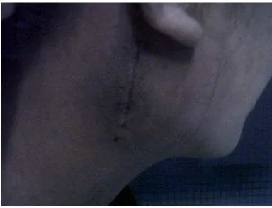
Figure 1.Single, well-defined, palpable, nontender mass of the right parotid and
homolateral neck region
Figure 2.Surgical excision of the mass
Figure 3.The wound was closed using non
resorbable sutures Figure 4.The surgical specimen
Figure 5.Postoperative healing Figure 6.Clinical follow-up
Discussion
Pleomorphic adenoma is the most common tumor of salivary gland origin, accounting for
the parotid gland (Peel and Seethala, 2007; Avecedo, et al., 2010). Bilateral occurrences of
pleomorphic adenoma are rare. Pleomorphic adenomas can occur in any decade, but the mean
age is 46 years with a slight female predilection. The typical presentation is that of a painless,
slowly growing mass (Peel and Seethala, 2007).In the present case, a 20-year-old female was
referred for diagnosis and treatment of "a lump in the right neck region" which was suspected as
NPC. The patient was presented with a 5-year history of a slowly enlarging painless mass in her
right preauricular and homolateral neck region. Her previous Fine Needle Aspiration Biopsy
(FNAB) revealed metastatic carcinoma prone to NPC but nasoendoscopic examination did not
show mass in the nasopharynx.
Actually,NPCs are epithelia neoplasms(Chan, Teo, and Johnson, 2004). NPC is the most
common primary malignant neoplasm of the nasopharynx(Sivanandan and Fee Jr., 2005). The
commonest presentation is a neck mass that is found on examination to represent a painless,
unilateral, cervical lymphadenopathy. In almost all cases, the superior cervical nodes are the first
group to be affected followed by enlargement of the mid and lower cervical nodes(van Hasselt
and Leung, 1999; Chan, Teo, and Johnson, 2004). In parts of Asia, NPC is endemic with
incidence rates of 15-50/100,000. NPC is the third commonest form of malignancy amongst
men.The median age at presentation is 40-50 years. The incidence rises after the age of 20 and
decreases after 60 years(Chan, Teo, and Johnson, 2004).
The imaging appearance of the pleomorphic adenoma deserves special attention.
pleomorphic adenomas frequently show gradual enhancement(Dubrulle and Souillard, 2006;
Curtin, 2007). Not truly lobulated, the margin of many pleomorphic adenomas is slightly
undulating or shows several slight bulges (Curtin, 2007). Due to an enhanced axial CT scan of
the present case, the cervical lymph node in the right and homolateral neck region showed to be
inhomogeneously enlarge. The margin of the tumor was not noted. Radiologically, it revealed
lymphoma or lymphadenopathy. A delayed scan frequently will show the delayed enhancement
typical of this tumor and demonstrate the margin. This has actually become more of a problem
with the most modern CT scan technology. Many scanning protocols attempt to emphasize
visualization of the arteries and veins and so the scan is done as the contrast is being injected and
at that time the tumor may not be visible against the background of the normal parotid gland. The
delayed enhancement phenomenon does not tend to be a problem with Magnetic Resonance
visualize the tumor. Also, several sequences are done after the injection of intravenous contrast
so there would be time for gradual enhancement to occur. Consistent reliable visualization of
lesions is the primary reason that most head and neck radiologists prefer MRI to CT for
investigation of salivary gland tumors(Curtin, 2007).
The gross appearance of the tumor is smooth and lobular with a well-defined capsule (Oh
and Eisele, 2006). They usually range from 2 to 5 cm in greatest dimension (Peel and Seethala,
2007). Needle biopsy may have its greatest utility in the diagnosis of a submandibular mass,
where it can help to distinguish neoplastic from more common inflammatory changes, which
may spare the patient unnecessary surgery. Aspiration cytology may also differentiate a reactive
lymph node adjacent to a salivary gland from a tumour within the gland itself (Spiro and Spiro,
2003). Histologically, pleomorphic adenomas show incomplete encapsulation with pseudopod
extensions. This case shows that the surgical specimen was of a size of 8 x 5 x 3 cm (Fig. 4). The
solid tumor mass of a yellowish-pinkish color was obvious. Due to the histological findings in
the primary tumor, the tumor appeared to be encapsulated. No malignant transformation was
identified in the specimen.
While pleomorphic adenoma is a benign tumor, it has the capacity to recur. Recurrence
rates range from 0.8-6.8% in large series with long-term follow-up(Peel and Seethala, 2007). In
the present case, the lesion was encapsulated, and couldcompletely be removed by blunt
preparation. Surgical management is the only recognized therapeutic alternative (Makeieff, et al.,
2010).The two main aspects of parotid surgery are recurrence of the tumor and damage of the
facial nerve (Papadogeorgakis, et al., 2011). Appropriate surgical therapy requires resection with
an adequate margin of normal tissue surrounding the tumor (Oh and Eisele, 2006). It is now
widely accepted that pleomorphic adenoma standard treatment must include at least partial
superficial parotidectomy allowing tumor removal with a generous cuff (minimum 2 cm margin,
except when abuts the facial nerve) of surrounding parotid tissue via en bloc resection
(Makeieff, et al., 2010). Enucleation or pseudocapsular violation may lead to tumor spillage and
localized tumor recurrence.
The factors that are thought to predispose to recurrence are poor surgical technique,
violation of integrity of the capsule, multicentric origin of the tumor, and benign tumors that
extend beyond the zygomatic arch (Papadogeorgakis, et al., 2011). While complete excision is
concern. In this case, postoperative facial nerve function revealed normal and wound healing was
uneventful with a satisfactory cosmetic outcome (Fig. 5).Since treatment of the relapsed tumor is
complicated with a high rate of secondary recurrence, this will result in a lifetime follow-up of
the patient who has to be informed on the possibility of further multiple recurrences(Peel and
Seethala, 2007).
Rarely, a histologically benign pleomorphic adenoma can metastasize and behave like a
low-grade malignancy. There are no features that can predict this rare occurrence. However,
many of these tumors metastasized after at least one initial local recurrence, suggesting the
possibility that altered anatomy secondary to surgery gave access to vascular and lymphatic
channels.Thackray and Lucas have estimated that left unresected, approximately 25% of
pleomorphic adenomas would eventually undergo carcinomatous change. These metastases seem
to show a particular affinity for lungs and bone especially the vertebral column. The average age
at diagnosis is 50–60 years (Peel and Seethala, 2007). Misdiagnosis is common because the
residual PA component may be small, and because various carcinoma subtypes may be present
(Zhao et al., 2013).Eneroth and Zetterberg undertook a microspectrophotometric DNA analysis
of pleomorphic adenomas and demonstrated a difference in the DNA content between
morphologicallybenign pleomorphic adenomas of short duration and those of long duration. This
finding supports the hypothesis that the risk of carcinomatous transformation in a pleomorphic
adenoma increases with the age of the tumor(Peel and Seethala, 2007).Therefore, follow-up of
the patient is needed.
Conclusion
Pleomorphic adenoma is the most common tumor of the parotid gland.Consistent reliable
visualization of lesions is the primary reason that most head and neck radiologists prefer MRI to
CT for investigation. Needle biopsy may have its greatest utility in the diagnosis of the tumor.
The two main aspects of parotid surgery are recurrence of the tumor and damage of the facial
nerve.While pleomorphic adenoma is a benign tumor, it has the capacity to recur and to undergo
References
Acevedo, J. L., Nolan, J., Markwell, J. K., David, T. (2010) ‘Pleomorphic Adenoma of The
Nasal Cavity: A Case Report’ Ear, Nose & Throat Journal. 89 (5) pp. 224.
Chan, A. T. C., Teo, P. M. L., and Johnson, P. J. (2004) ‘Nasopharyngeal Cancer’ In Brockstein,
B., Masters, G. (eds.) Head and Neck Cancer. New York: Kluwer Academic Publishers. pp.
275-7.
Curtin, H. D. (2007) ‘Imaging of the Salivary Glands’ In Myers, E.N. and Ferris, R.L. (eds.)
Salivary Gland Disorders. New York: Springer Berlin Heidelberg. pp. 19-22.
Downer, J., Fryer, E., Capper, J., Woo, E. K. (2011) ‘Pleomorphic Adenoma of The
Nasopharyngeal Mucosal Space with Locally Aggressive Appearance’ European Radiology. 21
() pp. 443-6.
Dubrulle, F. and Souillard, R. (2006) ‘Parotid Gland and Other Salivary Gland Tumors’ In
Hermans,R. (eds.) Head and Neck Cancer Imaging. New York:Springer Berlin Heidelberg. pp.
221-2.
Hasselt, C. A. V. and Leung, S. F. (1999) ‘Clinical Picture’ In Hasselt, C. A. V. and Gibb, A. G.
(eds.) Nasopharyngeal Carcinoma. Hong Kong: The Chinese University Press. pp. 105-7.
Makeieff, M., Pelliccia, P., Letois, F., Mercier, G., Arnaud, S., Ce´sar, C., Garrel, R., Crampette,
L., and Guerrier, B. (2010) ‘Recurrent Pleomorphic Adenoma: Results of Surgical Treatment’
Annals Surgical Oncology. 17 () pp. 3308–3313.
Oh, Y. S. and Eisele, D. W. (2006) ‘Salivary Gland Neoplasms’ In Bailey, B. J., Johnson, J. T.,
Newlands, S. D. (eds.) Head & Neck Surgery – Otolaryngology. 4th
Papadogeorgakis, N., Kalfarentzos, E. F., Petsinis, V., Parara, E., Kopaka, M. E. (2012)
ed. Lippincott: Williams &
Wilkins. pp. 1516-7.
‘Multinodular Neck Recurrence of Parotid Gland Pleomorphic Adenoma: A Case Report’
Oral Maxillofacal Surgery. 16 () pp. 137–140.
Peel, R.L. and Seethala R.S. (2007) ‘Pathology of Salivary Gland Disease’ In Myers, E.N. and
Sivanandan, R. and Fee Jr., W. E. (2005) ‘Benign and Malignant Tumors of The Nasopharynx’
In Cummings,C. W., Flint,P. W., Haughey,B. H., Robbins,K. T.,Thomas,J. R., Harker,L. A.,
Richardson,M. A., Schuller,D. E. (eds.) Cummings Otolaryngology—Head & Neck Surgery.
Philadelphia; Mosby.pp. 1669.
Spiro, J. D. and Spiro,R. H. (2003) ‘Salivary Gland Neoplasms’ In Evans,P. H. R., Montgomery,
P. Q., and Gullane, P. J. Principles and Practice of Head and Neck Oncology. London and New
York: Martin Dunitz. pp.678.
Zhao, J., Wang,J., Yu, C., Guo,L., Wang,K., Liang, Z., and Lou, J., (2013) ‘Prognostic factors
affecting the clinical outcomeof carcinoma ex pleomorphic adenoma in themajor salivary gland’