I
SUPERVISOR DECLARATION
“I hereby declare that I have read this thesis and in my opinion this report is sufficient in terms of scope and quality for the award of the degree of Bachelor of
Mechanical Engineering (Structure & Materials)”
Signature : ………
Supervisor : DR MOHD JUZAILA BIN ABDUL LATIF
II
BIOMECHANICAL CHARACTERISATION OF ARTICULAR CARTILAGE ACROSS SYNOVIAL JOINT
LIEW GUO REN
This report was submitted in fulfillment of the requirement for the award of Bachelor of Degree of Mechanical Engineering with Honours (Structure &
Materials)
Faculty of Mechanical Engineering
Universiti Teknikal Malaysia Melaka
III
DECLARATION
“I hereby declare that the work in this report was my own except for summaries and quotations which have been duly acknowledged.”
Signature : ……… Author : LIEW GUO REN
IV
ACKNOWLEDGEMENT
I am using this opportunity to express my gratitude to everyone who supported me throughout my Final Year Project. I am thankful for their aspiring guidance, invaluably constructive criticism and friendly advice during the project work. I am sincerely grateful to them for sharing their truthful and illuminating views on a number of issues related to the project.
Among others, I would like to express my warm thanks to my final year project supervisor, Dr. Mohd Juzaila Bin Abdul Latif, whose contribution in stimulating suggestion and encouragement, helped me to coordinate my project especially in writing this report. I am indebted to him for all the knowledge, wisdom and experience he shared during my studies.
Furthermore, I would like to express my appreciation to the master students: Yusra and Hikmah; Research fellow: Kevin, Mean Yee, Firdaus and Hajar, who have always sharing their knowledge and idea with me.
V
ABSTRAK
VI
ABSTRACT
VII
TABLE OF CONTENT
CHAPTER CONTENT PAGE
SUPERVISOR DECLARATION I
DECLARATION III
ACKNOWLEDGEMENT IV
ABSTRAK V
ABSTRACT VI
TABLE OF CONTENT VII
LIST OF FIGURES X
LIST OF TABLES XII
LIST OF SYMBOLS XIII
LIST OF ABBREVIATION XIV
CHAPTER 1 INTRODUCTION 1
1.1 Introduction 1
1.2 Problem Statement 3
1.3 Objective 4
1.4 Scope 4
CHAPTER 2 LITERATURE REVIEW 5
2.1 Osteoarthritis 5
2.1.1 Causes 6
2.1.2 Diagnosis 8
2.1.3 Treatment 9
2.1.3.1 Non-pharmacologic 10 2.1.3.2 Pharmacologic 10 2.1.3.3 Intra-articular injection 11 2.1.3.4 Surgical 11 2.2 Synovial joint-anatomy and physiology 12
VIII
CHAPTER CONTENT PAGE
2.3.1 Structure and composition 15 2.3.1.1 Collagen 17 2.3.1.2 Proteoglycans 17 2.3.1.3 Water 18 2.4 Characterization of biomechanical properties
of articular cartilage 19 2.4.1 Material theory 19 2.4.2 Specimen preparation 20 2.4.3 Experimental testing 20
2.4.4 Thickness 24
2.4.5 Computational methods 26
2.5 Summary 28
CHAPTER 3 METHODOLOGY 30
3.1 Introduction 30
3.2 Material and specimen preparation 31 3.2.1 Phosphate Buffered Saline 31 3.2.2 Bovine articular cartilage 31 3.3 Indentation test apparatus 32
3.4 Calibration test 34
3.4.1 Calibration procedure 34 3.5 Creep indentation test 35 3.6 Cartilage thickness measurement 36
3.7 Computational methods 37
3.7.1 Implementation of contact dependent
flow 37
3.7.2 Model development: Repeat of previous
study 38
3.7.3 Model development: Simulation of
IX
CHAPTER CONTENT PAGE
CHAPTER 4 RESULT AND DISCUSSION 42
4.1 Introduction 42
4.2 Cartilage Thickness 42
4.3 Computational results for idealized
Axisymmetric model 43
4.3.1 Implementation of contact dependent
flow 43
4.4 Biomechanical behavior of articular cartilage
Across synovial joint 44 4.4.1 Contact pressure 44 4.4.2 Pore pressure 46
4.5 Discussion 47
CHAPTER 5 CONCLUSION 52
5.1 Conclusion 52
5.2 Recommendation 53
X
LIST OF FIGURES
FIGURE TITLE PAGE
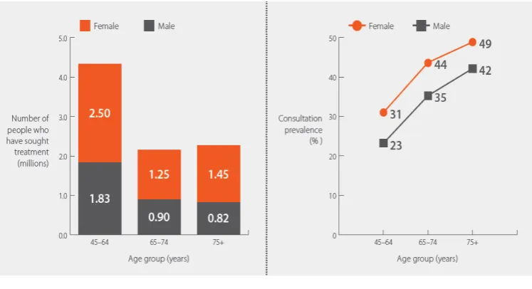
1.1 Estimated patients in UK who have sought treatment for
osteoarthritis 2
2.1 The schematic diagram of OA affects a joint 7 2.2 Examples of image of knee joint by X-ray 8 2.3 Examples of image of knee joint by MRI 9 2.4 Structure of synovial joint in the knee 13 2.5 Schematic presentation of a knee joint 14 2.6 Schematic diagram of articular cartilage 15 2.7 Schematic diagram indicating the extracellular matrix of
articular cartilage 18
2.8 Schematic diagram of Confined compression test 22 2.9 Schematic diagram of unconfined compression test 23 2.10 The schematic diagram indentation test 24 2.11 Indentation test to determine cartilage thickness 25 2.12 Cartilage model developed by using ANSYS 28
3.1 Specimen preparation 32
3.2 Schematic diagram of the indentation test rig 33
3.3 Indentation test apparatus 33
3.4 Graph of calibration 34
XI
FIGURE TITLE PAGE
3.8 Pawaskar’s model used for validate the implementation of contact dependent flow detection algorithm.
(Pawaskar,2006) 38
3.9 Axisymmetric FE model of cartilage specimen 40 4.1 Comparison graph of Contact pressure distribution at
cartilage surface between current study with Pawaskar’s
result 44
4.2 Compress pressure distribution on articular cartilage
surface 45
4.3 Contact pressure distribution at cartilage surface for three
difference thickness 45
4.4 Compress pressure distribution on articular cartilage
surface 46
4.5 Pore pressure distribution on cartilage surface 47
4.6 Location point of specimen 48
4.7 Direction of cartilage deformation after loading 50 4.8 Direction of fluid flow for step load , 2 s 50 4.9 Direction of fluid flow after constant load, 1000 s 50 4.10 Direction of fluid flow of constant load during 0 mm radial
XII
LIST OF TABLES
TABLE TITLE PAGE
2.1 Linear biphasic biomechanical properties of articular cartilage in human synovial joints.
21
2.2 Cartilage properties of the Mature tibial plateau determined from indentation test
26
3.1 Formulation of PBS tablet 31
3.2 Cartilage material properties used for FE validation model 39 4.1 Cartilage thickness for 3 difference points of articular
cartilage across synovial joint
XIII
LIST OF SYMBOLS
E – Young’s Modulus H – Aggregate modulus k – Permeability
mm – millimeter Pa - Pascal s – Second (time) v – Poisson’s ratio V – Voltage
XIV
LIST OF ABBREVIATION
ECM- Extracellular matric GAG- Glycosaminoglycans
LVDT – Linear variable differential transformer MRI – Magnetic resonance imaging
NSAID – Nonsteroidal anti-inflammatory drugs OA- Osteoarthritis
PBS - Phosphate buffered saline PGs- Proteoglycans
1
CHAPTER 1
INTRODUCTION
1.1 Introduction
2
Figure 1.1: The estimated patients in UK who have sought treatment for osteoarthritis, by gender and age group. a) By number of people, b) By
proportion of people. Adapt from Arthritis Research UK, 2013.
There are various joints in human body that may be affected by OA. However, this disease is most likely to affects the hands, hips, knees, lower back and neck (Nordqvist, 2014). Knee has one of the most complex and largest joint in the body which is known as synovial joint. With the unique structure, synovial joints allow very complex movements between adjacent bones like allow the leg to bend, straighten and carries the body weight. The ends of the bone are covered by the articular cartilage which are lubricated by the synovial fluid. The articular cartilage act as a protective surface that cushions the ends of bone in the joints and allows the joint to move smoothly.
There are two main causes of the OA, which are traumatic mechanical destruction and progressive mechanical degeneration (tear and wear). The traumatic mechanical destruction is due to the abnormal use and injury of the joint while the progressive mechanical degeneration is due to aging. During OA, the smooth surface of the cartilage will becomes rough and causing irritation. If the cartilage wears down completely, thus exposing the underlying subchondral bone and the bone in the joint may be rubbing against another bone, causing damage and pain. There is no cure for OA as yet, but the pain normally relieved by either
3
therapeutic or operational methods. The degradation of cartilage manifests itself in the variations of its mechanical and biological properties (Carter et al., 2004). This in turn causes the cartilage susceptible to structural failures, which degrades it further.
There are plenty of researchers from multiple fields such as medicine, engineering, chemistry, material science, etc have been studying the articular cartilage at nano-, micro- and macro- levels. Most of these studies are centred around to investigate about the biomechanical properties of the cartilage. A lot of experiment had been carried out to characterize the biomechanical properties and to study the behavior of the cartilage under different experimental and physiological conditions (Mow et al., 1989; Kwan et al., 1990). Similarly, computational models have been postulated to give firm theoretical foundations to the understanding of the mechanical behavior of the cartilage (Mow et al., 1980; Lai et al, 1991). However, the lack of mechanical property information on the synovial joint cartilage posed a problem for computation modeling. Previous studies have used the data get by the small specimen but it is not known either the mechanical behavior of the specimen can represent the whole cartilage in a joint.
Therefore, the aim of this study was to develop a method to characterize the thickness and biomechanical behavior of the articular cartilage across the synovial joint using indentation test. This is to identify that the biomechanical properties of the small specimen either can represent the whole joint.
1.2 Problem statement
4
1.3 Objective
The objective of this study is to study the biomechanical behavior of articular cartilage across the synovial joint.
1.4 Scope
The methods used for this project are the computational modeling method and the experimental method (indentation test). The scope includes:
1 To determine the thickness of cartilage across synovial joint.
2 To establish the method to study biomechanical behaviors of cartilage across synovial joint.
5
CHAPTER 2
LITERATURE REVIEW
2.1 Osteoarthritis
Osteoarthritis (OA) are the most common type of arthritis especially for those who reach middle age in United Kingdom. According to the National Health Service, UK, approximately 8.5 million people are affected by this disease. The Arthritis Foundation, USA, said that about 27 million Americans are affected in the year 2013. The women are more likely to be affected by osteoarthritis than men after the age of 50. This symptoms typically start after 40 years of age, and progress slowly (Arthritis Research UK, 2013). In America, arthritis and related conditions, such as osteoarthritis cost the country almost $128 billion annually in medical care and indirect expenses, including lost income and productivity. Between 1990 and 2010, disability due to osteoarthritis in the UK had increased by 16% and expected continue to grow (Nordqvist C., 2014).
6
even eating when the situation going worst. Osteoarthritis in the hips and knees will restrict mobility, limit walking, climbing, and bathing. In serious cases, osteoarthritis is a huge obstacle to people’s mobility and independence, and their tone of liveliness.
2.1.1 Causes
7
a. b.
c.
Figure 2.1: How osteoarthritis affects a joint a) A normal joint, b) A joint with mild osteoarthritis, c) A joint that has been deformed by severe osteoarthritis.
Adapted from Arthritis Research UK
The causes of OA are still not yet fully understood. However, some factors that can increase the risk of getting OA are known. The common risk factors include:
Age : The chance of getting OA rises after reach age 45 because the ability to heal decreases as a person get older
Gender : women taking more risk to get OA compared to men Obesity: Being overweight will cause excessive pressure on the knee Previous joint injury or disease: improper joint surgery and joint
diseases may taking risk of OA
Heredity: genetic mutations will make a person more likely to develop OA. It may also due to inherited abnormalities in the shape of the bones that surround the knee joint
8
2.1.2 Diagnosis
Diagnosing arthritis early can prevent permanent damage. It is not always easy to diagnose and can take several weeks or months to get a definitive diagnosis. Various diagnostic tests are employed in the evaluation of OA. Basically, the GP (general practitioner, primary care physician) will ask the patient about the symptoms, as well as carrying out some physical examination and check their medical history (Sinusas et al., 2012). Nowadays, there is no current and definitive test that can diagnose OA yet. If the possible conditions are suspected, imaging diagnostics will be use such as plain radiography (x-rays) and magnetic resonance imaging (MRI). However, the diagnostic tests are restricted for pregnant women although the risk of radiation exposure to the fetus are small.
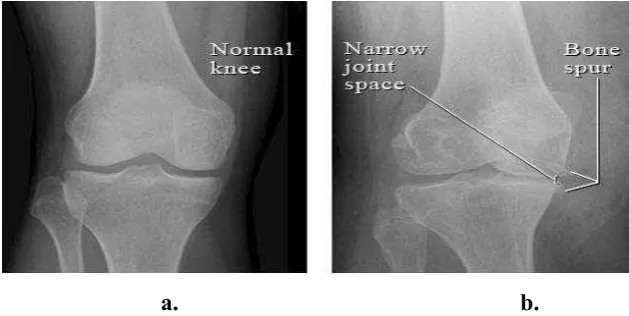
X-ray is the most common use by the doctor to diagnose OA. An x-ray will generate a very small amount of radiation to produce an image of the bones and tissues surrounding a joint as shown in Figure 2.2. The GP will uses the X-rays to evaluate arthritis. From the x-rays film, the GP can:
Rule out injury or other diseases of the joint
Have a baseline film for comparison while having treatment Look at the structures of the joint
a. b.
Figure 2.2: Examples of image of knee joint by X-rays. a) Normal knee b) bone spurs and narrowed joint space caused by osteoarthritis. Adapted from
9
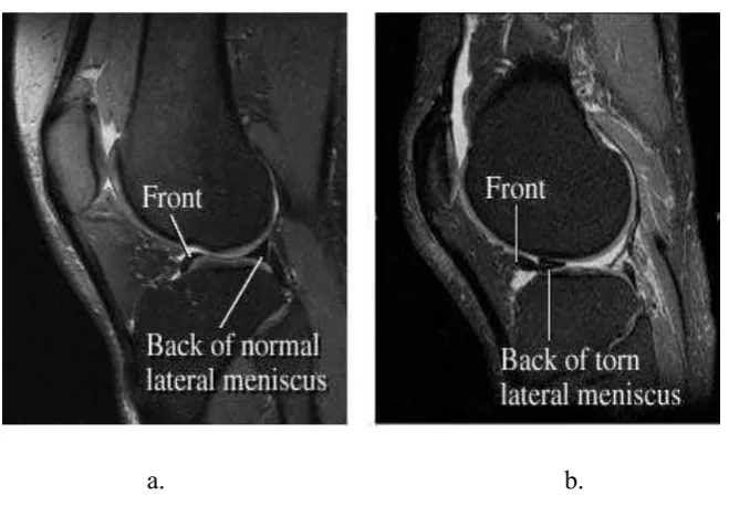
MRI scans may be used when X-rays do not show a clear reason for joint pain or when the X-rays suggest that other types of joint tissue could be damaged. MRI is a test that uses a magnetic field and pulses of radio wave energy to make pictures of organs and structures that are inside the body, as shown in Figure 2.3.
a. b.
Figure 2.3: Examples of image of knee joint by MRI. a) side view of the normal knee. b) Side view of knee joint with a piece of meniscus that has been torn and
moved. Adapted from Intermountain Medical Imaging, Boise
2.1.3 Treatment
10
2.1.3.1 Non-pharmacologic
Non-pharmacologic therapy often starts with exercise. The researchers found that the land-based exercise can relief knee pain and improve the physical function within 24 months (Thomas et al,2002 ; Frasen & McConnell, 2008) but some researchers found that the effects decline over time and finally disappear (Baar et al, 2001).
Therapeutic ultrasound is one of the non-pharmacologic treatment which is a physical therapy modality that often used in OA treatment. A Cochrane review of this modality concluded that although ultrasound therapy is almost certainly useful for some patients, but it is not a reliable or evidence-based therapy (Rutjes AW, 2010).
Another types of non-pharmacologic treatments include bracing and splinting to help support painful or unstable joints. A cane can help to support the body weight of OA patients, but it needs to be properly fitted.