• Tidak ada hasil yang ditemukan

Hubungan antara Infeksi Chlamydia trachomatis dengan oklusi tuba yang dideteksi dengan HSG pada wanita infertil

N/A
N/A
Protected

Academic year: 2016

Membagikan "Hubungan antara Infeksi Chlamydia trachomatis dengan oklusi tuba yang dideteksi dengan HSG pada wanita infertil"

Copied!
116
0
0

Teks penuh

(1)

HUBUNGAN ANTARA INFEKSI CHLAMYDIA

TRACHOMATIS DENGAN OKLUSI TUBA YANG

DIDETEKSI DENGAN HSG PADA WANITA INFERTIL

TESIS

OLEH :

REYNANTA

DEPARTEMEN OBSTETRI DAN GINEKOLOGI

FAKULTAS KEDOKTERAN UNIVERSITAS SUMATERA UTARA

RSUP. H. ADAM MALIK MEDAN

(2)

PENELITIAN INI DI BAWAH BIMBINGAN TIM-5

Pembimbing :

Dr. Binarwan Halim, Sp.OG(K)

Dr. M. Fidel Ganis Siregar, Sp.OG

Penyanggah :

Dr. Yusuf R. Surbakti, Sp.OG(K)

Dr. Ichwanul Adenin, Sp.OG(K)

Dr. Deri Edianto, Sp.OG(K)

Diajukan untuk melengkapi tugas-tugas dan memenuhi

salah satu syarat untuk mencapai keahlian dalam

(3)
(4)

K upe rse m ba hk a n K e pa da ya ng T e rk a sih da n T e rsa ya ng

Aya ha nda Dr. H . Rust a m Effe ndi Y S, SpPD-K GEH , FI N ASI M

da n

(5)

KATA PENGANTAR

Dengan nama Allah Yang Maha Pengasih lagi Maha Penyayang.

Segala Puji dan Syukur saya panjatkan kepada Allah SWT, berkat rahmat dan karunia-Nya penulisan

tesis ini dapat diselesaikan.

Tesis ini disusun untuk melengkapi tugas-tugas dan memenuhi salah satu syarat untuk

menyelesaikan Program Pendidikan Spesialis Obstetri dan Ginekologi. Sebagai manusia biasa, saya

menyadari bahwa tesis ini banyak kekurangannya dan masih jauh dari sempurna, namun demikian

besar harapan saya kiranya tulisan sederhana ini dapat bermanfaat dalam menambah perbendaharaan

khususnya tentang :

“HUBUNGAN ANTARA INFEKSI CHLAMYDIA TRACHOMATIS DENGAN OKLUSI TUBA YANG DIDETEKSI DENGAN HSG PADA WANITA INFERTIL”

Dengan selesainya laporan penelitian ini, perkenankanlah saya menyampaikan rasa terimakasih dan

penghargaan yang setinggi-tingginya kepada yang terhormat:

1. Rektor Universitas Sumatera Utara dan Dekan Fakultas Kedokteran Universitas Sumatera

Utara, yang telah memberikan kesempatan kepada saya untuk mengikuti Progaram Pendidikan

Dokter Spesialis Obstetri dan Ginekologi di Fakultas Kedokteran Universitas Sumatera Utara,

Medan.

2. Prof. Dr. Delfi Lutan, MSc, SpOG(K) Ketua Departemen Obstetri dan Ginekologi FK – USU

Medan; Dr. M. Fidel Ganis Siregar, SpOG Sekretaris Departemen Obstetri dan Ginekologi FK

– USU Medan; Dr. Henry Salim Siregar, SpOG(K) Ketua Program Studi Dokter Spesialis

Obstetri dan Ginekologi FK USU; Dr. M. Riza Tala, SpOG(K) Sekretaris Program Studi

Dokter Spesialis Obstetri dan Ginekologi FK USU; Prof. Dr. M. Jusuf Hanafiah, SpOG(K);

Prof. Dr. Djaffar Siddik, SpOG(K); Prof. DR. Dr. H.M. Thamrin Tanjung, SpOG(K); Prof. Dr.

Hamonangan Hutapea, SpOG(K); Prof. Dr. R. Haryono Roeshadi, SpOG(K); Prof. Dr. T. M.

Hanafiah, SpOG(K); Prof. Dr. Budi R. Hadibroto, SpOG(K); Prof. Dr. M. Fauzie Sahil,

SpOG(K) dan Prof. Dr. Daulat H. Sibuea, SpOG(K) yang telah bersama-sama berkenan

menerima saya mengikuti pendidikan spesialis di Departemen Obstetri dan Ginekologi FK

(6)

3. Dr. Binarwan Halim, SpOG(K) dan Dr. M. Fidel Ganis Siregar, SpOG yang telah memberikan

bimbingan kepada saya dengan penuh kesabaran telah meluangkan waktu yang sangat berharga

dalam melakukan penelitian ini; memeriksa; dan melengkapi penulisan hingga selesai. Kepada

Dr. Yusuf R. Surbakti, SpOG(K); Dr. Ichwanul Adenin, SpOG(K) dan Dr. Deri Edianto,

SpOG(K) selaku penyanggah dan nara sumber untuk membimbing, memeriksa, dan

melengkapi penulisan tesis ini hingga selesai.

4. Ucapan terima kasih yang tidak terhingga kepada Dr. Binarwan Halim, SpOG(K) beserta

seluruh staf, para medis dan karyawan/karyawati Halim Fertility Center Medan yang telah

mengizinkan saya dan banyak sekali memberikan bantuan kepada saya selama saya melakukan

penelitian di Halim Fertility Center Medan. Semoga Tuhan membalas kebaikan anda semua.

5. Dr. Deri Edianto, SpOG(K) sebagai bapak angkat saya selama menjalani masa pendidikan

yang telah banyak mengayomi, membimbing, dan memberikan nasehat yang bermanfaat

kepada saya selama dalam pendidikan.

6. Dr. Christoffel Tobing, SpOG(K) selaku pembimbing Mini Refarat Feto Maternal saya yang

berjudul “Percutaneus Umbilical Blood Sampling”; Dr. T. M. Ichsan Ibrahim, SpOG selaku

pembimbing Mini Refarat Fertilitas Endokrinologi dan Reproduksi saya yang berjudul

“Abortus berulang akibat infeksi” dan kepada Prof. Dr. M. Fauzie Sahil, SpOG(K) selaku pembimbing Mini Refarat Onkologi saya yang berjudul “Terapi Konsolidasi pada Penderita

Kanker Ovarium”

7. Dr. Surya Dharma, MPH dan DR. Ir. Erna Mutiara, M.Kes yang telah meluangkan waktu dan

pikiran untuk membimbing saya dalam menyelesaikan analisa data dan uji statistik pada tesis

ini.

8. Seluruh staff pengajar Departemen Obstetri dan Ginekologi FK USU/RSUP H. ADAM

MALIK; RSUD Dr. PIRNGADI; Rumkit KESDAM TK II; RS PTPN II Tembakau Deli; RSU

HAJI MINA; RSU SUNDARI yang secara langsung telah banyak membimbing dan mendidik

saya sejak awal hingga akhir pendidikan.

9. Direktur RSUP H. Adam Malik Medan yang telah memberikan kesempatan dan sarana untuk

bekerja sama selama mengikuti pendidikan Spesialis Obstetri dan Ginekologi di Departemen

Obstetri dan Ginekologi FK USU Medan.

10. Direktur RSUD Dr. Pirngadi Medan dan Kepala SMF RSUD Kebidanan dan Penyakit

Kandungan Dr. Pirngadi Medan Dr. Rushakim Lubis, SpOG beserta staff yang telah

memberikan kesempatan dan sarana untuk bekerja sama selama mengikuti pendidikan

(7)

11. Direktur RS PTPN II Tembakau Deli Medan; Dr. Sofian Abdul Ilah, SpOG dan Dr.

Nazaruddin Jaffar, SpOG(K) beserta staff yang telah memberikan kesempatan dan bimbingan

selama saya bertugas menjalani pendidikan di rumah sakit tersebut.

12. Ka. Rumkit Tk. II Puteri Hijau KESDAM II/BB Medan dan Kepala SMF Obstetri dan

Ginekologi Rumkit Tk. II Puteri Hijau KESDAM II/BB Medan Dr. Gunawan Rusuldi, SpOG

beserta staff yang telah memberikan kesempatan dan bimbingan selama saya bertugas di rumah

sakit tersebut.

13. Direktur RSU Haji Medan dan Kepala SMF Obstetri dan Ginekologi RSU Haji Medan Dr. H.

Muslich Perangin-angin, SpOG beserta staff yang telah memberikan kesempatan dan

bimbingan selama saya bertugas di rumah sakit tersebut.

14. Direktur RSU Sundari Medan dan Kepala SMF Obstetri dan Ginekologi RSU Sundari Medan

Dr. H. M. Haidir, MHA, SpOG dan Ibu Sundari, Am.Keb beserta staff yang telah memberikan

kesempatan dan bimbingan selama saya bertugas di rumah sakit tersebut.

15. Kepada Dr. H. Syafii Siregar, SpOG dan seluruh staff di bagian kebidanan dan kandungan

RSUD Panyabungan, Kab. Mandailing Natal yang telah memberikan kesempatan kepada saya

untuk menjalani pendidikan; memberikan bimbingan dan segala bantuan moril kepada saya

selama saya bertugas di rumah sakit tersebut.

16. Kepala Bagian Anestesiologi dan Reanimasi FK – USU Medan beserta staff atas kesempatan

dan bimbingan selama saya bertugas di bagian tersebut.

17. Kepala Departemen Patologi Anatomi FK – USU Medan beserta staff atas kesempatan dan

bimbingan selama saya bertugas di bagian tersebut.

18. Kepada senior-senior saya selama saya menjalani pendidikan : Dr Adek Novita Dayeng,

SpOG, Dr. Rillie Ritonga SpOG; Dr. Ade Taufik, SpOG; Dr. Samson Chandra, SpOG; Dr.

Miranda Diza, SpOG; Dr. Johny Marpaung, SpOG; Dr. Melvin N. G. Barus, SpOG; Dr. Roy

Yustin Simanjuntak, SpOG; Dr. Anandia Yuska, SpOG; Dr. Rony Ajartha Tarigan, SpOG; Dr.

Wahyudi Gani, SpOG, Dr. Maria N. Pardede SpOG; Dr. M. Aswin SpOG; Dr. M. Oky P,

SpOG; Dr. Dudy Aldiansyah, SpOG; Dr. Hayu Lestari H, SpOG; Dr. Juni Hardi Tarigan,

SpOG; Dr. David Leo, SpOG; Dr. Rachma B, SpOG; Dr. T. Rahmat Iqbal, SpOG; Dr. Nismah

SpOG; Dr. Muara P. Lubis, SpOG; Dr. Sukhbir Singh, SpOG; Dr. John N, SpOG; Dr. Simon

Saing, SpOG; Dr. Ferry Simatupang, SpOG; Dr. Yusmardi, SpOG; Dr. Dessy S. Hasibuan,

SpOG; Dr. Dwi Faradina, SpOG; Dr. Alim Sahid, SpOG; Dr. Nur Aflah, SpOG; Dr. Benny

Johan Marpaung, SpOG; Dr. Anggia M. Lbs, SpOG; Dr. David Luther Lubis, SpOG; Dr. Maya

(8)

Dr. Edward M, SpOG; Dr. Zillyaddein Rangkuti, SpOG; Dr. Riza Hendrawan Nasution; Dr.

Ari Abdurrahman Lubis, SpOG; Dr. Lili Kuswani, SpOG, Dr. Sri Jauharah Laily, SpOG, Dr.

Boy Rivai Pandapotan Siregar; Dr. M. Jusuf Rachmatsyah, SpOG; Dr. Yuri Andriansyah; Dr.

M. Rizki Yaznil, SpOG; Dr. T. Jeffrey Abdillah, SpOG; Dr. Made Surya Kumara, SpOG; dan

senior yang namanya tidak saya sebutkan diatas terima kasih atas bimbingan, arahan, dan

kebersamaan kita selama menjalani pendidikan.

19. Teman-teman seangkatan saya : Dr. Alfian Z. Siregar, SpOG; Dr. Andri Aswar, SpOG; Dr.

Firman Alamsyah,SpOG, Dr. Aidil Akbar, SpOG, Dr. Hatsari Marintan; Dr. Errol Hamzah,

SpOG, Dr. Rizka Heriansyah; terima kasih untuk kebersamaan dan kerjasama kita selama

menjalani pendidikan ini.

20. Kepada tim jagaku Dr. Riske Eka Putri; Dr. Sri Damayana Harahap; Dr. Hiro Hidaya Danial

Nst; Dr. Ika Sulaika; Dr. Johan Ricardo; Dr. Arvitamuriany T. Lubis; Dr. Andrian Octora

Sinuhaji, Dr. Tri Sugeng Hariadi; terimakasih saya ucapkan atas kerjasama dan bantuannya

selama pendidikan ini.

21. Kepada sejawat saya Dr. Hedy Tan; Dr. Hendry Adi; Dr. Elvira Muthia S; Dr. Ulfah Wijaya

Kusumah; Dr. Janwar S; Dr. M. Arief S; Dr. Fatin Atifa dan Dr. Dani Ariyani; Dr. Tigor; Dr.

Heika Silitonga; Dr. T. Johan; Dr. Arjuna Saputra, Dr. Ali Akbar; Dr. Aries Misrawany; Dr.

Rizky Pratama Yudha, Dr. Ray Christy Barus; Dr. Hendri Ginting; Dr. Eka Handayani; Dr.

Morel S; Dr. Meity Elvina; Dr. Ismail Usman; Dr. Yudha Sudewo; Dr. Pantas Saroha; Dr.

Novrial, Dr. M. Wahyu Wibowo; Dr. Edward S Manurung; Dr. Kiko Marapaung; Dr. Anindita;

Dr. Hotbin Purba; Dr. Edward Sugito; Dr. Abdur Rohim; Dr. Julita Lubis; Dr. Ivo Fitrian; Dr.

Chandran Saragih; Dr. Apriza Prahatama; Dr. Dona Wirniaty; Dr. Yasmien Hasby; Dr.

Juhriyani Malahayati, Dr. Daniel Simbolon; Dr. Hilma Lubis; Dr. Eva Maya; Dr. Yusrizal; Dr.

Dalmy Iskandar; Dr. Ratih Puty; Dr. Lydia Lubis; Dr Irliansyah Putra; Dr Mario Hutagalung

dan seluruh rekan sejawat yang tidak bisa saya sebutkan satu persatu, terima kasih saya

ucapkan atas bantuan dan kerjasama kita selama menjalani pendidikan ini.

22. Teman sejawat asisten ahli dari departemen lainnya, dokter muda, bidan, paramedis,

karyawan/karyawati, dan pasien-pasien yang telah ikut membantu dan bekerjasama dengan

saya dalam menjalani Program Pendidikan Dokter Spesialis Obstetri dan Ginekologi di

Departemen Obstetri dan Ginekologi FK – USU/RSUP H. Adam Malik; RSUD Dr. Pirngadi;

RS PTPN II Tembakau Deli; Rumkit TK. II Puteri Hijau KESDAM II/BB; RSU Haji dan RSU

(9)

Sembah sujud, hormat dan terima kasih yang tidak terhingga saya haturkan kepada kedua

orang tua saya yang tercinta dan tersayang Ayahanda Dr. H. Rustam Effendi YS,

SpPD-KGEH, FINASIM dan Ibunda Dr. Hj. Chairul Rahmah, SpPK tiada kata terindah yang dapat saya ucapkan melainkan rasa syukur dan terima kasih saya kepada Allah SWT yang

tidak terhingga karena telah menitipkan saya kepada kedua orang tua terhebat yang pernah

saya temui, yang telah membesarkan, membimbing, mendoakan, serta mendidik saya dengan

penuh kasih sayang dari masa kanak-kanak hingga saat ini, memberikan keteladanan yang baik

kepada saya dalam menjalani hidup, serta memberikan motivasi dan bantuan kepada saya

dalam menjalani pendidikan ini.

Kepada saudara-saudaraku tersayang Dr. Imelda Rey, SpPD dan suami; Dr. M.Khadafi dan

istri; Amido Rey; M. Archie Amanta; serta kepada keponakanku tersayang Astrid Beauty

Clarissa Nst, saya mengucapkan terima kasih atas kasih sayang yang kita bagi bersama, doa dan motivasi yang kalian berikan selama saya menjalani pendidikan ini.

Kepada seluruh keluarga besar saya, handai tolan yang tidak dapat saya sebutkan satu persatu,

baik secara langsung maupun tidak langsung, yang telah memberikan banyak bantuan,

dukungan dan doa, saya ucapkan terima kasih yang sebesar-besarnya. Semoga Allah SWT

senantiasa memberikan berkah-Nya kepada kita sekalian.

Wassalam

Medan, Oktober 2011

(10)

DAFTAR ISI

Halaman

KATA PENGANTAR ... i

DAFTAR ISI ... vi

DAFTAR SINGKATAN ... ix

DAFTAR TABEL ... ... x

DAFTAR GAMBAR ... xi

ABSTRAK ... xiii

BAB I PENDAHULUAN 1.1.Latar Belakang... ... 1

1.2. Rumusan Masalah... 4

1.3. Hipotesis Penelitian... 4

1.4. Tujuan Penelitian... 4

1.5. Manfaat Penelitian... 4

B A B II TINJAUAN PUSTAKA 2.1. Infertilitas 2.1.1. Defenisi... 5

2.1.2 Etiologi... 5

2.2. Chlamydia Trachomatis
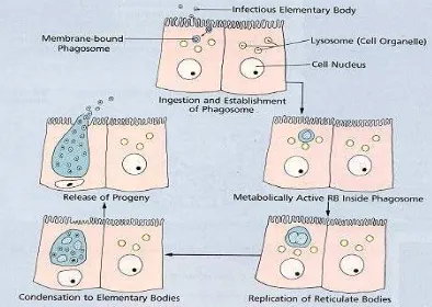
 2.2.1 Sejarah ... 6

2.2.2. Morfologi... 7

2.2.3. Epidemiologi... 8

2.2.4. Siklus Hidup... 9

2.2.5. Patogenesis... 11

2.2.6. Manifestasi Klinis ... 13

2.2.7. Penatalaksanaan ... 13

2.2.8. Faktor resiko ... 15

(11)

2.2.10. Prognosis... 16

2.3. Tes Diagnostik 2.3.1. Kultur ... 18

2.3.2. Pengujian Sitologi Langsung danEIA ... 19

2.3.3. Pendeteksian Asam Nukleat... 20

2.3.4. Serologi ... 21

2.4. Hubungan Chlamydia dengan Infertilitas Tuba ... 22

2.5. Pemeriksaan Patensi Tuba 2.5.1. Histerosalpingography... 28

2.5.2. Laparoskopi ... 33

2.5.3. Hysterosalpingocontrastsonography (HyCoSy) ... 37

BAB III METODE PENELITIAN 3.1. Desain Penelitian... 41

3.2. Tempat Dan Waktu Penelitian... 41

3.3. Sampel Dan Besar Sampel Penelitian... 41

3.4. Cara Kerja... 44

3.5. Alur Penelitian ... 45

3.6. Kerangka Konsep... 45

3.7. Batasan Operasional... ... 46

3.8. Pengolahan Data ... 46

BAB IV HASIL DAN PEMBAHASAN 
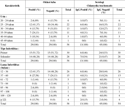
4.1. Karakteristik Peserta Penelitian ... 47

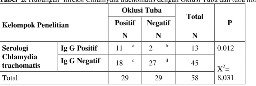
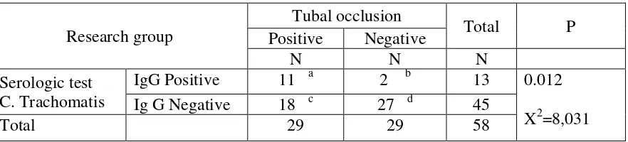
4.2. Hubungan Chlamydia trachomatis dengan Kelainan (Oklusi) Tuba ... 51

(12)

BAB V KESIMPULAN DAN SARAN

5.1. Kesimpulan ... ... 55

5.2. Saran ... 55

(13)

DAFTAR SINGKATAN

ATP : Adenosine Triphosphate.

CDC : Centers for Disease Control and Prevention.

DFA : Direct Fluorescent Antibody.

DNA : Deoxyribonucleic Acid.

EB : Elementary Body.

EIA : Enzyme Immuno Assay.

ELISA : Enzyme-Linked Immunosorbent Assay.

hCG : Human Chorionic Gonadotropin.

HSG : Hysterosalpingography.

Hycosy : Hysterosalpingo Contrast Sonography.

IgG : Immunoglobulin G.

IVF : Invitro Fertilization.

MOMP : Major Outer Membrane Protein.

NSAID : Non Steroidal Anti Inflammatory Drug.

PCR : Polymerase Chain Reaction.

PID : Pelvic Inflammatory Disease.

PRP : Penyakit Radang Panggul.

RB : Reticulate Body.

RNA : Ribonucleic Acid.

(14)

DAFTAR TABEL

Halaman

Tabel II.1. : Kategori Diagnostik pada Infertilitas... 6

Tabel II.2. : Rekomendasi Pengobatan untuk Chlamydia Trachomatis dari Centers for Disease Control and Prevention... 15

Tabel II.3. : Keuntungan dan Kerugian dari program skrining HyCoSy... 40

Tabel IV. 1. Karakteristik Peserta Penelitian Berdasarkan pada Kelainan(oklusi) Tuba dan

Serologi Chlamydia trachomatis ... 47

(15)

DAFTAR GAMBAR

Gambar II.1. : Skema Penyebab Infertilitas ... 6

Gambar II.2. : Chlamydia Trachomatis ... 8

Gambar II.3. : Siklus Hidup Chlamydia Trachomatis ... 11

Gambar II.4.: Infeksi saluran kelamin bagian atas... 24

Gambar II.5.: Akut salpingitis. Dengan gabungan neutrofil, limfosit, dan sel plasma di tuba falopi kerusakan beberapa lapisan epitel ... 26

Gambar II.6. : Salpingitis akut menunjukkan dilatasi tuba falopi... 26

Gambar II.7.: Gambar salpingitis akut pada transvaginal sonografi... 27

Gambar II.8.: hidrosalping ... 27

Gambar II.9.: Laparoskopi PID akut dan tuboovarian abses ... 27

Gambar II.10.: Laparoskopi kavum Douglas dengan pus dari PID akut ... 27

Gambar II.11. : Histerosalpingographi ... 31

Gambar II.12.: Gambaran histerosalpingogram normal.Sebuah rongga rahim halus berbentuk segitiga dan tumpahan dari kedua tuba ... 31

Gambar II.13. : Gambaran HSG normal (muncul gambaran hitam dan rongga normal dan tuba fallopi) ... ... 32

(16)

G a m b a r I I . 1 5 . :Gambaran histerosalpingogram menunjukkan adhesi intrauterin

minimal ... 3 2

Gambar II.16.: laparoskopi hidrosalping ... 36

Gambar II.17.: Septum uterus ... 37

Gambar II.18. : Investigasi untuk subfertilitas... . 39

Gambar II.19. : Prosedur operasi subfertilitas sebelum (1996) dan setelah (1997)

(diagram)... 39

Gambar II.20. : Kontras Levovist mengalir keluar melalui tuba fallopi ... 40

Gambar II.21. : Tuba fallopi normaldengan kontras Levovist dan tiga dimensi

rekonstruksi... 40

Gambar II.22. : Doppler HyCoSy tiga dimensi berwarna ... 40

(17)

The association of Chlamydia trachomatis infection in infertile women

with the occurence of tubal occlusion detected with HSG

Hubungan antara Infeksi Chlamydia trachomatis dengan oklusi tuba yang dideteksi dengan HSG pada wanita infertil

Reynanta, Binarwan Halim, M Fidel Ganis Siregar

Deparment of Obstetrics and Gynecology Medical Faculty of North Sumatera University – Haji Adam Malik Central General Hospital / Halim Fertility Centre Medan

Abstract

Objective: to assess the association between the C trachomatis infection with the tubal occlusion among the

infertile woman and to investigate the proportion of Chlamaydia trachomatis with tubal occlusion in infertile women and the proportion of Chlamaydia trachomatis with normal tube.

Material and methods: The comparation analytic study with cross sectional method was conducted during March 2010 until January 2011. Consecutive method was used tocollect the sample from any reproductive age of infertile woman who presented to the gynecology clinic at RSU H Adam Malik, RSU Putri Hijau, RSU Sundari and Halim Fertility centre Medan. Women of reproductive age refers to all women aged 15-49 years. Infertility is defined as ≥ 1 year of unprotected intercourse without pregnancy. The inclusion criteria : infertile women in reproductive age, no surgical history of ectopic tubal pregnacy, appendix, pelvic or tube. Exclusion criteria : The patient with uterin myoma , endometriosis cyst, abnormal uterus. This research had get approval from the Health Research Ethical Committee of North Sumatera. Tubal patency of the woman was determined with HSG. Patients were devided into two groups, normal tube and tubal occlusion.

Laboratory test for serum IgG antibody to C trachomatis were done with ELISA method at Laboratory Thamrin Medan. Serum IgG positive(+) indicating chlamydia’s infection. Statistical analysis with Chi-Square test with CI 95% or (p<0.05) and OR evaluation.

Results :Statistically, Chlamydia trachomatic antibody were significantly higher in the group of tubal occlusion than those women with normal tube. There was significant association statistically between the C trachomatis infection and tubal occlusion among the infertile women (p<0.05).The proportion of Chlamydia infections from patients with tubal occlusion were 11 of 29 (0.38) that more common than Chlamydia infection in the group of normal tube, 2 out of 29 (0.07).The infertile woman with positive serologic test IgG anti C trachomatis lead to the occurence of tubal occlusion, that were 11/13 cases (85%) while the infertile women with negative result of serologic test, most of them would not lead to the occurence of tubal occlusion, that were 27/45 cases (60%). The Odd Ratio (OR)value was 8.25. OR value > 1 indicated that C trachomatis infection can be used as a predictor for tubal abnormality.

Conclusion: There was significant association statistically between the C trachomatis infection and tubal

occlusion among the infertile women (p<0.05).The risk of 8.25 times for C trachomatis infection higher than those without infection.Chlamydia serology is an inexpensive and non invasive test, as a screening test before using HSG for the likelihood of tubal occlusion in infertile woman and may facilitate decisions on which women should proceed with further investigations without delay.

(18)

Abstrak

Tujuan :Melihat apakah ada hubungan infeksi Chlamydia trachomatis pada wanita infertil dengan terjadinya

oklusi tuba. Melihat proporsi chlamydia trachomatis pada wanita infertil dengan oklusi tuba dan proporsi chlamydia trachomatis pada wanita infertil dengan tuba normal.

Bahan dan cara kerja :Penelitian studi analitik komparatif, potong lintangdilaksanakan sejak Maret 2010

sampai dengan Januari 2011. Pengambilan sampel secara konsekutif dengan subjek penelitian wanita usia reproduksi infertil, yang berobat ke RSUP H Adam Malik, RSU Haji, RSU Putri Hijau, RSU Sundari dan Klinik Halim Fertility Centre, Medan. Wanita usia reproduksi adalah wanita pada umur 15-49 tahun. Infertil adalah wanita yang dalam satu tahun atau lebih telah melakukan hubungan seksual tanpa alat pelindung namun tidak mendapat kehamilan. Kriteria inklusi: wanita infertil dalam masa usia reproduksi, tidak ada riwayat operasi kehamilan ekstra tuba, appendix, pelvis atau tuba. Kriteria eksklusi : Pasien dengan mioma uteri, kista endometriosis, kelainan uterus. Penelitian telah mendapat persetujuan dari Komite etik penelitian kesehatan Sumatera Utara.Pemeriksaan histerosalpingografi dilakukan untuk melihat keadaan tuba pada sampel penelitian dan dibagi atas kelompok tuba normal dan tidak normal.Pemeriksaan laboratorium dilakukan pada kedua kelompok dengan memeriksa antibodi dalam serum IgG anti Chlamydia trachomatis antibody secara ELISA, di laboratorium klinik Thamrin Medan.IgG(+) menunjukkan adanya infeksi C trachomatis. Analisa statistik memakai Chi-Square dengan tingkat kepercayaan 95% dan batas kemaknaan dengan p< 0,05 serta penentuan rasio Odds.

Hasil :Secara statistik, antibodi chlamydia trachomatis lebih tinggi secara signifikan pada grup dengan

oklusi tuba dibandingkan dengan tuba normal. Didapati hubungan bermakna antara infeksi Chlamydia trachomatis dengan terjadinya oklusi tuba (p<0,05).Proporsi infeksi Chlamydia trachomatis pada kelompok oklusi tuba adalah 11/29 (0,38) dibanding pada kelompok tuba normal 2/29 (0,07). Wanita infertil dengan serologi IgG positif umumnya mengalami oklusi tuba (85%), sebaliknya wanita infertil dengan serologi IgG negative sebagian besar tidak mengalami oklusi tuba (60%). OR= 8,25. Dengan nilai OR>1, maka infeksi C.Trachomatis dapat dijadikan sebagai predictor untuk menilai oklusi tuba.

Kesimpulan :Didapati hubungan bermakna antara infeksi Chlamydia trachomatis dengan terjadinya oklusi

tuba (p<0,05).Resiko seorang ibu yang menderita infeksi Chlamydia trachomatis untuk mengalami oklusi tuba adalah 8,25 kali lebih besar dibandingkan dengan ibu yang tidak terinfeksi Chlamydia trachomatis. Pemeriksaan serologi chlamydia trachomatis tidak mahal dan tes yang tidak invasif, sebagai tes skrining sebelum pemeriksaan HSG untuk melihat oklusi tuba pada wanita infertil untuk mendukung tindakan selanjutnya.

(19)

Introduction

Infertility is a major problem, that influences 1 of 10 couples. Infertility affects about 10% to 15% of

reproductiveage couples in the United States. Despite an increased awareness of available therapies,

only 43% of infertile couples seek treatment, and only 24% seek specialized care.In the general

population, conception is expected to occurin 84% of women within 12 months and in 92% within

24months (te velde et al. 2000).Data from population-basedstudies suggest that 10–15% of couples in the westernworld experience infertility (templeton et al. 1990; evers,2003). Half of them (8%) will subsequently conceive withoutthe need for specialist advice and treatment. Of theremaining 8% who

require input from fertility clinics, half(4%) comprise couples with primary infertility (no historyof a

previous pregnancy) while the other half have secondaryinfertility (difficulties in conceiving after an

initialpregnancy). 1-3

Women of reproductive age refers to all women aged 15-49 years. Infertility is defined as 1 year of

unprotected intercourse without pregnancy. This condition may be further classified as primary

infertility, in which no previous pregnacies have occured, and secondary infertility, in which a prior

pregnancy, although not necessarily a live birth has occured. The main causes of infertility include

male factor, decreased ovarian reserve, ovulatory disorders, tubal injury , uterine factors, cervical and

immunologic factors and unexplained factors. Chlamydia trachomatis is the most common bacterial

sexually transmitted infection worldwide, especially among young adults. Chlamydia infections

remain often underdiagnosed, as they are asymptomatic in the majority of patients. Undiagnosed and

untreated chlamydia infections can ascend to the upper genital tract, where they colonize the

endometrial mucosa and the fallopian tubes, leading to pelvic inflammatory disease (PID).

Chlamydial PID can cause tubal occlusion and subsequent infertility. However, most women who

have tubal infertility have never been diagnosed with C.trachomatis PID because their infections

have been asymptomatic or subclinical. The United States Centres for Disease Control and

Prevention recommend annual screening of all sexually active women aged 25 or less, as is screening

of older women with risk factors (for example, those who have a new sexual partner or multiple sex

partners).

Chlamydia trachomatis is a small Gram-negative bacterium that is an obligate Intracellular parasite.

Seroepidemiological studies have indicated that chlamydia infections account for a large proportion

of asymptomatic genital tract infections by demonstrating a strong link between tubal abnormality

and the presence of chlamydia antibodies. Thus, Chlamydia IgG antibodies are associated with the

(20)

previous chlamydia infection. In chronically infected patients negative for endocervical

c.trachomatis, a positive serological test may be the only indication of chlamydia involvement. 25-36

The assessment on the tubal occlusion to the infertile woman can be implemented by any method

such as HSG, Hysterosalpingocontrastsonography(HyCoSy) or laparoscopic examination.

Laparoscopy with tubal patency testing remains the most accurate method of diagnosing

tuboperitoneal pathology. Laparoscopy is an expensive method and invasive to the patient.HSG is

used for screening purposes. Patients tend to experience this procedure as a painful and annoying

test. Furthermore, HSG has an infection-risk of 1-3 %. The Chlamydia trachomatis antibody test

(CAT), on the other hand, is a simple blood test,quick, suitable for screening, sensitivity is 91.1%,

specificity 98.5%, minimum invasive and and causes little inconvenience for patients. We

investigated the value of the C trachomatis test in screening for tubal factor infertility.

Materials and methods

37-52

In the study, we evaluated whether prior exposure to c.trachomatis, as determined by IgG chlamydia

antibody, was associated with tubal infertility.

This study aimed to assessed the association of c.trachomatis infection with tubal occlusion, and to

assessed the proportion of Chlamydia trachomatis infection in infertile woman among tubal

occlusion compared with infertile woman with normal tube.

This study is a comparison analytic study conducted at Adam Malik hospital, Haji hospital, Putri

Hijau hospital, Sundari hospital and Halim fertility centre Medan during March 2010 until January

2011. This research had get approval from the Health Research Ethical Committee of North

Sumatera. Consecutive method was used for collecting sample. This study is a comparison analytic

study between infertile case with tubal occlusion and normal tube to the infection of Chlamaydia

trachomatis, by the cross sectional study approach. In this study the infertile women’s age range

from 20 to 49 years. The informed consent was performed as a protocol. The patient give a written

permit.Patient with infertility problem who undertake the infertility treatment as a subject of this

research.The inclusion criteria : reproductive age of infertile woman with menstrual cycle (not

amenore) for one year that do sexual activity without having the protection of birth control method,

no surgical history of ectopic tubal pregnacy, appendix, pelvic or tube. The patient with uterinfibroid,

endometriosis cyst, abnormal uterus were not involved in this research. Tubal occlusion of the

infertile woman were determined with HSG. Patients were devided into two groups, i.e. tubal

occlusion and normal tube. After the examination of HSG, blood test were done. The venous blood

(21)

laboratory, the specific serum antibody was assessed to the Chlamydia trachomatis by Enzyme

Linked Immunosorbent Assay (ELISA),and then the sample is classified into 2 group, i.e.

seropositive group of Chalmydia trachomatis and group of seronegative Chlamydia trachomatis.

Statistical analysis was performed using the Statistical Package for Social Sciences (SPSS) software

version 17.0 for Windows. Association between the C trachomatis infection with tubal occlusion

among the infertile women were made by using the chi-square test.

A p value of less than 0.05 was considered statistically significant.

Results

Table 1. Baseline Characteristic

Characteristic Tubal occlusion Serology Ctrachomatis.

Positive (%) Negative (%) Total IgG (+) (%) IgG (-) (%) Total

A total 58 infertile women who underwent HSG investigation for infertility were identified. The

women’s age ranged from 20 to 49 years. The number of cases with tubal occlusion (positive) was

found more common in the group of age 25 – 29 yearsold, that was 12of 29 and the number of cases

(22)

29.The chlamydia antibody were more common found in women aged 25-29 years in this study. In

the group of age 25 – 29 years, 4 of 12 tubal occlusion cases, were positive serologic test, mean

while 2 of 10 cases with normal tube were positive serologic test.

In the group of primary infertile women, 6 of 15 tubal occlusion were positive serologic test for C

trachomatis, meanwhile there was no serologic positive from normal tube cases. In the group of

secondary infertile women 5 of 14 tubal occlusion cases were positive serologic test, meanwhile 2 of

14 normal tube were positive serologic test.

From the duration of infertility, the chlamydia antibody were more common found in 12 – 36 months

in this study. In the group of 12 to 36 months 4 of 12 tubal occlusion were positive serologic test,

meanwhile 2 of 14 normal tube were positive serologic test.

Table.2.The comparison of Chlamydia trachomatis infectionamong patients with tubal occlusion and

normal tube.

Our study showed that the proportion of serologic test for Chlamydia trachomatis infection among

the group of tubal occlusion were 11/29 (0.38) compared with the group of normal tube were 2/29

(0.07).

The table showed the infertile woman with positive serologic test IgG anti C trachomatis lead to the

occurence of tubal occlusion, that were 11/13 cases (85%) while the infertile women with negative

result of serologic test, most of them would not lead to the occurence of tubal occlusion, that were

27/45 cases (60%). Statistically, Chlamydia trachomatic antibody were significantly higher in the

group of tubal occlusion than those women with normal tube. There was significant association

statistically between the C trachomatis infection and tubal occlusion among the infertile women

(p<0.05).

The Odd Ratio (OR)value was 8.25. It means that a risk for a woman withthe infection of

Chlamydia trachomatis to the occurence of tubal occlusion was 8.25 times higher than women

without the infection. OR value > 1 indicated that C trachomatis infection can be used as a predictor

(23)

Discussion

The chlamydia antibody were more common found in women aged 25-29 years in this study. The

studies was similar to studies performed in Women’s Hospital in Iran. No significant correlation was

found between the participants’ age and the results of Chlamydia test in health care centers in Iran.

The prevalence was slightly higher in patients within the age group 25-29 (17,68%) than in the age

group 20-24 (15,24%).

In this study, the chlamydia antibody were more common found in those women who had conceived

previously as compared with primary infertile women. Women with primary infertility had a

statistically insignificant reduction in prevalence of antibody to chlamydia compared with women

who had secondary infertility in the Reproductive Medicine Clinic in Bristol, UK, the infertility

clinic in Iran, gynaecology clinic in Gambia. Secondary infertility may be a sequela of postpartum

sepsis, which is commonly observed at hospitals and clinics in the Gambia and which, given the

association between cervical chamydial infection and endometritis, may be due in part to

chlamydiae.

53,54,55,56,57

53,58,61

From the duration of infertility, the chlamydia antibody were more common found in 12 – 36 months

in this study. The studies was similar to studies performed in The Netherlands. There was not a

significant relation between the Chlamydia antibody and duration of infertility in infertility clinic in

Iran.53,60

The infection of Chlamydia trachomatic were significantly higher in the group of tubal occlusion

than those women with normal tube in this study. The results indicate that infertil Gambian woman

with tubal occlusion have a higher prevalence and higher levels of circulating IgG antibody against

C. Trachomatis than controls and that these differences are statistically highly significant. Tubal

occlusion was associated with chlamydia trachomatis antibody, which is in agreement with Tiitinen

et al (2006) that C.trachomatis-specific IgG antibodies were found more frequently (43,2 versus

13,5%), and the antibody levels were higher in the tubal factor infertility cases than in the controls

(p<0,001). The studies was similar to studies performed in Bristol, UK, clinic specialized in Brazil,

infertility clinic in Cairo University and clinic in The Netherlands.

Chlamydia serology is an inexpensive and non invasive test, as a screening test before using HSG

for the likelihood of tubal occlusion in infertile woman and may facilitate decisions on which women

should proceed with further investigations without delay.

58,59, 61,62, 63, 64

The limitation in this study concerns its inability to distinguish between various micro-organisms

other than C.trachomatis, for example : N. gonore, haemophilusinfluenzae, grup A streptococci,

(24)

Pendahuluan

Infertilitas merupakan suatu masalah umum, mempengaruhi sekitar 1 dari 10 pasangan. Infertilitas

mempengaruhi sekitar 10% sampai 15% dari pasangan usia reproduktif di Amerika Serikat.

Walaupun terdapat peningkatan pengetahuan mengenai infertilitas, hanya 43% pasangan infertil

mencari terapi, dan hanya 24% yang mencari terapi spesialistik. te Velde et al. (2000)dari kepustakaan 3, mendapatkan, pada populasi umum, konsepsi diperkirakan terjadi pada 84% wanita dalam 12 bulan

dan pada 92% dalam 24 bulan. Templeton et al. 1990; Evers (2003) dari kepustakaan 3mendapatkan data dari studi berbasis-populasi menunjukkan bahwa 10-15% pasangan di Barat mengalami infertilitas.

Setengah di antaranya (8%) selanjutnya akan mencapai konsepsi tanpa membutuhkan nasehat dan

pengobatan spesialis. Dari 8% lainnya yang membutuhkan penanganan dari klinik fertilitas,

setengahnya (4%) terdiri dari pasangan dengan infertilitas primer (tidak ada riwayat kehamilan

sebelumnya) sementara setengah lainnya mengalami infertilitas sekunder (kesulitan dalam mencapai

konsepsi setelah kehamilan awal).1-3

Wanita usia reproduktif mengacu pada semua wanita usia 15–49tahun. Infertilitas secara umum

didefenisikan sebagai tidak terjadinya kehamilan dalam setahun hubungan seksual tanpa proteksi.

Infertilitas diklasifikasikan menjadi infertilitas primer (tidak pernah hamil) dan infertilitas sekunder

(pernah hamil, walau kehamilan tidak harus dengan kelahiran hidup).1, 21

Faktor penyebab infertilitas dapat digolongkan menjadi : Faktor pria, Penurunan cadangan ovari, gangguan ovulasi (faktor ovulasi), faktor tuba, Faktor servikal, Faktor uterus, Kondisi seperti penyimpangan imunologis, Faktor yang tak dapat dijelaskan. Chlamydia trachomatis adalah

bakteri transmisi seksual yang paling umum dijumpai diseluruh dunia, khususnya orang

dewasa muda. Infeksi chlamydia sering tidak terdiagnosis, sebagaimana asimptomatik pada

kebanyakan pasien. Sekali bakteri pathogen seperti chlamydia trachomatis memasuki serviks dan

infeksi tidak ditangani, dapat menyebar ke uterus dan tuba fallopii dan akan menyebabkan penyakit

radang panggul. PID Chlamydia dapat menyebabkan oklusi tuba dan menyebabkan infertilitas.

Bagaimanapun, kebanyakan wanita yang mempunyai infertilitas tuba jarang terdiagnosa PID

C.trachomatis karena infeksinya asimptomatik atau subclinical. Pusat Pengendalian dan Pencegahan

Penyakit merekomendasikan screening gadis remaja yang aktif secara seksual pada pemeriksaan

ginekologik tahunan rutin usia 25 atau lebih muda, atau yang lebih tua dengan faktor resiko (untuk

contoh, yang mempunyai pasangan seksual baru atau banyak mitra seksual). 4-24

Chlamydia adalah mikroorganisma intrasel parasit yang mempunyai dinding sel yang serupa dengan

dinding sel bakteri gram-negatip. Studi seroepidemiologi menunjukkan bahwa infeksi Chlamydia

(25)

hubungan yang kuat antara tuba yang abnormal dan keberadaan antibodi Chlamydia. Oleh karena

itu, antibodi Chlamydia IgG dihubungkan dengan sekuel dan marker untuk keterpaparan

sebelumnya atau reaktivasi endogen dari infeksi Chlamydia sebelumnya. Pada pasien infeksi kronik

yang negatif untuk c.trachomatis endocervik, test serologi positif boleh menjadi satu-satunya

indikasi keterlibatan chlamydia. 25-36

Mengevaluasi patensi tuba pada wanita infertil diterapkan melalui metode pemeriksaan HSG,

Hysterosalpingocontrastsonography(HyCoSy) dan laparoskopi. Laparoskopi dengan tes patensi tuba

metode yang paling akurat untuk diagnosis patologi tuboperitoneal. Laparoskopi adalah metode yang

invasif dan mahal untuk pasien. HSG digunakan untuk tujuan penyaringan. Pasien cenderung

mengalami nyeri dan tidak mengenakkan pada prosedur tes ini. Lagipula, HSG mempunyai risiko

infeksi 1-3%. Tes antibodi Chlamydia trachomatis, disisi lain, adalah sebuah tes darah sederhana,

cepat, cocok untuk skrining, sensitivitas 91,1%, spesifisitas 98,5%, minimal invasif, dan

menyebabkan sedikit tidak menyenangkan untuk pasien. Kita menyelidiki nilai tes chlamydia

trachomatis dalam skrining untuk infertilitas faktor tuba. 37-52

Studi ini merupakan studi analitik komparatif yang dilaksanakan pada RS Adam Malik, RS Haji, RS

Putri Hijau, RS Sundari dan Halim Fertility Center Medan selama Maret 2010 sampai Januari 2011.

Penelitian ini mendapatkan persetujuan dari Health Research Ethical Committee Sumatera Utara.

Metode Consecutive digunakan dalam pengambilan sampel. Studi ini membandingkan wanita infertil

antara kasus infertil dengan tuba abnormal(oklusi) dan tuba normal dihubungkan dengan infeksi

Chlamydia trachomatis, dengan cara studi cross sectional. Pada studi ini usia wanita infertil 20-49

tahun. Inform konsen dilakukan sebagai protokol. Pasien menandatangani surat izin penelitian.

Pasien dengan masalah infertil yang ingin berobat masalah infertil dimasukkan sebagai subjek

penelitian. Kriteria inklusi : wanita usia reproduksi infertil yang tidak amenore untuk setahun

hubungan seksual tanpa proteksi dari metode pembatasan kelahiran, tidak ada riwayat operasi

kehamilan ektopik, apendik, pelvis atau tuba. Pasien dengan mioma uteri, kista endometriosis, uterus

abnormal tidak dimasukkan dalam penelitian ini. Tuba yang oklusi dari wanita infertil diperiksa

Pada studi ini, kita mengevaluasi apakah keterpaparan Chlamydia trachomatis, sebagaimana

ditentukan dengan antibodi IgG Chlamydia, dihubungkan dengan infertilitas tuba.

Studi ini bertujuan untuk menilai hubungan infeksi C.trachomatis dengan tuba yang

abnormal(oklusi), dan untuk menilai proporsi infeksi chlamydia trachomatis pada wanita infertil

dengan tuba abnormal(oklusi) dibandingkan dengan wanita infertil tuba normal.

(26)

dengan HSG. Pasien dibagi menjadi dua grup; tuba oklusi dan tuba normal. Setelah pemeriksaan

HSG, tes darah dilakukan. Sampel darah vena, 3 ml diperoleh dari pasien dan dikirim ke

laboratorium untuk diperiksa. Serum antibodi spesifik Chlamydia trachomatis oleh Enzyme Linked

Immunosorbent Assay (ELISA), dan kemudian sampel diklasifikasi dalam 2 grup, seropositif

Chlamydia trachomatis dan seronegatif Chlamydia trachomatis.

Analisa statistik menggunakan SPSS versi 17 Windows. Hubungan antara infeksi C.trachomatis

dengan oklusi tuba pada wanita infertil dengan menggunakan tes chi-square.

Nilai p < 0,05 dianggap signifikan secara statistik.

Hasil

Tabel 1. Karakteristik Peserta Penelitian

(27)

58 wanita infertil yang dilakukan HSG untuk infertilitas diidentifikasi. Wanita berusia 20-49 tahun.

Jumlah oklusi tuba lebih banyak ditemukan pada usia 25-29 tahun, dimana 12 dari 29 kasus dan

jumlah tuba normal lebih banyak dijumpai pada usia 25-29 tahun, dimana 10 dari 29 kasus. Antibodi

Chlamydia dijumpai lebih banyak pada wanita usia 25-29 tahun. Pada grup usia 25-29 tahun, 4 dari

12 kasus oklusi tuba, ditemukan positif tes serologi, sementara 2 dari 10 kasus dengan tuba normal

ditemukan positif tes serologi.

Pada grup infertil primer, 6 dari 15 oklusi tuba ditemukan positif tes Chlamydia trachomatis,

sementara tidak ada serologi positif dari tuba normal. Pada grup dengan infertil sekunder, 5 dari 14

tuba yang oklusi ditemukan positif tes serologi, sementara 2 dari 14 tuba normal ditemukan positif

tes serologi.

Dari durasi infertil, antibodi chlamydia ditemukan lebih banyak pada lama infertil 12-36 bulan pada

studi ini. Pada grup lama infertil 12-36 bulan, 4 dari 12 oklusi tuba ditemukan positif tes serologi,

sementara 2 dari 14 tuba normal ditemukan positif tes serologi Chlamydia.

Tabel 2. Hubungan Infeksi Chlamydia trachomatis dengan Oklusi Tuba dan tuba normal

Kelompok Penelitian

Dari tabel diatas juga dapat dihitung bahwa proporsi infeksi Chlamydia trachomatis pada oklusi tuba

dalam penelitian ini adalah 11/29 (38%) lebih tinggi dari proporsi infeksi Chlamydia trachomatis

pada tuba normal yaitu 2/29 (7%).

Dari tabel diatas dapat diketahui bahwa wanita infertil dengan serologi IgG positif umumnya

mengalami oklusituba, dengan didapati 11/13 (85%), sebaliknya wanita infertil dengan serologi IgG

negative sebagian besar tidak mengalami oklusi tuba, 27/45 (60%). Dari uji statistic, antibodi

Chlamydia dengan signifikan lebih tinggi pada grup yang oklusi tuba dibandingkan dengan tuba

normal. Adanya hubungan yang bermakna secara antara infeksi Chlamydia trachomatis dengan

terjadinya oklusituba pada wanita infertil (p<0,05).

Didapatkan nilai OR adalah 8,25. Hal ini berarti resiko seorang ibu yang menderita infeksi

(28)

dengan ibu yang tidak terinfeksi Chlamydia trachomatis. Dengan nilai OR>1, maka infeksi

C.Trachomatis dapat dijadikan sebagai predictor untuk menilai kelainan tuba.

Diskusi

Antibodi Chlamydia ditemukan lebih banyak pada usia 25-29 tahun pada penelitian ini. Penelitian ini

serupa dengan penelitian yang dilaksanakan di Women’s Hospital di Iran. Tidak ada hubungan

signifikan ditemukan antara usia dan hasil tes Chlamydia pada Health Care Centers di Iran.

Prevalensi ditemukan lebih tinggi pada usia 25-29(17,68%) daripada grup usia

20-24(15,24%).53,54,55,56,57

Pada studi ini, antibodi chlamydia ditemukan lebih banyak pada wanita yang pernah hamil

dibandingkan dengan wanita infertil primer. Wanita dengan infertil primer secara signifikan

berkurang dalam prevalensi antibodi Chlamydia dibandingkan dengan wanita dengan infertil

sekunder pada Reproductive Medicine Clinic in Bristol,UK, Klinik Infertilitas di Iran, Klinik

Ginekologi di Gambia. Infertil sekunder boleh disebabkan sekuel sepsis post partum, yang mana

umum dijumpai pada Rumah Sakit dan Klinik di Gambia dan yang mana, memberikan hubungan

antara infeksi Chlamydia cervix dan endometritis, merupakan bagian dari Chlamydia.

53,58,61

Dari durasi infertil, antibodi Chlamydia ditemukan lebih banyak pada 12-36 bulan pada penelitian

ini. Penelitian ini serupa dengan penelitian di Belanda. Tidak ada hubungan signifikan diantara

antibodi Chlamydia dengan lamanya infertil pada Klinik Infertil di Iran.

53,60

Infeksi Chlamydia trachomatis ditemukan lebih tinggi pada grup dengan oklusi tuba dibandingkan

wanita dengan tuba normal pada penelitian ini. Hasil menunjukkan bahwa wanita infertil di Gambia

dengan oklusi tuba lebih tinggi prevalensinya dan level lebih tinggi sirkulasi antibodi IgG terhadap

Chlamydia trachomatis dibandingkan dengan kontrol dan perbedaan ini lebih tinggi signifikan secara

statistik. Hubungan tuba oklusi dengan antibodi Chlamydia trachomatis, sesuai dengan Tiitinen

dkk(2006) bahwa antibodi IgG spesifik Chlamydia trachomatis lebih banyak ditemukan(43,2 versus

13,5%), dan level antibodi lebih tinggi pada oklusi tuba dibandingkan kontrol (p<0,001). Ini sesuai

dengan studi yang dilaksanakan di Bristol, UK, klinik spesialis di Brazil, Klinik Infertil di

Universitas Kairo dan klinik di Belanda. 58,59, 61,62, 63, 64

Keterbatasan dalam penelitian ini adalah tidak dilakukannya pemeriksaan kemungkinan infeksi lain

selain infeksi chlamydia trachomatis untuk contoh : N.gonore Haemophilus influenzae, group A

(29)

Tes Serologi Chlamydia tidak mahal dan tidak invasif, sebagai skrining sebelum menggunakan HSG

untuk kemungkinan oklusi tuba pada wanita infertil dan bisa memudahkan keputusan untuk

pemeriksaan selanjutnya pada wanita dengan segera.

Reference

1. Burney RO, Schust DJ, Yao MWM. Infertility. In: Berek, Jonatan S, eds. Berek and Novak’s Gynecology. 14th

ed. Lippincott Williams & Wilkins. 2007. p. 1187-1276.

2. Jelovsek FR. Severe pelvic infection and infertility. Women’s health resource [Online]. 2008 Jun 16 [cited 2009

Oct 14]; Available from: URL

3. Bhattacharya S. Infertility. In: Edmonds DK, ed. Dewhurst’s textbook of obstetrics & gynaecology. 7th ed.

Blackwell Publishing London. 2007. p. 440-60.

4. Westhoff CL. Chlamydia screening and fertility prevention. Preserved at the National STD Prevention

Conference 2002 Mar 6: 120-30.

5. Irene Su H, Sondheimer SJ. The infertile couple. In: Pfeifer, Samantha M, eds. NMS obstetrics and gynecology.

6th ed. Lippincott Williams & Wilkins. 2008. p. 295-305.

6. Garcia JE. Chlamydia and tubal factor infertility [Online]. 2004 Oct [cited 2009 Jun 6];Availablefrom:

URL

7. Vorvick LJ. Chlamydia Trachomatis infection. Medline Plus USA [Online]. 2008 Jan 14 [cited 2009 Sept 9];

Available from: URL

8. Black CM. Current methods of laboratory diagnosis of Chlamydia Trachomatis infection. Clin. Microbiol. Rev.

1997 Jan; 10(1) : 160-84.

9. Smith RP. Sexually transmissible infection in obstetrics and gynecology principles for practise. Duff P

International Ed; 2001 p. 928-31.

10. Centers for Disease Control and Prevention. Chlamydia, CDC Fact Sheet, STD, Division of Sexually

Transmitted Disease [Online]. 2009 Aug 4 [cited 2010 Jan 20]; [2 screens]. Available from:

URL

11. Low N. Effectiveness of chlamydia screening: systematic review.

12. Aswad SG, Badrinat6h P, Osman N, Khaliq SA, Mc Ilvenny S, Sidky S. Prevalence of Chlamydia Trachomatis

infection among women in Middle Eastern community. BMC Woman’s Health, 2004, 4(3): 1-6.

13. Houry DE. Chlamydia in emergency medicine, Medscape reference [Online]. 2004 Sept 6 [cited 2009 Feb 14];

Available from: URL:http://www.emedicine.com/emerg/topic925.htm. Sept 2004

14. Aldeen T, Haghdoost A, Hay P. Urine based screening for asymptomatic/undiagnosed genital chlamydia

infection in young people visiting the accident and emergency department is feasible, acceptable, and can be

epidemiologically helpful. Sex.Transm. Infect. 2003; 79 : 229-33.

15. Chan PD, Johnson SM. Current clinical strategies gynecology and obstetrics: pelvic inflammatory disease.

(30)

16. Cohen CR, Sinei S, Bukusi EA, Bwayo JJ, Holmes KK, Brunham RC. Human leukocyte antigen class II DQ

alleles associated with Chlamydia trachomatis tubal infertility.Obstet Gynecol. January 2000; 95(1) :72-7.

17. Pernoll ML. Infertility and related issues. In: Pernoll ML, ed. Benson & Pernoll’s handbook of obstetric &

gynecology. 10th ed. New York: McGraw-Hill Companies; 2001. p.769-99.

18. Tanner E. Infections of the genital tract. In: Fortner KB, Szymanski LM, Fox HE, Wallach EE, eds. The John

Hopkins Manual of Gynecology and Obstetrics. 3rd ed. Lippincott Williams & Wilkins. 2007. p. 289-302.

19. World Health Organization. Reproductive health indicators: Reproductive Health indicators. Guidelines for their

generation, interpretation and analysis for global monitoring. Prevalence of infertility in women. WHO Library

Cataloguing-in-Publication Data; 2006. p. 49-50.

20. Speroff L, FritZ MA. Female infertility. In: Speroff Speroff L, Fritz MA, eds. Clinical gynecologyc

endocrinology & infertility. 7th ed. Lippincott Williams & Wilkins. 2005. p.1012-67.

21. Meszaros G, Sanders S. Endocrine and reproductive systems. Elsevier Inc 2007; 92 (1): 53-6.

22. Smith RP, Netter FH. Netter’s obstetrics, gynecology and women’s health. New Jersey: Icon Learning

System; 2002. p. 367-69.

23. Cahill D. Infertility. In: Baguley S, Kumar S, Persad R, eds. Key topics in sexual health. Taylor & Francis

Group, London and New York. 2006. p.146-50.

24. DeCherney AH, Nathan L. Current diagnosis & treatment obstetrics & gynecology. 10th ed. USA: McGraw-Hill

Companies; 2007. p. 759-61.

25. Brooks GF, Butel JS, Morse SA. Medical microbiology. 22nd ed. Buku 1. McGraw-Hill; 2005.

26. Advances in the diagnosis of sexually transmitted diseases caused by bacteria [Online]. 2008 Jan [cited 2009

Oct 10]; Available from: URL:http://www.roche.com/pages/facets/1/chlamyde.htm

27. NCQA Publication. Chlamydia screening in women: the state of managed care quality [Online] 2001 Oct 11

[cited 2009 Jul 23]: Available from: URL

28. Prodigy. Chlamydia-uncomplicated, genital (women): clinical knowledge summaries [Online]. 2007 March 1

[cited 2009 Aug 2]; Available from: URL:http://www.prodigy.nhs.uk

29. Pattman R, Snow, M, Handy P, Sankar KN, Elawad B. Oxford handbook of genitourinary medicine, HIV, and

AIDS. Chlamydia Trachomatis infections. 1st ed. Oxford University Press; 2005; p124-33.

30. Mumtaz, Mellars BJ, Ridgway GL, Oriel D. Enzyme immunoassay for detection of Chlamydia Trachomatis

antigen in urethral and endocervical swabs. J Clin Pathol 1985; 38: 740-42.

31. Brooks GF, Butel JS, Morse SA. Chlamydia. In: Jawetz, Melnick and Adelberg’s Medical Microbiology, 23 rd

ed. McGraw-Hill Co, Inc. 2004. Alih bahasa Hartanto H, Rachman C, Dimanti A, Diani A. Penerbit EGC

Jakarta. Cet. I: 2008. h. 366-76.

32. Porras. Epidemiology of genital Chlamydia Trachomatis infection among young women in Costa Rica. Journal

of The Sexually Transmitted Disease 2008 May; 35(5): 461-8.

33. Screening Chlamydia infection. US Preventive Services Task Forces [Onliine]. 2005 May [cited 2009 Jun 18]

Available from: URL:http://www.ahrg. gov/clinic/ uspstf/ uspschlm. htm

34. Magic D. Doxyxicline compared with Azithromycin for treating women with genital Chlamydia Trachomatis

(31)

35. Wisconsin Division of Public Health Disease Surveillance Manual. Chlamydia [Online] 2004 Jan [cited 2009

Nov 19]; Available from: URL

36. Microwell Elisa Chlamydia Trachomatis IgG: diagnostic automation inc. 2003.

37. Murtiastutik D. Buku ajarinfeksi menular seksual: Infeksi Chlamydia. Surabaya: Airlangga University Press,

Cetakan kedua; 2008. p. 381-84

38. Barnes RC. Laboratory diagnosis of human Chlamydial infection. Clin. Microbiol. Rev. 1989 April; 2(2): 119 –

36.

39. El-Mowafi DM, Diamond MP. Fallopian tube, Geneva Foundation for Medical Education and Research

[Online]. 2008 Sep 4[cited 2009 Dec 14]; Availablefrom:URL:

International_activities_En/El_Mowafi/Fallopian_tube.htm

40. Bodurka DC, Buckner F, Cheng E, Coleman RL, Davis, AJ, Dotters DJ, et al. Comprehensive gynecology:

Chapter 41 infertility. 5th ed. China: Mosby Elsevier; 2007.

41. Parsons AK, Fleischer AC, Londono JL. Sonohysterography and sonohystero salphi ngography: A Text atlas of

normnal and abnormal findings. In: Fleischer AC, Manning FA, Jeanty P, Romero R, eds. Sonography in

obstetrics &gynecology: principle and practice. 6th ed. McGraw-Hill Companies. 2001.p. 1110-61.

42. Hart DM, Norman J, Callander R, Ramsden I. Gynaecology illustrated. Infertility. 5th ed. China: Harcourt

Publishers Limited; 2000. p. 389-409.

43. Gabbe SG, et al. Gabbe obstetric: normal and problem pregnancies.Chapter 47 Maternal and Perinatal Infection.

5th ed. Elsevier Churchill Livingstone; 2007.

44. Advanced Fertility Center of Chicago. Tubal Factor Infertility [Online]. 2008 Feb 15 [cited 2010 Feb 14];

Available from:

45. Malpani A, Malpani A. The tubal connection, how to have a baby: overcoming infertility, Malpani infertility

clinic. [Online]. 2009 May 18 [cited 2010 Feb 14]; Available from

46. Advanced Fertility Center of Chicago. Hysterosalpingogram-HSG-fertility test for tubal patency and normalcy

of uterine cavity [Online]. 2009 April 17 [cited 2010 Aug 14]; Available from:

47. YourEpharmacy. Chlamydia [Online]. 2008 Nov 12 [cited 2009 Dec 14]; Available from:URL:

http:/

48. March CM. Hysterography for Infertility. Hysteroscopy and hysterosalphingography. In : Baggish MS, Valle,

RF, Guedj H, eds. Hysteroscopy: visual perspectives ofuterineanatomy, physiology andpathology. 3rded.

Lippincott Williams & Wilkins: 2007. p. 417-50.

49. Chudleigh T, Thilaganathan B. Obstetric ultrasound: how, why and when. Ultrasound and infertility. 3rd ed.

Edinburgh:Elsevier Science Limited; 2004. p.79-94.

50. Rebar RW. Problems with the fallopian tubes. The Merck Manual of Medical Information [Online]. 2008 Oct

[cited 2009 Oct]; Available from: URL:http://www.merck.com/mmhe/sec22/ch254/ch254d.html.

51. Killick SR. Hysterosalpingo contrast sonography as a screening test fot tubal patency in infertile woman. J

(32)

52. HyCoSy. Pelvic ultrasound guided procedures, ultrasound care, Sydney Adventist Hospital [Online]. 2009 Jan

12 [cited 2010 Mar 5]; Available from:

guided-procedures-hy-co-sy.html.

53. Peivandi S, Moslemizadeh N, Gharajech S, Ajami A. The role of Chlamydia Trachomatis IgG antibody testing

in predicting tubal factor infertility in Northern Iran. International Journal of Fertility and Sterility (IJFS), 2009

Nov-Dec; 3(3): 143-8.

54. Hadi N, Barazandeh F, Azad F, et al. The prevalence of Chlamydia trachomatis infection in women attending

health clinics in Shiraz. Islamic Republic of Iran, International Journal of Fertility and Sterility 2010 Oct – Dec;

4(3) : 128-33.

55. Mawak JD, Dashe N, Agabi YA, Panshak BW. Prevalence of genital Chlamydia trachomatis infection among

gynaecologic clinic attendees in Jos. Nigeria: Shiraz E-Medical Journal 2011 April; 12(2): 100-6.

56. Yazdi JZ, Khorramizadeh MR, Badami N, Kazemi B, Aminharati F, Eftekhar Z, et al. Comparative assesment

of Chlamydia trachomatis infection in Iranian women with cervicitis : A Cross – Sectional Study. Iranian J publ

Health 2006; 35 (2): 69-75.

57. Nazer M, Nowroozi J, Mirsalehian A, Kazemi B, Mousavi T, Mehdizadeh A. Determination of asymptomatic

Chlamydia Trachomatis infections by Omp1 gene based –PCR. Yakhteh Medical Journal, Spring 2008; 10(1):

41-6.

58. Mabey DCW, Ogbaselassie G, Robertson JN, Heckels JE, Ward ME. Tubal infertility in the Gambia :

Chlamydial and Gonococcal serology in women with tubal occlusion compared with pregnant controls. Bulletin

of the World Health Organization 1985; 63(6): 1107-13.

59. Machado ACS, Guimaraes EMB, Sakurai E, Fioravante FCR, Amarai WN, Alves MFC. High titers of

Chlamydia trachomatis antibodies in Brazilian women with tubal occlusion of previous ectopic pregnancy.

Infectious diseases in obstetrics and gynecology. Hindawi Pub. Corp. 2007, Article ID 2007, 24816.:1-6.

60. Veenemans LMW, van der Linden PJQ. The value of Chlamydia trachomatis antibody testing in predicting

tubal factor infertility. Human Reproduction 2002; 17(3): 695-8.

61. Akande VA, Hunt LP, Cahill DJ, Caul EO, Ford WCL, Jenksins JM. Tubal damage in infertile women :

prediction using chlamydia serology. Hum.Reprod 2003; 18(9): 1841-7.

62. den Hartog JE, Land JA, Stassen FRM, Kessels AGH, Bruggeman CA. Serological markers of persistent C.

trachomatis infections in women with tubal factor subfertility. Hum. Reprod. 2005; 20(4): 986-90.

63. Azab HE, Hassouna AA. Clinical findings and Chlamydia antibody titre as predictors of tubal factor infertility.

The Internet Journal of Gynecology and Obstetrics TM ISSN: 1528-8439 2009;11 (2).

64. Tiitinen A, Surcel HM, Halttunen M, Birkelund S, Bloigu A, Christiansen G, et al. Chlamydia trachomatis and

Chlamydial heat shock protein 60-specific antibody and cell-mediated responses predict tubal factor infertility.

(33)

The association of Chlamydia trachomatis infection in infertile women

with the occurence of tubal occlusion detected with HSG

Hubungan antara Infeksi Chlamydia trachomatis dengan oklusi tuba yang dideteksi dengan HSG pada wanita infertil

Reynanta, Binarwan Halim, M Fidel Ganis Siregar

Deparment of Obstetrics and Gynecology Medical Faculty of North Sumatera University – Haji Adam Malik Central General Hospital / Halim Fertility Centre Medan

Abstract

Objective: to assess the association between the C trachomatis infection with the tubal occlusion among the

infertile woman and to investigate the proportion of Chlamaydia trachomatis with tubal occlusion in infertile women and the proportion of Chlamaydia trachomatis with normal tube.

Material and methods: The comparation analytic study with cross sectional method was conducted during March 2010 until January 2011. Consecutive method was used tocollect the sample from any reproductive age of infertile woman who presented to the gynecology clinic at RSU H Adam Malik, RSU Putri Hijau, RSU Sundari and Halim Fertility centre Medan. Women of reproductive age refers to all women aged 15-49 years. Infertility is defined as ≥ 1 year of unprotected intercourse without pregnancy. The inclusion criteria : infertile women in reproductive age, no surgical history of ectopic tubal pregnacy, appendix, pelvic or tube. Exclusion criteria : The patient with uterin myoma , endometriosis cyst, abnormal uterus. This research had get approval from the Health Research Ethical Committee of North Sumatera. Tubal patency of the woman was determined with HSG. Patients were devided into two groups, normal tube and tubal occlusion.

Laboratory test for serum IgG antibody to C trachomatis were done with ELISA method at Laboratory Thamrin Medan. Serum IgG positive(+) indicating chlamydia’s infection. Statistical analysis with Chi-Square test with CI 95% or (p<0.05) and OR evaluation.

Results :Statistically, Chlamydia trachomatic antibody were significantly higher in the group of tubal occlusion than those women with normal tube. There was significant association statistically between the C trachomatis infection and tubal occlusion among the infertile women (p<0.05).The proportion of Chlamydia infections from patients with tubal occlusion were 11 of 29 (0.38) that more common than Chlamydia infection in the group of normal tube, 2 out of 29 (0.07).The infertile woman with positive serologic test IgG anti C trachomatis lead to the occurence of tubal occlusion, that were 11/13 cases (85%) while the infertile women with negative result of serologic test, most of them would not lead to the occurence of tubal occlusion, that were 27/45 cases (60%). The Odd Ratio (OR)value was 8.25. OR value > 1 indicated that C trachomatis infection can be used as a predictor for tubal abnormality.

Conclusion: There was significant association statistically between the C trachomatis infection and tubal

occlusion among the infertile women (p<0.05).The risk of 8.25 times for C trachomatis infection higher than those without infection.Chlamydia serology is an inexpensive and non invasive test, as a screening test before using HSG for the likelihood of tubal occlusion in infertile woman and may facilitate decisions on which women should proceed with further investigations without delay.

(34)

Abstrak

Tujuan :Melihat apakah ada hubungan infeksi Chlamydia trachomatis pada wanita infertil dengan terjadinya

oklusi tuba. Melihat proporsi chlamydia trachomatis pada wanita infertil dengan oklusi tuba dan proporsi chlamydia trachomatis pada wanita infertil dengan tuba normal.

Bahan dan cara kerja :Penelitian studi analitik komparatif, potong lintangdilaksanakan sejak Maret 2010

sampai dengan Januari 2011. Pengambilan sampel secara konsekutif dengan subjek penelitian wanita usia reproduksi infertil, yang berobat ke RSUP H Adam Malik, RSU Haji, RSU Putri Hijau, RSU Sundari dan Klinik Halim Fertility Centre, Medan. Wanita usia reproduksi adalah wanita pada umur 15-49 tahun. Infertil adalah wanita yang dalam satu tahun atau lebih telah melakukan hubungan seksual tanpa alat pelindung namun tidak mendapat kehamilan. Kriteria inklusi: wanita infertil dalam masa usia reproduksi, tidak ada riwayat operasi kehamilan ekstra tuba, appendix, pelvis atau tuba. Kriteria eksklusi : Pasien dengan mioma uteri, kista endometriosis, kelainan uterus. Penelitian telah mendapat persetujuan dari Komite etik penelitian kesehatan Sumatera Utara.Pemeriksaan histerosalpingografi dilakukan untuk melihat keadaan tuba pada sampel penelitian dan dibagi atas kelompok tuba normal dan tidak normal.Pemeriksaan laboratorium dilakukan pada kedua kelompok dengan memeriksa antibodi dalam serum IgG anti Chlamydia trachomatis antibody secara ELISA, di laboratorium klinik Thamrin Medan.IgG(+) menunjukkan adanya infeksi C trachomatis. Analisa statistik memakai Chi-Square dengan tingkat kepercayaan 95% dan batas kemaknaan dengan p< 0,05 serta penentuan rasio Odds.

Hasil :Secara statistik, antibodi chlamydia trachomatis lebih tinggi secara signifikan pada grup dengan

oklusi tuba dibandingkan dengan tuba normal. Didapati hubungan bermakna antara infeksi Chlamydia trachomatis dengan terjadinya oklusi tuba (p<0,05).Proporsi infeksi Chlamydia trachomatis pada kelompok oklusi tuba adalah 11/29 (0,38) dibanding pada kelompok tuba normal 2/29 (0,07). Wanita infertil dengan serologi IgG positif umumnya mengalami oklusi tuba (85%), sebaliknya wanita infertil dengan serologi IgG negative sebagian besar tidak mengalami oklusi tuba (60%). OR= 8,25. Dengan nilai OR>1, maka infeksi C.Trachomatis dapat dijadikan sebagai predictor untuk menilai oklusi tuba.

Kesimpulan :Didapati hubungan bermakna antara infeksi Chlamydia trachomatis dengan terjadinya oklusi

tuba (p<0,05).Resiko seorang ibu yang menderita infeksi Chlamydia trachomatis untuk mengalami oklusi tuba adalah 8,25 kali lebih besar dibandingkan dengan ibu yang tidak terinfeksi Chlamydia trachomatis. Pemeriksaan serologi chlamydia trachomatis tidak mahal dan tes yang tidak invasif, sebagai tes skrining sebelum pemeriksaan HSG untuk melihat oklusi tuba pada wanita infertil untuk mendukung tindakan selanjutnya.

(35)

Introduction

Infertility is a major problem, that influences 1 of 10 couples. Infertility affects about 10% to 15% of

reproductiveage couples in the United States. Despite an increased awareness of available therapies,

only 43% of infertile couples seek treatment, and only 24% seek specialized care.In the general

population, conception is expected to occurin 84% of women within 12 months and in 92% within

24months (te velde et al. 2000).Data from population-basedstudies suggest that 10–15% of couples in the westernworld experience infertility (templeton et al. 1990; evers,2003). Half of them (8%) will subsequently conceive withoutthe need for specialist advice and treatment. Of theremaining 8% who

require input from fertility clinics, half(4%) comprise couples with primary infertility (no historyof a

previous pregnancy) while the other half have secondaryinfertility (difficulties in conceiving after an

initialpregnancy). 1-3

Women of reproductive age refers to all women aged 15-49 years. Infertility is defined as 1 year of

unprotected intercourse without pregnancy. This condition may be further classified as primary

infertility, in which no previous pregnacies have occured, and secondary infertility, in which a prior

pregnancy, although not necessarily a live birth has occured. The main causes of infertility include

male factor, decreased ovarian reserve, ovulatory disorders, tubal injury , uterine factors, cervical and

immunologic factors and unexplained factors. Chlamydia trachomatis is the most common bacterial

sexually transmitted infection worldwide, especially among young adults. Chlamydia infections

remain often underdiagnosed, as they are asymptomatic in the majority of patients. Undiagnosed and

untreated chlamydia infections can ascend to the upper genital tract, where they colonize the

endometrial mucosa and the fallopian tubes, leading to pelvic inflammatory disease (PID).

Chlamydial PID can cause tubal occlusion and subsequent infertility. However, most women who

have tubal infertility have never been diagnosed with C.trachomatis PID because their infections

have been asymptomatic or subclinical. The United States Centres for Disease Control and

Prevention recommend annual screening of all sexually active women aged 25 or less, as is screening

of older women with risk factors (for example, those who have a new sexual partner or multiple sex

partners).

Chlamydia trachomatis is a small Gram-negative bacterium that is an obligate Intracellular parasite.

Seroepidemiological studies have indicated that chlamydia infections account for a large proportion

of asymptomatic genital tract infections by demonstrating a strong link between tubal abnormality

and the presence of chlamydia antibodies. Thus, Chlamydia IgG antibodies are associated with the

Gambar

Table.2.The comparison of Chlamydia trachomatis infectionamong patients with tubal occlusion and
Tabel 1. Karakteristik Peserta Penelitian
Tabel  2. Hubungan  Infeksi Chlamydia trachomatis dengan Oklusi Tuba dan tuba normal
Table.2.The comparison of Chlamydia trachomatis infectionamong patients with tubal occlusion and
+7

Referensi

Dokumen terkait