Journal of Reproduction and Development, Vol. 46, No. 5, 2000
—Original—
Lectin-Binding Patterns in the Testes of the Java Fruit Bat
(
Pteropus vampyrus
) and the Japanese Lesser Horseshoe Bat
(
Rhinolophus cornutus
)
Takashi MORIGAKI
1), Masamichi KUROHMARU
1), Yoshiakira KANAI
1),
Mitsuru MUKOHYAMA
2), Eiichi HONDO
3), Junzo YAMADA
3),
Srihadi AGUNGPRIYONO
4)and Yoshihiro HAYASHI
1)1)
Department of Veterinary Anatomy, The University of Tokyo, Tokyo 113-8657,
2)
Sannohe High School, Aomori 039-0141,
3)Department of Veterinary Anatomy,
Obihiro University of Agriculture and Veterinary Medicine, Hokkaido 080-8535, Japan and
4)Department of Veterinary Anatomy, Bogor Agricultural University, Bogor 16151, Indonesia
Abstract. Lectin binding patterns in the testes of the Java fruit bat, Pteropus vampyrus, and the Japanese lesser horseshoe bat, Rhinolophus cornutus, were investigated by light microscopy. Binding patterns were similar in both species, except for some slight differences. UEA-I, SBA, DBA and BSL-I revealed no reaction in the testes of either species. Con A and WGA gave a diffuse reaction all over the seminiferous epithelium in both species. In addition to this binding pattern, PHA-E exhibited a granular reaction within the cytoplasm of pachytene spermatocytes. PNA and RCA-I gave an intense reaction in the acrosomal region from Golgi to acrosome-phase spermatids in both species, but, in addition to this binding pattern, RCA-I reacted in the cytoplasm of spermatocytes and spermatids in the Java fruit bats. PSA revealed a granular reaction in the cytoplasm of spermatids in the Japanese lesser horseshoe bats. These binding patterns were similar to those of mammals studied before, except that a few specific bindings were detected in the bats.
Key words: Lectin, Japanese lesser horseshoe bat, Java fruit bat, Spermatid, Testis.
(J. Reprod. Dev. 46: 309–314, 2000)
ince lectins have a specific binding affinity for s u gar res idu es , th ey h a ve been us ed as h i s t o c h e m i c a l r e a g e n t s t o i n v e s t i g a t e t h e distribution of glycoconjugates in various tissues. Until now, several studies have been done on lectin-binding patterns in the testes of mammalian species, such as the man [1–4], rodents [5–9], insectivore [10], common tree shrew [11], bull [12] and goat [13], but, there are no reports on the Order Chiroptera. In this study, two kinds of bats were selected: one (Java fruit bat) belongs to the
Suborder Megachiroptera, and the other (Japanese lesser horseshoe bat) belongs to the Suborder Microchiroptera. The lectin-binding patterns in the testes of these two species were investigated by light microscopy. To detect the specificity in both species, the lectin binding patterns were compared with each other and with those of other mammalian species.
Materials and Methods
Animals
Two adult Java fruit bats obtained in East Java,
MORIGAKIet al.
310
I n do n e s ia a n d t h re e a du l t J a pa n es e l es s er horseshoe bats captured in Aomori prefecture, Japan were used in the present study. All of these bats showed signs of active spermatogenesis according to the histological analysis described b e l o w . B o d y w e i g h t a n d f o r e a r m l e n g t h information are shown in Table 1.
Light microscopy
Under pentobarbital anesthesia, the animals were perfused with 0.9% physiological saline followed by Bouin’s fixative through the left ventricle. The testes were excised, sliced into slabs and immersed in the same fixative overnight. They were dehydrated in a graded series of ethanol, infiltrated in xylene and embedded in paraffin wax. Sections (4 µm) were deparaffinized, stained with
periodic acid-Schiff (PAS)-hematoxylin and examined by light microscopy to confirm active spermatogenesis in these animals.
Lectin histochemistry
The following lectins were used in the present study: Ulex europaeus agglutinin I (UEA-I), soybean (Glycine max) agglutinin (SBA), peanut (Arachis hypogaea) agglutinin (PNA), Ricinus communis
agglutinin I (RCA-I), Dolichos biflorus agglutinin (DBA), Phaseolus vulgaris agglutinin (PHA-E),
Bandeiraea simplicifolia I (BSL-I), Concanavalin (Canavalia ensiformis) agglutinin (Con A), Pisum sativum agglutinin (PSA) and wheat germ (Triticum vulgaris) agglutinin (WGA).
The sections (4 µm) obtained from the paraffin block described above were deparaffinized, rehydrated and treated with 0.01 M phosphate-buffered saline (PBS) and 1% bovine serum albumin (BSA) -PBS. The sections were incubated with biotinylated lectins (Vector stain , CA, USA, 25
µg/ml) in 1% BSA-PBS for 60 min. After washing
with PBS, they were treated with 1% BSA-PBS and incubated with avidin-biotin peroxidase complex (ABC, Vector stain, CA, USA) for 30 min. They were then washed again with PBS, immersed in diami nobenzidine (D AB, 0.2 m g/m l)-H2O2
(0.005%) for 5 min and rinsed in distilled water. They were stained with hematoxylin and observed by light microscopy.
N eg a t i v e co n t r o l s w i t h o u t l ec t i n s w er e processed in parallel.
Results
Java fruit bat
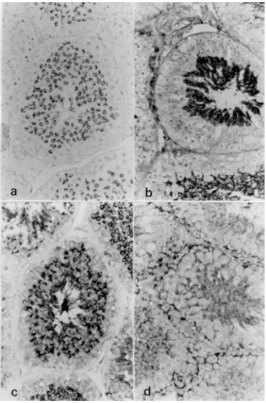
The spermatids of the Java fruit bats could be easily subdivided into 4 phases (Golgi, cap, acrosome and maturation-phases). Six lectins (PNA, PHA-E, Con A, WGA, RCA-I and PSA) exhibited a positive reaction in spermatogenic cells, whereas four lectins (UEA-I, SBA, DBA and BSL-I) revealed a negative reaction in the testes. PNA and RCA-I showed an intense reaction in the acrosomal region of spermatids. In short, PNA was intensely positive in the acrosomal region from Golgi to acrosome-phase spermatids (Fig. 1a). This reaction disappeared in maturation-phase spermatids. RCA-I gave an intense reaction in the acrosomal region from Golgi to acrosome-phase spermatids and also reacted in the cytoplasm of spermatocytes and spermatids (Fig. 1b). In addition to a diffuse reaction in the seminiferous epithelium, PHA-E exhibited a granular reaction within the cytoplasm of pachytene spermatocytes (Fig. 1c). Although Con A and WGA revealed a diffuse reaction all over the seminiferous epithelium, WGA showed an intense reaction in the cytoplasm of acrosome-phase spermatids and Sertoli cells (Fig. 1d). PSA showed a weak reaction in the interstitial region, Sertoli cells and elongate spermatids.
Japanese lesser horseshoe bat
The spermatids of the Japanese lesser horseshoe bats could also be subdivided into 4 phases (Golgi, cap, acrosome and maturation-phases). In most cases, the lectin binding patterns in the testes of the Japanese lesser horseshoe bats were similar to those of the Java fruit bats (Fig. 2a, b), but they were different in the RCA-I and PSA reactions. In the Japanese horseshoe bats, RCA-I was strongly positive in the acrosomal region from Golgi to acrosome-phase spermatids as well as in the Java
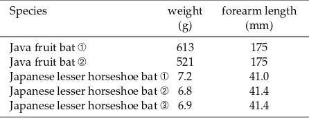
Table 1. The body weight and forearm length of the Java fruit bat and the Japanese lesser horseshoe bat
Species weight forearm length (g) (mm)
Java fruit bat ➀ 613 175
Java fruit bat ➁ 521 175
311 LECTIN-BINDING PATTERNS IN THE TESTES OF BATS