DOI: 10.12928/TELKOMNIKA.v12i1.1686 107
Endocardial Border Detection Using Radial Search and
Domain Knowledge
Yong Chen*, Dong C Liu
School of Computer Science, Sichuan University, ChengDu, P.R. China *Corresponding author, e-mail: [email protected]
Abstract
The ejection fraction rate is a frequently used parameter when treating patients who suffered from heart disease. However, the measurement of this ejection rate depends on manual segmentation of left ventricle cavity in the end-systolic and end-diastolic phases. This paper proposes a semi-automatic algorithm for the detection of left ventricular border in two dimensional long axis ultrasound echocardiographic images. First, we apply a preprocessing filter to the ultrasound for the sake of speckle reduction. Then the knowledge of the anatomical structure of human heart and local homogeneity of blood pool is being used to detect the border of left ventricle. The proposed method evaluates 80 ultrasound images from four healthy volunteers and the generated contours are compared with contours manually drawn by an expert. The measured Dice Metric and Hausdorff Distance recorded by the proposed algorithm are 85.1% ± 0.4% and 3.25 ± 0.46 mm respectively. The numerical results reported in this paper indicate that the proposed algorithm is able to correctly segment the left ventricle cavity and can be used as an alternative to manual contouring of left ventricle cavity from ultrasound images.
Keywords: border detection, radial search, domain knowledge, segmentation of left ventricle, echocardiography
1. Introduction
As compared to other medical imaging modalities such as Magnetic Resonance Imaging (MRI) or Computer Tomography (CT), ultrasound imaging is non-invasive, real-time, and more accessible. Therefore it has been widely used in various medical disciplines, such as echocardiography, breast ultrasound, etc. When treating patients who suffered from heart disease, clinicians often used the ejection fraction (EF), which is defined as the quotient “ejected volume” over “ventricular volume at the beginning of the systole phase”, to evaluate the heart condition of the patients.[1] At present, the ejection fraction rate depends on the manual contouring of the left ventricle (LV) cavity in the end-systolic and end-diastolic phases. However, manual contouring of LV in ultrasound images is an arduous task and can be rather time-consuming. So computer aided automatic segmentation of LV is preferred because an automation of the segmentation would prevent inter-observers and intra-observer variability and make the measurement of the EF more accessible.
orientation. They claimed that the proposed method can handle speckles and perform well on images with weak boundaries. In [6], George A Brock Fisher etc. proposed a contrast agent based method to detect the boundary between the blood and the tissue. The method first administered a contrast agent to the region of interest and then sent two ultrasound beams with different power levels in a row. The time segment values of the first beam and the second beam were phase-compared. The method declared that the boundary was located at the place where a phase change of echo returns occurred. Other studies by application of active contours based on intensity [7], Bayesian framework using deformable templates and Markov random field [8], level set method using intensity gradient [9], were also reported in the literature.
However, no segmentation technique is perfect and there is still room for improvement. We are interested in making use of the anatomical structure of human heart to facilitate the detection of endocardial border. In [10], Belohlavek proposed to capture expert knowledge of left ventricle cavity shapes by training a reference database. The information of the anatomic structure of human heart, i.e., the apex point and two mitra valve points identified by user, was utilized both in learning phase and performing phases to help locating the long axis left ventricle endocardium. Yet there are two problems with the method described in [11]. The first is that we need to train a large set of representative samples to get a robust reference database. The second is that it may fail to recognize the border correctly if the image under test has not been trained before. Therefore we propose to give up reference database training and to use local homogeneity based radial search method instead.
In this paper, a new semi-automatic method for LV segmentation is presented. It makes use of three user chosen points, one point at the apex of the heart and two others attaches to the left and right mitra valve respectively. These three points are used as the base for the emission of radial lines. The results of the semi-automatic segmentation are compared with the contours drawn by the expert using statistical analysis.
The rest of this paper is organized as follows. We begin in section 2 by describing the proposed method in detail. Then section 3 presents the experiment result and discussion. Finally, we conclude our work in section 4.
2. Research Method 2.1. Data Acquisition
An ultrasound image database consisting of 80 ultrasound images from four healthy volunteers were captured by an expert. All digital ultrasound images were obtained using Saset iMago C21 system (SASET Healthcare, San Francisco, CA) with a 4 MHz phased array transducer. When the expert was capturing long axis left ventricle images, he asked the volunteers to lying supine and placed the transducer over the tip of heart. The capturing resolution of the images was 0.3×0.3 mm2. Except for a few cases, the sonographic setting remained the same throughout the course of data acquisition.
2.2. Outline of the Proposed Method
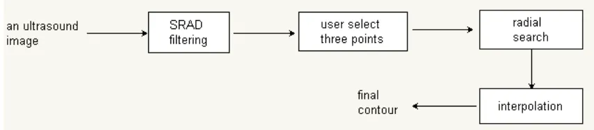
[image:2.595.84.511.637.731.2]An outline of the proposed LV segmentation algorithm is described in Figure 1, where the major components and data flow is illustrated. The detail information will be described in the remainder of section 2.
2.3. Preprocessing
Since speckle often degrades the quality of the ultrasound image and makes segmentation difficult, it is better to do some image preprocessing to attenuate the speckle noise before segmentation. In this work, the Speckle-Reducing Anisotropic Diffusion (SRAD) filtering algorithm [11] has been used. The essence of SRAD is to replace the gradient-based edge detector with the instantaneous coefficient of variation that is suitable for speckle filtering. The general update function of SRAD is formulated as:
, ,
( (
,)
,)
|
|
t t t t t
i j i j i j i j
s
t
I
I
div c C
I
(1)
in which the diffusion coefficient
C
i jt, is defined as2 2 2
, , 2 , 2 2 , , 1 1 | | ( )
2 1 6
1
( )
4
i j i j
i j
i j i j
I I C I I (2)
SRAD uses an iterative scheme to update the pixel value. In this work, the parameter lamda was set as 0.5 and the iteration number was set as 150.
2.4. Semi-automatic Segmentation
Before we describe the detail of the proposed method, let us introduce some notations first. As shown in [12], the homogeneity of a pixel at (i, j) is defined as
2 ij ij ij h
(3)
where σ2 ,ij and μij are the variance and mean of a neighborhood sub-window centered at (i, j), respectively. Since the distribution of intensities of blood pool is homogeneous whereas the distribution of intensities of endocardium is not, it is reasonable to assume that the local homogeneity of a pixel inside the blood pool is smaller than that of a pixel in the endocardium area. We will use this feature to discriminate the candidate points of final contour.
There are four steps in the proposed method.
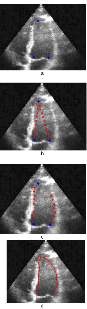
Step 1. User selects three points, i.e., the apex point and two mitra valve points, in the long axis left ventricle ultrasound image under test. One example is shown in Fig. 2a.
Step 2. Computer connects two lines between the apex point and the two mitra valve points, respectively. We denote the left line as line_l and the right line as line_r. Then it generates nine equidistant nodes along line_l and line_r, respectively. One example is shown in Fig. 2b.
Step 3. We describe how to find the candidate points of left part of the final contour here, whereas the candidate points of right part of the final contour can be found in the same way. The detection of the candidate points is performed using radial search. Starting from each node of line_l, say its pixel is at (u, v), the algorithm searches for its corresponding candidate point for the final contour along a horizontal line extending to the left. The local homogeneity of each pixel on this line is calculated and then compared with that of the pixel at (u, v). We claim that the candidate point, say its location is (i, j), is found if hij >= (ratio × huv) where ratio is an adjustable parameter. Since the selection of the ratio and the size of the neighborhood sub-window have an important effect on the final result, we must choose them carefully. In our experiments, the size of the neighborhood sub-window is set as 11 ×11 and the ratio is set as 1.8. One example of the result of step 3 is shown in Fig. 2c.
a
b
c
[image:4.595.214.386.77.687.2]
d
Figure 2. An illustration of the four steps of the semi-automatic method
3. Results and Discussion 3.1. Evaluation metric
The quality of our segementation method was validated using two measures: 1) Dice Metric (DM) and 2) Hausdorff Distance (HD) [13].
DM measures the similarity between two areas and is defined as:
2 * (
1
2)
*100
1
2
area
area
DM
area
area
(4)
The HD aims to evaluate the distance between two contours. If two contours are represented by a set of points A = {a1, a2, …, am} and B = {b1, b2, …, bm}, where each ai and bi is
an ordered point on the curve. The Distance to the closest point (DCP) for ai to B is defined as
( , )
imin ||
j i||
j
d a B
b
a
(5)and the HD is define as
( , )
max(max{
( , )}, max{
i( , )})
ji j
HD A B
DCP a B
DCP b A
(6)3.2. Experimental results and discussion
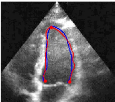
Figure 3 presented an example of the segmentation result. The result returned by the proposed method, which was shown in red color, was superposed on the manually drawn contour, which was shown in blue color. The result of measures (DM, HD) was (0.91, 2.7 mm) for this case.
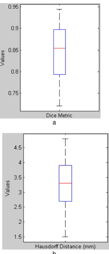
[image:5.595.206.390.503.667.2]A group of 80 long-axis four chamber ultrasound images from 4 different volunteers, classified by an expert as of average quality or above, were adopted to assess the performance of the proposed method. The proposed method yielded a DM of 85.1% ± 0.4% and a HD of 3.25 ± 0.46 mm in this dataset. As an additional statistical measure, Figure 4 reported the boxplot of DM and HD measurement. The boxplot of DM was shown in Figure 4a and the boxplot of HD was shown in Figure 4b.
a
[image:6.595.210.386.80.488.2]
b
Figure 4. An illustration of the boxplot of DM and HD
a. the boxplot of Dice Metric b. the boxplot of Hausdorff Distance
Our algorithm was implemented in Matlab 7.1 (The MathWorks, Natick,MA) on a 2.5 GHz Intel Pentium desktop PC. Once the three points were selected, the average computing time to calculate the final contour was 0.03 second.
If we look closer at the Figure 1, we can see that there are two candidate points missing on the right top part of the left ventricle cavity in Figure 1c, which is caused by the signal dropouts of ultrasound imaging. Nevertheless, we still get a reasonable robust final contour owing to the fact that the majority of the candidate points have been found. So we may conclude that our method is robust to the signal dropouts of the ultrasound left ventricle images.
4. Conclusion
locate the left ventricle contours that are close to the contour drawn by the expert. It may be used as an alternative to manual contouring of left ventricle from ultrasound images.
Future work would be focused on collecting more objects, especially images from those people who suffered from heart diseases, and testing our method on them.
Acknowledgement
The authors would like to thank the members of the Medical Imaging Laboratory at SiChuan University for volunteering for ultrasound scans; Dr. Zuo at SASET Healthcare for kindly providing the ultrasound data. This work was supported in part by the Foundation of China and Natural Science Foundation of Sichuan Province (Grant No. 2013GZX0147-3).
References
[1] Jerry J. Batzel, Franz Kappel, Daniel Schneditz, Hien T.Tran. Cardiovascular and Respiratory Systems: Modeling, Analysis, and Control, first edition. Philadelphia: Siam, 2007.
[2] A Sarti, C Corsi, E Mazzini, and C Lamberti. Maximum Likelihood Segmentation with Rayleigh Distribution of Ultrasound Images. Computers in Cardiology. 2004; 31: 329-332.
[3] Maria do Carmo dos Reis, Adson F. da Rocha, Daniel F. Vasconcels, Bruno L. M. Espinoza, Francisco A. de O.Nascimento, Joao L.A. de Carvalho, Sauro Salomoni, and Juliana F. Camapum.
Semi-Automatic Detection of the Left Ventricle Border. 30th Annual International IEEE EMBS Conference, Vancouver, British Columbia, Canada, August 20-24, 2008.
[4] Gustavo Carneiro, Jacinto Nascimento, and Antonio Freitas. Robust left ventricle segmentation from ultrasound data using deep neural networks and efficient search methods. Biomedical Imaging: From Nano to Macro, 2010 IEEE International Symposium on. 2010: 1085-1088.
[5] A.Belaid, D.Boukerroui, Y.Mainground, and J.Lerallut. Phased-based level set segmentation of ultrasound images. IEEE Transactions on Information Technology in Biomedicine. 2011; 15(1): 138-147.
[6] George A Brock Fisher, David M Prater. Acoustic Border Detection Using Power Modulation, US Patent 6,997,875 B2 (Patent), 2006.
[7] A. Mishra, P.K. Dutta, M.K. Ghosh. A GA based approach for boundary detection of left ventricle with echocardiographic image sequences. Image and Vision Computing. 2003; 21: 967-976.
[8] M.Mignotte, J.Meunier, J.C.Tardif. Endocardial boundary estimation and tracking in echocardiographic images using deformable template and Markov random fields. Pattern Analysis and Applications.
2001; 4(4): 256-271.
[9] J.Y, Yan, T.Zhuang. Applying improved fast marching method to endocardial boundary detection in echocardiographic images. Pattern Recognition Letters. 2003; 24(15): 2777-2784.
[10] M. Belohlavek, Fast cardiac boundary imaging, US Patent 5,871,019 (Patent), 1999.
[11] Yongjian Yu, and Scott T. Acton. Speckle Reducing Anisotropic Diffusion. IEEE Transactions on image processing. 2002; 11(11).
[12] Y. Chen, R.M. Yin, and P. Flynn. Aggressive region growing for speckle reduction in ultrasound images. Pattern Recognition Letters. 2003; 24(4-5): 677-69.