ISOLATION OF CHITINOLYTIC BACTERIA USED AS
BIOLOGICAL CONTROL OF SUSPECTED PATHOGENIC
FUNGI ON OIL PALM SEEDLINGS
AGUSTINUS HARYANTO
DEPARTMENT OF BIOLOGY
FACULTY OF MATHEMATICS AND NATURAL SCIENCE BOGOR AGRICULTURAL UNIVERSITY
STATEMENT ABOUT UNDERGRADUATE THESIS,
INFORMATION SOURCES, AND ACT OF SPILLING OVER
COPYRIGHTS
By this writing I clarify that the undergraduate thesis Isolation of Chitinolytic Bacteria Used as Biological Control of Suspected Pathogenic Fungi on Oil Palm Seedlings is my own work under the supervisions of the advising committee and hasn’t been proposed for any institution. Copied information source of published and unpublished writing of other author has been mentioned in the text and incorporated in the references at the last part of this thesis.
By this writing I hand over the copyright of my undergraduate thesis to Bogor Agricultural University.
ABSTRACT
AGUSTINUS HARYANTO. Isolation of Chitinolytic Bacteria Used as Biological Control of Suspected Pathogenic Fungi on Oil Palm Seedlings. Supervised by NISA RACHMANIA MUBARIK and SRI LISTIYOWATI.
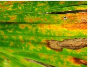
Oil palm (Elaeis guineensis Jacq.) is one of the main plantation commodities in Indonesia. Production of oil palm is influenced by several environmental condition such as rainfall, soil, climate, and pathogen outbreak. Pathogen outbreak such as anthracnose and leaf blight cause rotting on leaves of oil palm seedling which in the end will kill the plant. Prevention on pathogen outbreaks on oil palm which was caused by pathogenic fungi can be done by using chitinolytic bacteria which can produce chitinase enzyme as biological control. Chitin is one of the element in cell wall of the fungi. The aims of this experiments were to isolate chitinolytic bacteria and to investigate under in vitro test of their potential as biological control of suspected pathogenic fungi on oil palm. Results showed isolate SAHA12.08, SAHA12.10, and SAHA12.13 had antagonistic activity to the growth of Curvularia sp. and Colletotrichum sp. suspected pathogenic whereas isolate KAHN15.12 only had antagonistic activity to Curvularia suspected pathogenic.
Key words: antagonistic activity, chitinolytic, pathogenic fungi, oil palm
ABSTRAK
AGUSTINUS HARYANTO. Isolasi Bakteri Kitinolitik yang Berpotensi sebagai Biokontrol terhadap Cendawan yang Diduga Patogen pada Bibit Kelapa Sawit. Dibimbing oleh NISA RACHMANIA MUBARIK dan SRI LISTIYOWATI.
Kelapa sawit (Elaeis guineensis Jacq.) merupakan salah satu komoditas perkebunan utama di Indonesia. Produksi kelapa sawit sangat dipengaruhi oleh berbagai faktor lingkungan seperti curah hujan, tanah, iklim, dan serangan penyakit. Serangan penyakit seperti antraknosa dan bercak daun menyebabkan pembusukan pada daun kelapa sawit saat pembibitan yang pada akhirnya akan membunuh tanaman kelapa sawit tersebut. Upaya pencegahan serangan penyakit pada kelapa sawit yang disebabkan oleh cendawan patogen dapat dilakukan dengan penggunakan bakteri kitinolitik yang mampu menghasilkan enzim kitinase sebagai pengendali biologi. Kitin merupakan salah satu komponen penyusun dinding sel dari cendawan. Penelitian ini bertujuan untuk mengisolasi bakteri kitinolitik dan menguji potensinya sebagai pengendali hayati dalam keadaan in vitro terhadap cendawan patogen pada kelapa sawit. Hasil menunjukkan isolat SAHA12.08, SAHA12.10, dan SAHA12.13 memiliki aktivitas antagonis terhadap pertumbuhan cendawan yang diduga patogen Curvularia sp. dan Colletotrichum sp., sedangkan isolat KAHN15.12 hanya memiliki aktivitas antagonis terhadap pertumbuhan Curvularia sp. yang diduga patogen.
An Undergraduate Thesis Intended to Acquire Bachelor Degree In Faculty of Mathematics and Natural Science
ISOLATION OF CHITINOLYTIC BACTERIA USED AS
BIOLOGICAL CONTROL OF SUSPECTED PATHOGENIC
FUNGI ON OIL PALM SEEDLINGS
AGUSTINUS HARYANTO
DEPARTMENT OF BIOLOGY
FACULTY OF MATHEMATICS AND NATURAL SCIENCE BOGOR AGRICULTURAL UNIVERSITY
Thesis Title: Isolation of Chitinolytic Bacteria Used as Biological Control of Suspected Pathogenic Fungi on Oil palm Seedlings
Name : Agustinus Haryanto NIM : G34090116
Approved by
Dr Nisa Rachmania Mubarik, MSi Supervisor I
Dr Sri Listiyowati, MSi Supervisor II
Acknowledged by
Dr Ir Iman Rusmana, MSi Head of Department Biology
FOREWORDS
First of all, I would like to give thanks to Jesus Christ for all blessing I’ve got. This research is made through an experiment entitled Isolation of Chitinolytic Bacteria Used as Biological Control of Suspected Pathogenic Fungi on Oil Palm Seedlings which was conducted from January until July 2013 on IPB. This study was funded by Start Up Funding Collaborative Research Centre (CRC) 990 in 2012 and Directorate of Higher Education, Ministry of National Education Republic of Indonesia through Penelitian Unggulan Perguruan Tinggi Institut Pertanian Bogor in 2013 to Nisa Rachmania Mubarik.
Biggest acknowledgment is given to Dr Nisa Rachmania Mubarik, MSi and Dr Sri Listiyowati, MSi as my supervisor for the advice and supervisions. Acknowledgment is also given to name as Dr Achmad Farajallah, MSi for the advice and discussion. Special acknowledgment is given to my families and my friends for all their love and support.
At last, I hope this research will be helpful for all the readers.
TABLE OF CONTENTS
LIST OF TABLES vi
LIST OF FIGURES vi
LIST OF APPENDIXES vi
INTRODUCTION 1
Background 1
Aims 1
MATERIALS AND METHOD 1
Time and Place 1
Materials 2
Isolation and Identification of Chitinolytic Bacteria 2 Isolation and Identification of Leaf Spot Fungi from Oil Palm 2 In vitro Screening of Chitinolytic Bacteria against Suspected Pathogenic
Fungi 2
Identification of Chitinolytic Bacteria which has Antagonistic Activity 3 Bacterial Growth Curve from Selected Isolates and Antagonistic Activity 3
RESULTS 3
Isolation and Identification of Chitinolytic Bacteria 3 Isolation and Identification of Leaf Spot Fungi from Oil Palm 4 In vitro Screening of Chitinolytic Bacteria against Suspected Pathogenic
Fungi 5
Identification of Chitinolytic Bacteria which has Antagonistic Activity 6 Bacterial Growth Curve from Selected Isolates and Antagonistic Activity 7
DISCUSSION 8
CONCLUSION AND SUGGESTION 11
Conclusion 11
Suggestion 11
REFERENCES 11
APPENDIXES 16
LIST OF TABLES
1 Identification results of chitinolytic isolates 4 2 Antagonistic activity of ten highest chitinolytic index (CI) isolates 6 3 Antagonistic properties on suspected pathogenic fungi 6
LIST OF FIGURES
1 Symptoms of sick leaves caused by leaf spot fungi of oil palm 5
2 Cell morphology of suspected pathogenic fungi from oil palm. (a) Curvularia sp. (b) Colletotrichum sp. 5
3 Colony morphology of suspected pathogenic fungi from oil palm. (a) Curvularia sp. (b) Colletotrichum sp. 5
4 Growth curve of isolate SAHA12.08 and its antagonistic activity
toward Curvularia sp. 7
5 Growth curve of isolate SAHA12.08 and its antagonistic activity
toward Colletotrichum sp. 8
LIST OF APPENDIXES
1 Soil samples data from Taman Nasional Bukit Dua Belas (Jambi) 14
2 Isolates origin from soil samples data 15
3 Biochemical characteristics of isolates SAHA12.08 and SAHA12.13
1
INTRODUCTION
Background
Production of oil palm products increased approximately from 167.000 tones in 1967 to 18 million tones in 2009, or almost 107 times within 42 years. Areas of oil palms have increased from 105.000 hectares in 1967 to 7.8 million hectares (Ditjenbun 2009). Some of pathogens Botryodiplodia palmarum, Melanconium sp., Glomerella cingulata and Curvularia eragrostidis which are commonly found at Southeast Asia greatly damaged oil palm seedling (Aderungboye 1977). Therefore, by controlling the pathogen outbreaks, it will lead to increase in oil palm production itself.
Biological control using microorganism has been studied intensively since it means as an available environmental friendly alternative. Introduction of chitinolytic bacteria as antagonist agents is to control pathogenic fungi. As stated in many previous reports, the production of chitinase enzyme was related to fungal growth inhibition and the biological control of fungal pathogen was possible because of the ability of the chitinolytic bacteria to degrade fungal cell walls (Kamil et al. 2007; Suryanto et al. 2010; Gomaa 2012). Chitinolytic bacteria is capable to inhibit fungal activity because it can produce chitinase to degrade chitin which is one of the element in cell wall in the fungi (Peter 2005). Application of chitinolytic products from bacteria is one of environmental friendly alternatives which is safer to control pathogenic fungi than using synthetic fungicide. While using chitin colloidal as common substrate for chitinase enzyme inducer to screen chitinolytic bacteria, it probably lead to degradation of chitin structures of other chitin substrate in its application which has the same chitin structures with chitin colloidal (Haran and Chet 1995). Therefore, this study need further investigation in its application on the field.
Aims
The aims of this research were to isolate chitinolytic bacteria and to investigate under in vitro test of their potential as biology control of suspected pathogenic fungi on oil palm seedlings.
MATERIALS AND METHOD
Time and Place
2
Materials
Soil samples were obtained from Taman Nasional Bukit Dua Belas, Jambi (Appendix 1). Leaf spot fungi were obtained from infected leaves of oil palm from Indonesia Biotechnology Research Institute for Estate Crops, Bogor.
Isolation and Identification of Chitinolytic Bacteria
In total of five plots were used in chitinolytic bacteria screening with twice repetition from each plot. Every 3.0 g of soil sample is diluted in 30 mL of nutrient broth (NB) with 1% chitin colloidal and it was incubated at room temperature for 24 hours. Then, all cultures were done with serial dilution from 10-6 to 10-8 in NaCl 0.85 %. Suspension was spreaded on chitin agar (1% chitin colloidal, 0.1% MgSO4·7H2O, 0.02% K2HPO4, 0.1% yeast extract, and 1.5% agar) which was incubated at 37oC for 48 hours. Each colony of different bacteria is streaked on the new agar chitin until single colony of bacteria was found. Cultures were incubated at 37oC for 48 hours. Isolates were observed based on chitinolytic index (CI) which was hinted by clear zone. Every chitinolytic was identified by using Gram staining.
Isolation and Identification of Leaf Spot Fungi from Oil Palm
Isolation was began with preparation of infected leaves. Every three spots on infected leaves was cut into 1x1 cm covering half healthy leaf and half sick leaf. The leaves were rinsed with flowing water and then it was soaked into sodium hypochlorite (NaClO) 1% for a minute. The leaves then were rinsed with sterilized water and it was dried with sterilized tissue. Finally, the leaves were put on potato dextrose agar (PDA) containing chloramphenicol 0.5 %. Fungal cultures were incubated at room temperature for a week. Every hyphae were reinoculated into new PDA to get pure cultures. Cultures were identified by its morphological characteristic.
In vitro Screening of Chitinolytic Bacteria against Suspected Pathogenic
Fungi
3
Identification of Chitinolytic Bacteria which has Antagonistic Activity
Isolates which had antagonistic activity to all suspected pathogenic fungi were identified by using kit BioMérieux, USA. Isolates with antagonistic activity were streaked on chitin agar. Cultures were incubated at 37oC for 24 hours. Cultures then were stained with Gram staining for verification. Three loopful of the cultures of each isolates were diluted into kit API 50 CHB/E medium. In amount of 200 µl kit API 50 CHB/E medium was filled into the tube. In addition, the first tube was filled by kit API 50 CHB/E medium which had not been inoculated by isolates as control. Result which was showed was interpretated by using the apiweb identification software with the database (V4.0).
Bacterial Growth Curve from Selected Isolates and Antagonistic Activity
Inoculation of 2-4 loopful of selected isolates into 50 ml NB with 1% chitin colloidal as enzyme production medium which was incubated at 37oC for 15 hours. In amount of 1 ml culture was inoculated into 100 ml NB with 1% chitin colloidal. Enzyme production medium was incubated on incubator shaker at 120 rpm at 37oC. Optical density (λ = 600 nm) and antagonistic activity of the culture were measured every 12 hours. Antagonistic activity was tested by using culture and crude chitinase of the isolate with agar well diffusion method. Supernatant (crude chitinase) which contains extracellular metabolites was obtained by centrifugation on 12.000 rpm (Centrifuge MiniSpin with rotor F-45-12-11) for five minutes. Well which was containing 100 µl culture or crude chitinase was made 2 cm from margin of PDA plate. Opposite the well, at a distance of at least 3 cm, suspected pathogenic fungi was inoculated. After incubation for 6 days at room temperature, inhibition of the pathogen’s growth was assessed by the percentage of inhibition of radial growth [100% x (r1-r2)/r1). R1 is length of radial growth towards plate margin (4 cm) and R2 is length of radial growth towards antagonistic (Fokkema 1983).
RESULTS
Isolation and Identification of Chitinolytic Bacteria
4
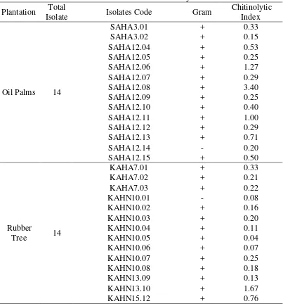
Table 1 Identification results of chitinolytic isolates Plantation Total
Isolate Isolates Code Gram
Chitinolytic
Isolation and Identification of Leaf Spot Fungi from Oil Palm
5
Figure 1 Symptoms of sick leaves caused by leaf spot fungi of oil palm
Figure 2 Cell morphology of suspected pathogenic fungi from oil palm (a) Curvularia sp. (b) Colletotrichum sp.
Figure 3 Colony morphology of suspected pathogenic fungi from oil palm (a) Curvularia sp. (b) Colletotrichum sp.
In vitro Screening of Chitinolytic Bacteria against Suspected Pathogenic
Fungi
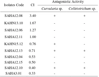
Ten chitinolytic bacteria with highest chitinolytic index were antagonistic tested against two isolates suspected pathogenic fungi. The result showed isolate SAHA12.08, SAHA12.10, and SAHA12.13 were able to inhibit the growth of two suspected pathogenic fungus at once (Colletotrichum sp. and Curvularia sp.) whereas KAHN15.12 was only able to inhibit the growth of Curvularia sp. suspected pathogenic (Table 2).
Isolate SAHA12.13 showed highest inhibition to Curvularia sp. in 58.75% and Colletotrichum sp. in 52.50% while isolate SAHA12.08 showed the lowest inhibition to Curvularia sp. in 36.25% and Colletotrichum sp. in 16.82% (Table 3).
a b
6
The result showed that isolate SAHA12.08 which had the highest chitinolytic index did not show stronger inhibition to all suspected pathogenic fungi than isolate SAHA12.13 which had chitinolytic index lower than SAHA12.08.
Table 2 Antagonistic activity of ten highest chitinolytic index (CI) isolates Isolates Code CI
Antagonistic Activity
Curvularia sp. Colletotrichum sp.
SAHA12.08 3.40 + +
+ antagonistic activity - no antagonistic activity
Table 3 Antagonistic properties on suspected pathogenic fungi Suspected
Identification of Chitinolytic Bacteria which has Antagonistic Activity
7
Bacterial Growth Curve from Selected Isolates and Antagonistic Activity
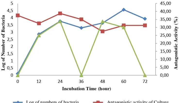
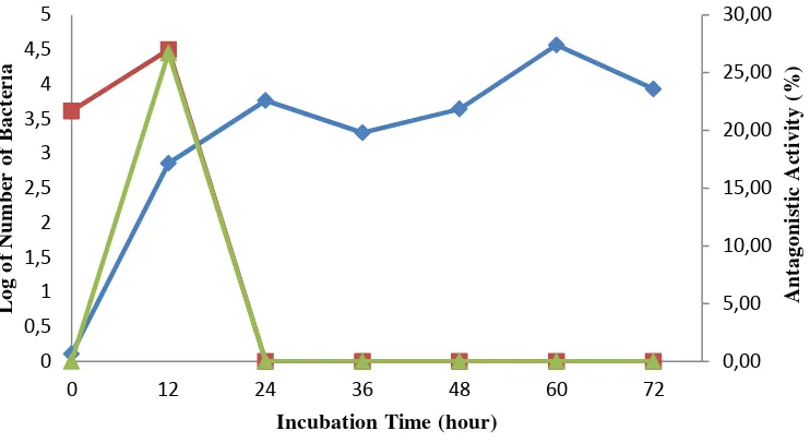
Two isolates of higher chitinolytic index with antagonistic activity to all suspected pathogenic fungi were measured its growth as well as antagonistic activity. Those isolates were SAHA12.08 and SAHA12.13. The results showed that isolate SAHA12.08 probably ended exponential phase at 24 hours. Stationary phase of isolated SAHA12.08 probably occurred within 24 hours to 60 hours while death phase was probably reached after 60 hours. Antagonistic activity of culture to Curvularia sp. showed that the highest inhibition of 38.75% occurred at 24 hours while decreasing occurred after 24 hours (Figure 4), whereas antagonistic activity of the culture to Colletotrichum sp. showed that the highest inhibition of 26.97% occurred at 12 hours while decreasing occurred after 12 highest inhibition of 26.67% occurred at 12 hours while decreasing occurred past 12 hours. Results also showed that antagonistic test with culture had stronger antagonistic activity than crude chitinase (Figure 4 and 5). Isolate SAHA12.13 was also observed its growth and antagonistic activity through culture and crude chitinase, however this isolate showed no activity at all to inhibit suspected pathogenic fungi.
Figure 4 Growth curve of isolate SAHA12.08 and its antagonistic activity toward Curvularia sp.
Log of numbers of bacteria Culture/Curvularia sp. Crude Chitinase/Curvularia sp.
Log of numbers of bacteria
Antagonistic activity of Crude Chitinase
8
Figure 5 Growth curve of isolate SAHA12.08 and its antagonistic activity toward Colletotrichum sp.
DISCUSSION
An amount of 28 isolates of chitinolytic bacteria were successfully isolated by using chitin agar containing chitin colloidal. Chitinolytic isolates can be detected through clear zone around bacterial colony. Low numbers of isolates which were isolated were caused by using enrichment and dilution techniques. Enrichment and dilution techniques were only capable in selecting those microorganism that are numerically abundant or those microorganism that show superior growth in a given medium but not the actual number of bacteria which were found in the sample (Jackson et al 1998). From 28 isolates of chitinolytic bacteria, isolate SAHA12.08 showed highest chitinolytic index in amount of 3.40. Chitinolytic index of isolate SAHA12.08 (3.40) was higher than the average of chitinolytic index of isolates from chili plant roots which was 1.00 (Nurdebyandaru et al. 2008). Isolate SAHA12.08 also showed higher chitinolytic index than the highest chitinolytic index of isolates from rubber tree which was 0.52 (Muharni and Widjajanti 2011).
In amount of 10 highest chitinolytic index isolates were used in antagonistic test against suspected pathogenic fungi. There were two suspected pathogenic fungi which were successfully isolated. One of them was Curvularia sp. which showed morphological characteristics including white to pinkish gray initial colony which turns to olive brown or black as the colony matures, brown hyphae, septate hyphae, and brown conidiophores which are simple or branched. Most species of Curvularia are facultative pathogens of plants in tropical or subtropical
0,00
Log of numbers of bacteria Culture/Botryodiplodia sp.
Crude Chitinase/Botryodiplodia sp.Antagonistic activity of Crude Chitinase
9
areas, while the remaining few are found in temperate zones (Larone 1995). Another suspected pathogenic fungus which was successfully isolated was Colletotrichum sp. It had sparse, cottony, white to pale gray colored colony. Spores are formed in acervuli, erumpent, cushion like masses of hyphae bearing conidiophores, hyaline, and oblong to fusoid conidia (Horst 2008). While Colletotrichum species cause serious plant disease that devastate crop plants worldwide, they also commonly isolated as endophytes from healthy plants and as saprobes on dead plant material (Photita et al 2005). The majority of the fungi including Curvularia sp. and Colletotrichum sp. have chitin but not cellulose in their cell walls, therefore its cell wall could be degraded by chitinolytic bacteria (Landecker 1996).
From 10 of highest chitinolytic index isolates, only 4 isolates showed antagonistic activity. Those isolates were SAHA12.08, SAHA12.10, SAHA12.13, and KAHN15.12. Isolate SAHA12.13 showed strongest inhibition towards suspected pathogenic fungus which was indicated by 58.75% of inhibition of radial growth to Curvularia sp. and 52.50% of inhibition of radial growth to Colletotrichum sp. Isolate SAHA12.08 and SAHA12.10 were also able to inhibit the growth of Curvularia sp. and Colletotrichum sp. however its antagonistic activity were not as strong as isolate SAHA12.13. Isolate SAHA12.10 showed inhibition of radial growth to Cuvularia sp. in amount of 57.50% and 46.97% of inhibition to the growth of Colletotrichum sp. Inhibition of radial growth to Curvularia sp. and Colletotrichum sp. by isolate SAHA12.10 was slightly lower than SAHA12.13, however SAHA12.10 showed low chitinolytic index and therefore this isolate was not tested any further. KAHN15.12 were only able to inhibit the growth of Curvularia sp. Variation in inhibition maybe was caused by specification enzyme to the species, differences chitinase activity, and chitin composition in fungi cell wall, also existence of secondary metabolites. The cell wall of fungi generally consist of not only chitin but also another type of sugars. For example, β-1,3 glucan which was bound to chitin. In that case, there are more than one enzyme which are responsible in degradation of cell wall (Anand and Reddy 2009).
Isolate KAHN13.10 showed the second highest chitinolytic index but surprisingly isolate KAHN13.10 was not able to inhibit suspected pathogenic fungi. On the other hand isolate SAHA12.10 and SAHA12.13 showed lower chitinolytic index than isolate KAHN13.10, but they were able to inhibit the growth of suspected pathogenic fungi especially isolate SAHA12.13 which had strongest inhibition to suspected pathogenic fungi.This results showed that compatibility of enzyme to the substrate is very essential in playing antagonistic activity as well as antifungal properties (Gohel et al. 2006).
10
Colletotrichum sp. past 12 hours. This might be caused by the decreasing production of hydrolytic enzymes and incapability of secondary metabolites which were produced on stationary phase to inhibit Colletotrichum sp.
The antagonistic test by using supernatant (cell-free-filtrate) which contains extracellular metabolites was aimed to observe whether hydrolytic enzyme such as chitinase was responsible in inhibiting suspected pathogenic fungal growth. The result showed that the filtrate was able to inhibit the growth of suspected fungi, although the inhibition was lower than inhibition by culture. Differences in ability to inhibit might be caused by differences in concentration of hydrolytic enzyme or even secondary metabolites on the filtrate (Prapagdee et al. 2008). Multiple chitinases as antagonistic properties also contribute important role in antagonistic activity. It is believed that multiple chitinases within a single organisms lead to a more efficient use of the respective substrate of chitin as a hydrolases based on amino acid sequence similarities (Henrissat 1999). In addition, another factor that affect the ability of the isolates to inhibit the growth of pathogenic fungi probably caused by the other bioactive compounds which were produced by the isolate other than extracellular chitinase. In order to prove either extracellular chitinase or other bioactive compounds which were responsible in antagonistic activity to pathogenic fungi, this study needs further investigation.
Identification using kit API 50 CH (BioMérieux, USA) showed isolate SAHA12.08 was most likely Geobacillus thermoglucosidasius, whereas isolate SAHA12.13 was most likely Bacillus cereus. Isolate SAHA12.08 was Gram-positive and rod shaped bacteria which was isolated from top soil around palm tree. Geobacillus thermoglucosidasius which was identified from SAHA12.08 is rod-shaped bacteria, occurring either singly or in short chains and motile by means of peritrichous flagella. The cell wall structure is Gram-positive, but the Gram-stain reaction may vary between positive and negative. It is aerobic or facultatively anaerobic bacteria and obligately thermophilic (Nazina et al. 2001). Identification using API 50 CHB system turned out to be inapplicable for isolate SAHA12.08 because discrepancies in the reaction patterns resulted in differing identifications. This misidentified was caused by limited database for the species, therefore the use of genomic methods such as 16S rRNA sequencing may be preferable (Boyd et al. 2005).
11
antifungal properties. Prokaryotes which were commonly found be able to degrade chitin were Pseudomonas, Vibrio, Photobacterium, Actinomycetes, Bacillus, Clostridium, and Enterobacter (Gooday 1990).
CONCLUSION AND SUGGESTION
Conclusion
An amount of 28 chitinolytic isolates was successfully isolated. Isolate SAHA12.08, SAHA12.10, and SAHA12.13 were able to inhibit the growth of Curvularia sp. and Colletotrichum sp. Whereas, isolate KAHN15.12 was only able to inhibit Curvularia sp. Isolate SAHA12.08 showed optimally antagonistic activity to all suspected pathogenic fungi at exponential phase. Identification of isolate SAHA12.08 which was most likely Geobacillus thermoglucosidasius by API 50 CHB turned out to be inapplicable. Identification of isolate SAHA12.13 by using API 50 CHB which was Bacillus cereus showed acceptable result.
Suggestion
There are some suggestions about this research. First, suspected pathogenic fungi should be explored for its actual pathogenic ability on oil palm. Exploration of the other abilities of all chitinolytic isolates also needs to be investigated. Last, characterization of chitinase molecule and also molecular identification of chitinolytic isolates and suspected pathogenic fungi isolates should be researched in the future.
REFERENCES
Aderungboye FO. 1977. Disease of the oil palm. PANS. 23(3):305-326.doi:10.1080/09670877709412457.
Anand S, Reddy J. 2009. Biocontrol potential of Trichoderma sp. against plant pathogens. Int J Agric Sci. 1(2):30-39.
Boyd MA, Antonio MAD, Hillier SL. 2005. Comparison of API 50 CH strips to whole-chromosomal DNA probes for identification of Lactobacillus species. J Clin Microbiol. 43(10):5309-5311. doi:10.1128/JCM.-43.10.5309-5311.2005.
[Ditjenbun] Direktorat Jenderal Perkebunan. 2009. Statistik Perkebunan Indonesia. Jakarta (ID): Ditjenbun.
Fokkema NJ. 1973. The role of saprophytic fungi in antagonism against Drechslera sorokiniana (Helminthosporium sativum) on agar plates and on rye leaves with pollen. Phys Plant Pathol. 3:195-205.
12
Gohel V, Singh A, Vimal M, Ashwini P, Chhatpar HS. 2006. Bioprospectiong and antifungal potential of chitinolytic microorganisms [review]. Afr J Biotechnol. 5(2):54-72.
Gomaa EZ. 2012. Chitinase production by Bacillus thuringiensis and Bacillus licheniformis: their potential in antifungal biocontrol. J Microbiol. 50(1):103-111.doi:10.1007/s12275-012-1343-y.
Gooday GW. 1990. The ecology of chitin degradation advance. Microbiol Ecol. 11:387-430.
Haran S, Chet I. 1995. New components of the chitinolytic system of Trichoderma harzianum. Mycol Rev. 94: 441-446.
Henrissat B. 1999. Classification of Chitinases Modules. Jollѐs P, Muzzarelli RAA, editor. Basel (CH): Birkhӓuser Verlag.
Horst RK. 2008. Westcott’s Plant Disease Handbook. 7th ed. New York (US): Springer.
Huang CJ, Wang TK, Chung SC, Chen CY. 2005. Identification of an antifungal chitinase from a potential biocontrol agent, Bacillus cereus 28-9. J Biochem Mol Biol. 38(1):82-88.
Jackson CR, Roden EE, Churchill PF. 1998. Changes in bacterial species composition in enrichment cultures with various dilutions of inoculums as monitored by denaturing gradient gel electrophoresis. Appl Environ Microbiol. 64(12):5046-5048.
Kamil Z, Rizk M, Saleh M, Moustafa S. 2007. Isolation and identification of rhizosphere soil chitinolytic bacteria and their potential in antifungal biocontrol. Global J Mol Sci. 2(2):57-66.
Landecker EM. 1996. Fundamentals of the Fungi. 4th ed. New Jersey (US): Prentice Hall.
Larone DH. 1995. Medically Important Fungi – A Guide to Identification. 3rd ed. Washington (US): ASM Pr.
Muharni, Widjajanti H. 2011. Skrining bakteri kitinolitik antagonis terhadap pertumbuhan jamur akar putih (Rigidopus lignosus) dari rizosfer tanaman karet. J Penel Sains. 14(1):51-56.
Nazina TN, Tourova TP, Poltaraus AB, Novikova EV, Grigoryan AA, Ivanova AE, Lysenko AM, Petrunyaka, Osipov GA, Belyaev SS et al. 2001. Taxonomic study of aerobic thermophilic bacilli: descriptions of Geobacillus subterraneus gen. nov., sp. nov.and Geobacillus uzenensis sp.nov. from petroleum reservoirs and transfer of Bacillus stearothermophilus, Bacillus thermo-catenulatus, Bacillus thermoleovorans, Bacillus kaustophilus, Bacillus thermoglucosidasius and Bacillus thermodenitrificans to Geobacillus as the new combination G. stearothermophilus, G. thermocatenulatus, G. thermoleovorans, G. kaustophilus, G. thermoglucosidasius and G. thermodenitrificans. Int J Syst Evol Microbiol. 51:433-446.
Nurdebyandaru N, Mubarik NR, Prawasti TS, 2008.Chitinolytic bacteria isolated from chili rhizophere: chitinase characterization and application as biocontrol for Aphis gossypii. J Microbiol Indones. 4(3):103-107.doi:10.5454/mi.4.3.1.
13
Photita W, Taylor PWJ, Ford R, Hyde KD, Lumyong S. 2005. Morphological and molecular characterization of Colletotrichum species from herbaceous plants in Thailand. Fungal Div. 18: 117-133.
Prapagdee B, Kuekulvong C, Mongkolsuk S. 2008. Antifungal potential of extracellular metabolites produced by Streptomyces hygroscopicus against phytopathogenic fungi. Int J Biol Sci. 4(5): 330-337.
Suryanto D, Patonah S, Munir E. 2010. Control of fusarium wilt of chili with chitinolytic bacteria. Hayati J Biosci. 17(1):5-8.doi:10.4308/hjb.17.1.5. Svitil AL, Chadhain SMN, Moore JA, Kirchman DL. 1997. Chitin degradation
proteins produced by the marine bacterium Vibrio harveyi growing on different forms of chitin. Appl Environ Microbiol. 63(2):408-413.
14
Appendix 1 Soil samples data from Taman Nasional Bukit Dua Belas (Jambi)
Plot Subplot Soil Code Plantation
15
Appendix 2 Isolates origin from soil samples data
Plot Soil Code Isolates Code
16
17