CONTROL OF SUBCLINICAL MASTITIS USING
CERTAIN
HOMEOPATHIC COMBINATION
DRUGS
ITS
INFLUENCE
ON MILK
QUALITY
AND
PRODUCTION
AGATHA
WINNY
SANJAYA
THE GRADUATE
PRUGRAM
BOGOR A G ; R l i C U L T W
IJNWEMITY
BOGOR
Just as my non-violence will never fail homaeopathy never fails But the followers of homeopathy may fail owing to faulty application of the principles.
ABSTRAK
AGATHA WINNY SANJAYA. Upaya Pencegahan Mastitis Subklinik dengan Menggunakan Homeopatikum secara Kombinasi dan Pengaruhnya terhadap Kualitas dan Produksi Susu: Dibimbing oleh 1 WAYAN TEGUH WIBAWAN, MIRNAWATI SUDARWANTO, HEINRICH ENBERGS, SETYO WIDODO, MASDUKI PARTADIREDJA (aim), HElNER SOMMER (alm)
Tigapuluhtiga ekor sapi perah penderita mastitis subklinis dikelompokkan dalam grup A ( I 0 ekor), grup B (12 ekor) dan grup C (1 1 ekor). Sapi diobati dengan homeopatika dalam bentuk kombinasi dan plasebo, diobati pada minggu ke-4 dan 3 sebelum partus (a-p.) dan setiap minggu selarna 4 kali berturut-turut setelah partus (p.p.). Grup A diobati dengan Coenzyme
camp@
(minggu ke-4 dan 3 a.p.), Lachesis comp@ dan ~raumeep (minggu ke-I dan 2 p.p.), Coenzymecamp@
dan Carduus comp@ (minggu ke-3 dan 4 p.p.). Grup B diobati dengan ~ r a u m e e l ~ dan Mucosa comp@ (minggu ke-4 dan 3 a.p.), Lachesiscamp@
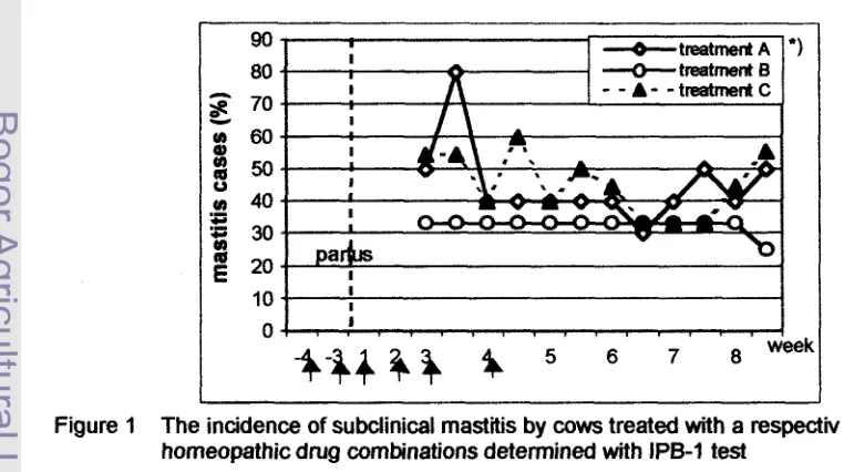
dan iraumeel@(minggu ke-1 dan 2 p.p.), Coenzyme comp@ dan Carduus comp@ (minggu ke-3 dan 4 p.p.). Grup C adalah plasebo.Pengujian dengan reagens IPB-1 di Lapangan memperlihatkan bahwa insidensi mastitis subklinis pada grup B adalah sebanyak 33,3% dan bertahan konstan sampai akhir pengamatan, sedangkan grup A dan C kasus mastitis sangat bervariasi. Kenaikan jumlah sel somatik susu dari grup A dan B satu minggu setelah terapi terakhir selama 2 minggu (minggu ke 5 - 7 p.p.) menuryukkan adanya suatu respon yang nyata terhadap terapi homeopathy kemudian diikuti penurunan jumlah somatik sel mendekati nilai normal.
Respon setelah disuntik Coenzyme comp. (minggu ke-3 a$.) memperlihatkan adanya peningkatan jumlah sel darah merah (RBC) untuk grup A (17,8%), selanjutnya diikuti penurunan RBC sampai akhir pengamatan. Gambaran haemoglobin dan pack cell volume tidak mengalami perubahan (konstan). Limfosit grup A dan C meningkat menjelang proses kelahiran, ha1 sebaliknya terjadi bagi grup B. Kadar haptoglobin (Hp) pada fase peripartal meningkat bagi grup B 0,86
mglml dan bagi grup A maupun plasebo 1,44 mglml serta 1,42 mglml. Pengamatan
5 minggu p.p. grup B mengalami penurunan jumlah Hp menjadi 0,09 mglml sampai akhir pengamatan adalah 0,07 mglml. Kadar Hp darah grup A menurun secara perlahan, kadar terendah dicapai pada minggu ke-5 p.p. yakni 0,18 mglml.
ABSTRACT
AGATHA WlNNY SANJAYA. Control of Subclinical Mastitis Using Certain Homeopathic Combination Drugs and Its Influence on Milk Quality and Production. Under the direction of I WAYAN TEGUH WIBAWAN, MlRNAWATl SUDARWANTO, HEINRICH ENBERGS, SETYO WIDODO, MASDUKI PARTADIREDJA (late), HEINER SOMMER (Late).
A total of 33 dairy lactating cows suffered from subclinical mastitis were classified into group A (10 cows), group B (12 cows) and group C (11 cows) which were treated with the combination of homeopathic drugs and placebo, applied at the 4'h and 3* week antepartum (a.p.) and every week for four times postpartum (p.p.). Group A received Coenzyme compQD (given in the 4'h and 3'C' week a.p.), Lachesis compQD with ~raumeel' (I& and 2nd week p.p.) and
Coenzyme compQD with Carduus compQD (3rd and 4'h week p.p.). Group B received ~ r a u m e e l ~ with Mucosa compQD (4" and 3rd week a.
i?
.), Lachesis comp@ with ~raurneel~(1" and 2nd week p.p.) and Coenzyme comp with Carduus compQP (3* and 4'h week p.p.) and group C as a placebo.Incidence of mastitis in group A and C appeared irregularly. In contrast, group B showed a constant percentage (33.3%). Group A and B showed significant response to the homeopathic drugs, expressed as an increasing of the somatic cell count value after the last therapy for two weeks (5'h
-
week p.p.) and at the last observation decreased nearly to the normal value.At the puerperal phase, the red blood
cell
of group A increased (17.8%) after the first therapy. Hemoglobine and pack cell volume showed no comparable results. Lymphocyte of group A and placebo increased during puerperal phase, contrary happened to group B. At peripartal phase haptoglobin increased for group B 0.86 mglml and group A as well as placebo 1.44 mglml. After birth, group B expressed a constant value (0.09-
0.07 mg/ml), while group A and placebo raised significantly.Statement of originality
The studies described in this dissertation were performed in accordance with the
regulations for the degree of Doctor in
the
Bogor Agricultural University.This dissertation consists entirely of original research which has not been
submitted in whole or in part for any other degree at this university or at any other
CONTROL OF SUBCLINICAL MASTITIS USING
CERTAtN HOMEOPATHIC COMBLNATION DRUGS AND
ITS INFLUENCE ON MILK QUALITY AND PRODUCTION
AGATHA WlNNY SANJAYA
Doctoral Dissertation
submitted in fulfilment of the requirements to achieve a Doctor degree in Veterinary Sdence Study Program
THE GRADUATE PROGRAM
BOGOR AGRICULTURAL UNIVERSITY
B O G O R
CURRICULUM VITAE
Agatha W~nny Sanjaya was born in MEDAN, June
7*
1946 as the fifth child from the five children of lgnatius Dyan Kumiatan and Veronica Sukawati. The authormarried with Dominicus Setyasusila Sanjaya and has two children Benedictus
Adhi and Martina Citra.
In 1964 entered the Faculty of Veterinary Medicine Bogor Agricultural
University and inaugurated as doctor of Veterinary Medicine in 1973. Master
degree was achieved in Veterinary Science in 1990 and in 1997 February,
entered Doctor Program at the Graduate Program in Bogor Agricultural University
and was funded by the Beasiswa Pendidikan Pascasarjana. A part of the
research was funded by the Project of University Research for Graduate
Title of Dissertation : CONTROL OF SUBCLINICAL MASTITIS USING CERTAIN HOMEOPATHIC COMBINATION DRUGS AND ITS INFLUENCE ON MILK QUALITY AND
PRODUCTION
Name of Student : Agatha Winny Sanjaya
Registration Number : 965104
Study Program : Veterinary Science
Approved by: 1. Advisory Committee:
Dr.drh.1 Wavan Tenuh Wibawan M.S. Prof.Dr.drh.Hi.Mimawati Sudawanto
chairman member
Prof. Dr. Heinrich Enberns member
2. Head of Study Program: 3. Director of the Graduate Program:
Acknowledgements
I would like to express my gratitude to my chairman advisor, Dr. drh. I
Wayan Teguh Wibawan, MS, Department of Animal Diseases and Veterinary
Public Health, member advisory, Prof. Dr. drh. Hj. Mimawati Sudawanto, Prof.
Dr. H. Enbergs, Dr. drh Setyo Widodo, for all his invaluable input, assistance and
support throughout the course of this work. Special my honour are due to the
late: Prof. Dr. Heiner Sommer and Prof. Dr. drh. H. Masduki Partadiredja for their
encouragement.
I am very grateful to the Beasiswa Pendidikan Pascasarjana, Project of
University Research for Graduate Education, for their sponsorship which enabled
me to study in Germany. My thanks also go to the Rector IPB, Director of PPS-
IPB, Dean of Veterinary Medicine Faculty, the head of Department of Animal
Diseases and Veterinary Public Health at IPB, Bogor, lnstitut fiir Anatomie,
Physiologie und Hygiene der Haustiere der Rheinischen-Friedrich-Wilhelms-
Universiet at Bonn, Fa. HEEL Baden-Baden Germany and the chief of Animal
Quarantine Service, Jakarta International Airport, for their support.
I would also like thank to the director of TAURUS Dairy Farm, Ir. Nugroho
and his crew, all my co-workers, who were very helpful and sympathetic during
the completion of the work in the field and laboratory.
I am also grateful to the staff members of Veterinary Public Health
Laboratory, University of Bonn where I had done my research work and to all of
my colleagues who supported me in various ways: Dr D.W. Lukman; Drh R.K.
Achyadi MS; Ir E. Sudamika M.Si; Dr Fachrudin; Drh E.S. Pribadi, MS; Drh Lukas
Tonga, Ir Agustine Santosa MS, Dr Linscheid; Dr U. Miiller; Dr S. Knura and my
friend Fr. Rosemarie Schattevoy. Last but not least, I would like to thank my
LlST OF CONTENTS
LIST OF TABLES LIST OF FIGURES LlST OF APPENDICES LlST OF ABBREVIATION
1. INTRODUCTION 1.1 Background
1.2 General Objectives of this Study 1.3 Hypothesis
2. LITERATURE STUDY 2.1 Milk
2.2 Defence Mechanism of Mammary gland 2.3 lrnmunological Defence at Peripartal Phase 2.4 Subclinical Mastitis
2.4.1 Prevalence and Characteristic of Subclinical Mastitis 2.4.2 Diagnosis of Subclinical Mastitis
2.5 Homeopathy as an Alternative Therapy
2.6 Advantageous of using Combination Form of Homeopathic Drugs 2.7 Control of Subclinical Mastitis by Homeopathic Treatment
2.8 Homeopathica Substances 2.8.1 Coenzyme compositum 2.8.2 Traumeel
2.8.3 Mucosa compositum
3. MATERIAL AND METHODS
3.1 Materials
3.1.1 Milk and Blood Samples 3.1.2 Instruments
3.1.3 Chemicals. 3.2 Animal contingent 3.3 Methods
vii vi i ix
3.3.1 Determination of Health Status of Cows Concerning to Subclinical Mastitis
3.3.2 Milk Quality Analysis
3.3.3 Blood Analysis
3.3.4 Clinical Chemistry
3.4 Data Collection
3.5 Research Design
3.6 Data Analysis
4. RESULTS AND DISCUSSION
4.1 Milk
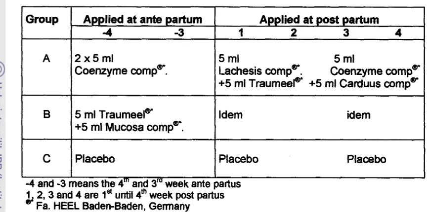
4.1.1 Incidence of Subclinical Mastitis
4.1.2 Somatic Cell Count
4.1.3 pH of Milk
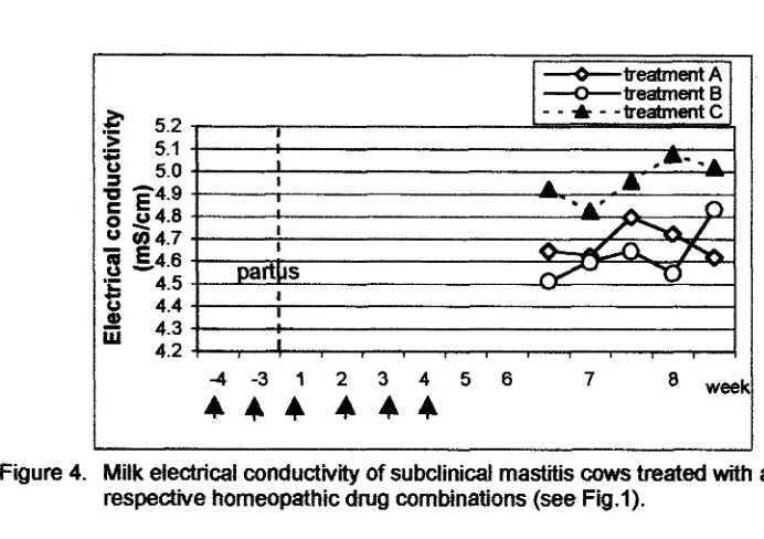
4.1.4 Milk Electrical Conductivity
4.1.5 Fat Content
4.1.6 Protein Content
4.1.7 Lactate dehydrogenase Enzyme in the Milk
4.2 Blood Profile
4.2.1 Red Blood Cells and Pack Cell Volume
4.2.2 Hemoglobin
4.2.3 Leukocyte Profiles.
4.2.4 Lactate dehydrogenase and Aspartate Aminotransferase Enzymes
4.2.5 Cholesterol
4.2.6 Urea in Blood
4.3 Haptoglobin
4.4 Milk Yield
5. CONCLUSIONS AND RECOMMENDATIONS
A. Conclusions B. Recommendations
LlST OF TABLE
1. Relationship between somatic cell count-score, somatic cell count and daily production loss of dairy cows
2. Estimated infection prevalence and looses in milk production Associated with elevated bulk tank somatic cell counts
3. Determination of CMT-score associated with an average somatic cell count
4. A comparison of methods used in subclinical mastitis test and their correlation with somatic cell counts
5. An overview of the use homeopathic drugs in mastitis control
6. Combination of homeopathic drugs used in the treatments
7. Application schedule of respective homeopathic combination
(A, B and C) and time of milk and blood sampling
LlST OF FIGURE
I. The incidence of subclinical mastitis by cows treated with a respective homeopathic drug combinations determined with IPB-I test
2. The effect of homeopathic treatments to the profile of somatic cell counts with a respective homeopathic drug combinations
3. The pH value of milk from subclinical mastitis treated with a respective homeopathic drug combinaiions
4. Milk electrical conductivity of subclinical matitis cows treated with a respective homeopathic drug combinations
5. Fat contents of milk from subclinical mastitis cows which pretreated with difference homeopathic combinations
6. Protein content of milk from subclinical mastitis cows treated with a respective homeopathic drug combination
7. Lactate dehydrogenase in milk of subclinical mastitis cows before and after treatments
8. Red blood cell profile of milk from subclinical mastitis cows treated with a respective combination of homeopathic drugs
10. The leukocyte profile of subclinical mastitis cows treated with respective
combinations of homeopathic drugs 48
I I. The neutrophil profile of subclinical mastitis cows treated with
respective combination of homeopathic drugs 49
12. The lymphocyte profile of subclinical mastitis cows treated with
respective combination of homeopathic drugs 49
13. The eosinophil profile of subclinical mastitis cows treated with
respective combination of homeopathic drugs 50
14. The LDH profiles of blood from subclinical mastitis cows treated with
respective combination of homeopathic drugs 52
15. The ASAT profiles of blood from subclinical mastitis cows treated with
respective combination of homeopathic drugs 52-
16. The cholesterol profile of blood from subclinical mastitis cows treated
with respective combination of homeopathic drugs 53
17. The urea profile of blood from subclinical mastitis cows treated with
respective combination of homeopathic drugs 54
18. Haptoglobin profile of subclinical mastitis cows treated with respective
combination of homeopathic drugs 56
19. Milk yield observed in normal lactation at the 3'Cl-p month before
,LIST OF APPENDICES
1. Mean values of somatic cell count in milk of subclinical mastitis cows treated with respective combination of homeopathic drugs
2. Mean values of pH milk of subclinical mastitis cows treated with respective combination of homeopathic
3. Mean values of electrical conductivity of subclinical mastitis cows treated with respective combination of homeopathic drugs
4. Mean values of milk fat from subclinical mastitis cows treated with respective combination of homeopathic drugs
5. Mean values of milk protein from subclinical mastitis cows treated
with respective combination of homeopathic drugs 68
6. Mean values observations of red blood cell, pack cell volume and
hemoglobin from subclinical mastitis cows 69
7. Mean values observation of leukocyte and its differentiation picture from subclinical mastitis cows
8. Mean values observation of blood enzyme and metabolite from
subclinical mastitis cows 71
9. Mean values of haptoglobin concentration in blood plasma from
subclinical mastitis cows 72
10. Total 5 months production from cows before and after treatment
with respective combination of homeopathic drugs 72
11. Statistical Analysis of Varians of 5 months milk production in normal
LIST OF ABBREVIATION APRP C CMT comp. CRP de n o w D DHlA Dl DNA et a!.
Fig. Hb HP IU LDH min. mg ml mS NAD PCV p.a. pH PMN rpm RBC
=
degree Celsius=
registered=
weighheight=
unitllitre=
ante partum=
post partum=
Aspartate Aminotransferase enzyme= Acute Phase-Reactant Protein
=
centesimal scale=
California Mastitis Test=
compositum=
C-Reactive Protein=
remedy made from the products of a disease=
decimal scale=
Dairy Herd Improvement Association= decilitre
=
Desoxyribonucleic acid=
et alia= Figure
=
hemoglobin= haptoglobin
=
international unit=
Lactate dehydrogenase=
minute=
milligram=
millilitre= millimetre Siemens
=
Nicotine Adenine Denucleotide=
Pack Cell Volume=
pro analyse=
hydrogen ion concentration=
Polymorphonuclear Neutrophil=
rotation pro minute [image:15.593.81.443.48.781.2]sec SCC
VLDL WMT WST
IJL
= second
= Somatic
cell counts contains leukocyte cells and mammary epithel cell debris= very low density lipoprotein
= Wisconsin Mastitis Test
=
Whiteside TestCONTROL OF SUBCLINICAL MASTITIS USING CERTAIN HOMEOPATHIC COMBINATION DRUGS AND ITS INFLUENCE ON MILK QUALITY AND PRODUCTION
1. INTRODUCTION
Subclinical mastitis is one of the most important diseases in dairy cattle in
lndonesia with the prevalence of 85-90% (Sudarwanto, 1995). It shows no
significant changes of udder and in milk appearance but causes a significant
economic loss (10-18%) mainly because of losses of milk production (15-40%)
per day, poor quality of milk, milk discard, drug costs and veterinary fee (Kirk et
a/., 1994; Lee, 1996; Sudarwanto, 1999). Similar results had also shown by Hirst
et a/. (1 984)
.
The decrease of milk production is felt to be more important than that
caused by clinical mastitis. The recent data show that the amount of dairy cattle
in Indonesia is 343,000 heads from which 260,000 are in lactation. They
produced 405,000 tons of milk/ lactation period. By assumption that 60% of the
lactated group suffer from subclinical mastitis, the economic loss due to the
decrease of milk production is approximately 108 billion rupiahs per year
(Sudarwanto, 1999).
1 .I Background
Antibiotics have been used in controlling mastitis. The use of inappropriate
antibiotics cause bacterial resistance, residue problems, allergy, metabolic
disorders, toxicity and influenced in milk processing (Kiehvein, 1976). Previous
Jakarta, Bogor and Bandung contained significantly high antibiotic residues
(Sudarwanto et a/. 1992).
Nowadays, greater emphasis has been placed on antibiotic residue
avoidance to ensure a residue-free milk for the consuming public. The major
change in this direction is the reduction in the use of antibiotics in the treatment
of mastitis.
As an alternative, homeopathic drugs are to be taken into consideration.
Their advantages are that they cause no toxic side effects and no residues in
animal's products. Missing corresponding placebo-controlled studies it was the
aim of the following studies to test the effect of certain homeopathic drugs in
prevention of subclinical mastitis in dairy cows.
1.2 General Objectives of this Study
1). To study the most effective combination of the homeopathic drugs in
prevention and therapy of subclinical mastitis.
2). To study the effect of the homeopathic drugs in enhancing milk production
and quality.
3). To study the influence of homeopathic drugs to the blood profile of subclinical
mastitis-treated cows.
1.3 Hypothesis
1). Homeopathic drugs depress the incidence of subclinical mastitis.
2). Homeopathic drugs enhance the milk quality and milk yield.
3). The effective combination of homeopathic drugs for subclinical mastitis
2.
LITERATURE STUDY2.1 Milk
Milk is composed of water, carbohydrate (lactose), fat, protein, minerals
and vitamins and is secreted as a complex mixture of these components. The
properties and importance of milk are greater and more complex than the sum of
its individual component parts.
Water content of milk is about 87% and dependent upon the synthesis of
lactose. If water is added to the milk, the additional water is easily detectable by
several methods. These methods are based upon changes in freezing point of
the milk (cryoscopic method) or on changes in refraction light of the whey
component of milk after precipitation and removal of the casein and fat
components. These methods and other standard methods of milk testing are
described in the Association of Official Agricultural Chemists (AOAC), Official
Laboratory Analytical Procedures (National Academy of Science).
Carbohydrate substance of milk is mostly lactose. Lactose is a disaccha-
ride composed of the monosaccharides D-glucose and D-galactose, joined in a
13-1,4-glycosidic linkage. The chemical name of lactose is 4-0-13-D-galacto-
pyranosyl-D-glucopyranose and is essentially unique to milk. Lactose plays an
important role in milk synthesis. Lactose is responsible for maintaining the
osmolarity of milk and for drawing water in milk synthesis. Lactose is not as
sweet as other disaccharides such as sucrose or monosaccharides fructose or
glucose. The lactose cleaved to glucose and galactose in the neonate intestine
is done by an enzyme activity called lactase or 13-galactosidase. Other carbo-
hydrates are found in milk at low concentrations, such as free glucose (0.1 mM)
The fat component of milk is composed of a complex mixture of lipids.
Triglycerides or triacylglycerides are the major type of lipid in milk fat, 98% of the
total milk fat (wtw). Triglycerides are composed of 3 fatty acids covalently bound
to a glycerol molecule by ester bonds. Milk fat is secreted from mammary
epithelial cells as fat globules which are primarily composed of a globule of
triglyceride surrounded by a lipid bilayer membrane similar to the apical
membrane of the epithelial cells. This fat globule membranes helps to stabilize
the fat globules in an emulsion within the aqueous environment of milk. Other
milk lipids include diacylglycerides (0.25-0.48%), monoacylglycerides (0.02-
0.04%), phospholipids (0.6-1.0%), cholesterol (0.2-0.4%), glycolipids (0.006%)
and free fatty acids (0.1-0.4%).
The fatty acids used for the synthesis of triglycerides arise from the break
down of blood lipids and from de novo synthesis within the mammary epithelial
cells. About 40-60% of the fatty acids come from the blood. These are primarily
derived from very low density lipoproteins (VLDL), which are synthesized in the
intestine or liver. VLDL are composed of 90-95% on the inner core and 5-10%
protein at the outer surface. Chylomicrons, containing ingested fatty acids from
the intestine, also can act as a source of blood-derived fatty acids for the
mammary gland. Synthesis of short or medium chain of fatty acids occurs by de
novo synthesis in the cytoplasm of the mammary epithelial cells. The sources of
carbon the acetate and 13-hydroxybutyrate are mainly used.
The total protein component of milk is composed of numerous specific
proteins. The primary group of milk proteins are the caseins. Caseins have an
appropriate amino acid composition which is important for growth and develop-
ment of the nursing young. This high quality protein in cow milk is one of the key
reasons why milk is such an important human food. Casein is one of the most
other protein component of milk is whey or plasma phase of milk. The major
whey protein in cow milk are a-lactalbumin and 13-lactoglobulin. Alpha lactal-
bumin is an important protein in the synthesis of lactose and its presence is
central to the process of milk synthesis. Other whey proteins are immuno-
globulins (especially high in colostrum), serum albumin, enzymes, hormones,
growth factors, nutrien transporters, and disease resistance factors.
The major minerals found in milk are calcium and phosphorus. These
minerals are required in large quantities by the rapidly growing neonate for bone
growth and development of soft tissues. They are both mostly associated with
the casein micelle structure. Milk also contains most other minerals found in the
body.
Milk contain all the major vitamins. The fat soluble vitamins A, D, E, and K
are found primarily in the milk fat, and has limited amounts of vitamin K. The B
vitamins are found in the aqueous phase of milk. Milk always contains leukocyte
cells and mammary epithelial cell debris, also known as somatic cells in cow
milk. The concentration of leukocytes in milk varies with the species, infection
status of the gland and the stage of lactation.
2.2 Defence Mechanism of Mammary gland
Physical barriers of mammary glands play an important role in the defence
mechanisms of the mammary glands. This included the teat shape, teat
sphincter, streak diameter, streak canal, the length and the diameter of streak
canal and the keratin lining in streak canal, which contains factors that seem to
be bacteriostatic. Keratin is a meshlike substance, formed from descquamated
epithelial cells, fatty acids and cationic proteins and play as a physical
desquamated during milking which removes bacteria in the streak canal. The
keratin's fatty acids are bactericidal and bacteriostatic and it has proteins which
bind to and cause lysis of Gram positive bacteria. Furstenberg's rosette is
situated at the internal end of the streak canal, contain a protective leukocytes
population, which are thought to leave teat wall and enter the cistern via
Furstenberg's rosette. This rosette contains also ubiquitin a cationic protein
which has bactericidal activities.
lmrnunological defence of mammary glands includes all physiological
mechanisms allowing the body to identify and neutralise foreign bodies and is
comprised of: (1) cellular Immunity and (2) humoral Immunity. The immune cells
include the role of leukocytes such as granulocytes (neutrophils, basophils and
eosinophils) and lymphocytes, especially T-killer cells, and monocytes or macro-
phages. These cells are mainly responsible for the phagocytosis process in milk
and are considered as a second line of defence in the mammary gland. Humoral
immunity includes the role of antibody as opsonin, neutralisation of antigens or
activation of the complement system.
2.3 lmrnunological Defence at Peripartal Phase
lmmunological defence must be understood as an integrated system of
non-specific and specific immune responses, which has an important function to
maintain the physiological homeostasis. Peripartal period (about 4 weeks before
and after birth) is considered as a critical point and unstable physiological
condition of the dairy cows due to the sudden changes or switch over of many
processes including hormonal, metabolic, stress and cytologic performance.
Especially during the early lactation stage problems may arise to maintain the
cow in a physiological balance. This might influence the whole system of
(1) exhaustion of reserves of carbohydrates, (2) conversion of the body's protein into sugars, (3) mobilisation of lipid depots and decomposition of the lipids to produce energy with a risk of a fatty fiver and fmlly (4) increase of so called aceton-bodies. Interference between aceton bodies and leukocyte function decreased the interferon (IFN-Y) production. Interferon involved in the regulation of T cells, 6 cells, macrophages and granulocytes, so that the disturbance of IFN production caused the reduction of phagocytic activity of the -phages as well as of the granulocytes which are considered as the first line of defense of the bovine mammary gland against infections. This unstable physiological condition might lead to the incidence of mastitis facilitated by postpartal immunosupression (Kandefer-Szerszen et a/.
,
1992; Klucinski etal.,
1988). The stress-situation and damages of tissues connected with the birth process, especially accrued in the soft tissue of the birth ways.2.4 Subclinical Mastitis
2.4.1 Prevslence and Charadetfstics of Subclinical MasWs
Mastitis from the Greek mastos-breast and itis is a general term used to refer to any inflammation of the mammary gland The previous studies indicated that subclinical mastitis in dairy cattle is thought to be a multicomplex disease.
infectious subclinical mastitis the Streptococcus agalaciiae, Staphyilocioccus aueus and Escherichia mli are well known as the main causal agents (Wibawan et al., 1999). Bacteria could enter the mammary glands and multiply sufficiently to trigger the inflammatory response. Vasodilatation occurred and results in increased blood flow to the mammary glands. Inflammatory products such as prostaglandins, leucotrienes, pmteases, and oxygen radicals increased the vascular permeability. Leukocytes, initially polymorphonuclear neutrophils (PMN) leaved the blood vessels and diapedicaly enter the surrounding tissue, after which macrophages predominate.
Subclinical mastitis has remained the most economically important problem in dairy cattle in Indonesia with high prevalence ranging fmm 85 to 90%
(Sudarwanto, 1995; Wibawan et a1.,1999). The information about the pathogene- sis of subclinical mastitis is very limited. In subclinical mastitis, the milk appears to be normal. Bacteria usually, but not always, can be isolated in milk. Milk yield is depressed, and composition may
be
altered. No pathological changes of mammary gland could be observed but the histopathological examination showed that the inflammation of the mammary gland had occurred. Inflammation of mammary gland caused by colonisation as well as by invading pathogens is common among lactating dairy cows and is a major cause of economic losses.contributes to the loss of income in the following ways. 70% from lost of milk production, 14% from death and premature culling, 8% miUc discarded at treatment and 8% for drugs and veterinary (Bailey, 1996).
S. aureus and S. agalactiae as the main causal agents of infectious subclinical mastitis often start with an acute phase and generally become chronic and subclinical. In contrast, E. coli caused an extensive damage of mammary gland tissue. This opposite characteristics are likely to results from the difference of pathogenic mechanisms used by those bacteria. By S. agaladiae and S, aureus the adhesion is thought to be the most important and critical step of infection and followed by the colonisation of bacteria on the surface of the mammary epithelial cells wbawan et al., 1999). The differences in the pathogenesis and clinical reaction of causal agents offer the opportunity to contrast the inflammatory responses and to study the early host responses to the respective causal agents by measuring the inflammatory markers such as haptoglobin, C-reactive protein (CRP) or other inflammatory mediators which might be used for the early screening test of subclinical mastitis
in
dairy cattle (Sandholm et a/., 1995). In case of acute mastitis, the clinical symptomes areobvious and laboratory tests are not required to detect inflammation. Detection of subclinical mastitis is a challenging problem. Subclinical mastitis reduces milk production, milk composition and quality of milk and also maintains infection foci in the herd (reservoir). The extent to which various compositional changes occur depends on the inflammatory response. This depends on the virulence factor of the pathogen and the amount of affected tissue within the mammary gland. Inflammation should be analysed by monitoring various inflammatory mediators.
Ca, Mg concentration. Disturbance of the milk synthesis caused the decreasing
of casein-,
p
ladoglobulin-, a lactalbumine-, lactose concentration and fat in milk.The high concentration of Na and CI in the milk caused a salty taste and alkalic
property of milk. The milk becomes heat unstable and was not completely
coagulated by rennin and caused failure in cheese ripening. According to Munm
in Hamann and Rund (1994) mastitis effect on milk and milk product are : (1)
fresh milk changes very easy in a rancid taste and the protein are heat unstable,
(2) pasteurised milk has an unfavourable flavour and with an unstable quality and (3) effect on making cheese are: a reduction in starter activation and
delayed coagulation time, breaking firmness decrease, lost of fat and casein in
milk at last there is a low profit and (4) problem in processing butter would
influenced the taste with an oxidation taste, present a weak aroma, prolonged
rippening butter time and inhibition of diacetyl product.
2.4.2 Diagnosis of Subclinical Mastitis
2.4.2.1 Somatic Cell Count (SCC)
Somatic cell counts (SCC) from a day's milk is the best indicator of the
extent to which the gland is involved in fighting a mastitis infection. Milk contains
cells which derive from the cow, these are somatic cells. The number of somatic
cells is immediately high after calving, but it drops rapidly during the first week of
lactation and increases again towards end-lactation. Therefore, the number of
cells depends on two factors: 1). a real increase in their quantity as a
consequence of udder initation and attraction to the inflammatory site or 2). The
dilution factor, i.e. the daily milk yield shows an inverse relation with the somatic
cell count Somatic cells are present in the milk in high number only when and
(Sandholm et a/., 1995). The quality of raw milk produced in Germany is
guaranteed by a milk quality control scheme conducted in accordance with
European Committee standards. In addition, because of its effect on the price
paid for milk, the somatic cell count in bulk milk is an indicator of udder health.
Udder health can only be maintained by strict adherence to accepted control
measures during milking, hygiene and prophylactic treatment (Fehlings and
Deneke, 2000). The cells found in milk consist of about 75% leukocytes and
about 25% epithelial cells. Normally SCC are 60-70% epithelial cell, 10%
lymphocyte and 2030% leukocytes (Sinell and Neuschulz, 1965). Leukocyte
numbers increase in response to bacterial infection, tissue injury and stress.
Increasing somatic cell numbers is a result of an increase in the number of
leukocytes.
The somatic cell count of milk serves as an indirect method to measure the
level of irritation in the mammary gland. There are 3 methods of evaluating SCC:
(1) Bulk Tank SCC (BTSCC), (2) Weighted SCC (VVTSCC) and Somatic Cell
Count Score (SCCS). The BTSCC is usually based on single sample of milk
from the bulk tank. The WTSCC is calculated on samples from individual cows
and is weighted or adjusted based
on
each cow's level of milk production. TheWTSCC for all milking cows in a herd should approximate the BTSCC. The
SCCS is based on the logarithmic conversion of the actual SCC to a linear score
as shown in Table 1.
The loss in milk production between scores 2 and 3 is estimated at 1.5
Ibs/cow/day based on an actual SCC difference of about 50,000 cells. Cows with a SCCS of 0-3 a= generally considered uninfected while cows with SCCS of 7
-
Table I . Relationship between somatic cell count-score, somatic
ceH count
rangeand daily production loss of
dsmy
cows(Shook
and
Saeman in Smith et a/. 2000)The value of SCCS is associated with the ladation period, milk yield, the dens@ of cows
in a herd and the season. The first
lactation have a Iower SCCS with a h i r percentage inthe
0-3 category compared to older cows. This is not directty associated with theage
but
mused by increased rate ofudder
infections. Unmfectedcows,
regardless of age generally have low SCC (Ebehart et a/.,1979). High producing cows as well as high density herds trends to have a
higher SCC compared to low producing cows
or
lower dens@ herds. A high SCCS are generally observed duringthe
summer time, in the months of July,Herds with butk tank SCC above
200,000
wilt have varying degreesof
subclinical mastitis present.In the Table 2, data from the National Mastitis Council (1987)
show
that 6%of the quarters in a herd could be expected to be infected in a herd with a bulk tank SCC of 200,000. At 500,000 SCC, 16% of tho quarters may be infected
with a 6% reduction in milk produdion. Ttds information lead
to
the conclusion that the monitoring of udder heal# is very importantin
order to control andreduce the level of mastitis in a herd. Somatic
d l
count measurement providethe
opportunity
to monitor and evaluate udder health status.Tabk 2. Estimated infedion prevalence
and
t m s in milk produdion associatedwith elevated bulk tank somatic
ceH
counts* Pmduction loss cabMed as a percent d production expected at 200,000 cell1 ml. National M a s t i Council (1 981).
Somatic celt count under 400,000
dldmt
are typicalof
herds that have good management practices, but no partiwlar emphasison
mastitis control. Incontrast, somatic cell caunts greater than 500,000 cellslml indicates that one
third
of
the mammary gtands are infectedand the
lost of milk due to subclinical2.4.22 California Mastitis Test ( C m
and
Mliscmin
Mast&% Test (WIUT)The CMT test is a simple method to estimate the DNA
content
of milk. This test bases on an anionic detergent, Na-iauryt sulphate (Dodecyl Sulfate Sodium),which dissolves eel membranes
and
nuclei. Consequently DNA is released andit forms a transient gel with
the
detergent The more DNA in the milksample,
the higher theviscosity of
the get (Sandholm etaL,
1995). The v a w ~ o nof
scoring of CMT, dependupon
the skill of the person reading the result and the methodused to conduct the test. Uniformity in techniques are necessary if results are to
be comparable. The use of the CMT on the entire herd
at
monthly intervals canbe
extremely useful asan
aid in detecting herd mastitis problems. individual andtotal quarter infedions
can
be determined and, with proper records,the
tevel of herd mastitis can be monitored. The relation between CW-Score and theaverage somatic cell count is presented in Table 3
Table 3. Determination of CMT-Score associated with an average somatic cell count.
The advantages of
the
CMT are: (I) fairty accurate in measuring somaticceH
concentratin in mitk, meiating well with athertest,
(2) it is sensitive andcan
be used for samplingquafters,
bucketand
buk tank milk samples, (3)foreign material does not interfere with the
test,
(4) simple and a little equipment CMT-ScoredNegative Trace
2
3
Average somatic cell
count
(cells/ml)300,OOO
m,m
~,~
2,700,000
[image:30.564.73.514.470.640.2]is needed, (5) environmentat temperature changes have only a lime effed on the CMT as Long as the milk has been refrigemfed and is not over two days OM and
(5) herd mastitis levels can be estimated from bulk €ank A CMT
d
2 or 3 in tank milk indicates a probable percentof
infected cows.The Wisconsin Mastitis Test (WMT) is an other
method,
which frequently is used as a screening test for subclinical mastitis. The principleaf
the WAM is the same as that of the CMT except that the amount of gel formed is measured in a tube calibrated in miltimeters (mm) instead of being visually scmed. Theadvantageous of WMT is conducted under precise procedures and standard
temperatwe conditions. A more accurate and
precise
method of measuringsomatic cetls in milk is
the
ekctronically m - dsomatic
cell countsused
by Dairy Herd Improvement Association (DHIA).Somatic cell counts is expressed in thousands of cells per milliliter, sometimes referred to as raw score, and converted
to
a Enear score rangingTaMe 4. A comparison of methods used in subdinical mastitis
test
and their correlation with somatic cellcounts
(Eastridge and HoMet, 1992).2.4.23 IPE1
Pest
The determinatk#1
of
mastitis irt the btdcan
bedone
by using diagnostical reagent. Recently, fieid subdical mastitis tests which are availablesuch
as CMT are rarely donedue
to theprice
and the avaitabifi of the reagents.Another reagent called IPB-1
reagent,
is devetoped for a field test that isrelatively easy to use, inexpensive, and consttucted of ingredients readily are
available in domestic market. The reliability value of IP8-I is better than CMT or
WST, the
sensitivrty
of IPB-I is 0.99and
CMT 0.92, the spectficity 0.92 with 0.36and
the
prect'~dive value 0.95 with 0.97 respectively. The prdictive value still2.4.2.4 Lac- dehydrogenase
Lactate dehydrogenase (LDH) is an enzyme widely distributed among the
various species of the animal kingdom and ubiquitous in the tissue of man and
other vertebrates. This enzyme catalyses the oxidation of lactate to pyruvate and
the reverse reaction, the reduction of pyruvate to lactate. LDH plays an important
role in the intermediary metabolism of various tissues. For the catalytic activities
of LDH the presence of a coenzyme nicotine amide adenine denucleotide (NAD)
is essential.
The LDH activity in milk might be used as an indicator to determine the
early stage of inflammation in the mammary glands and is recommended for the
screening technique (Hambitzer and Sommer, 1987). The value of this enzyme
in the milk is not more than 85 UA and when this value exceeded, mastitis is
present (Andersson, 1991). Accoding to Sommer et a/. (1986), LDH activity in
milk is higher in samples containing pathogenic bacteria (S. aureus) compared to
the non infectious mastitis milk. A high positive correlation was found between
cell count and LDH activity in milk containing pathogenic bacteria. The LDH
activlty increased from about 130 to 260 Un as cell count increased from
250,000 to 1.5 millionlml. No comparable results were shown by the samples
containing non-pathogenic bacteria, where the activity of LDH remained fairly
constant about 100 UII as cell count increased from about 250,000 to 1.25
millionlml.
2.4.2.5 Acute P h s e Response and Subclinical Mastitis
The maintenance of a physiologic homeostasis during ups and downs of
daily living is assured by a number of physiological mechanism. Tissue injury
and infection which represent a threat to the integrity of the organism, and
eqicilibrium. The local response of tissue
to
suchinjury
or infection is acute inflammation. It's major clinical maniksWons resuit from changes in vascular caliber and flow, increased vascular permeability and attraction of leukocytes. Durtng the first few days €&tawing insult, a vast numberof
systemic andmetabolic changes occur, that is the acute response. The acute response should
be understood as a value in helping to pemtit survival during
the
period immediately foflowing iniuty and in achieving the same goalsas
We localinflammatory response does, containment or destruction of infedious agents,
removal of damaged tissue and repair of the affected organ.
In mammals, tissue damage or inflammation at a localized site lead to
systemic changes known as the acute phase response. The acute phase reaction is an earty body defense mechanism tfiggered in response to cellular
injury manifested through a variety of adverse conditions,
such
as infection,inflammation and advanced matignancies. This reaction provides substantial
immediate protection before humoraCmedited immunity becomes effective.
Among the varied physiological atteratian, which together produce
this
response,is a
change
in the circulating levels of a numberof
liver derived proteins, collectively knownas acute
phase reactant p m f e h (APRP) (Koj, $974; Saini eta/.,
1998). Acute phae-pmtetns are serum proteins, whose concentrationsincrease during
the
acute phase response to inflammationor
infedion. Therespanse occurs in alt animals, but it is s ~ i m t t y different in various species (Eckersall et a/., 1988). APRP in man
and
animals indude C-reactive protein(CRP), serum amyloid A (SAA), haptogtobi (Hp), cenrioplasmin, orosomucoid, al-antitrypsin, a2-moglobuIirtl comptement and coagulation proteins (Peppys
100 kDa find in blood and fomred in the Cver
of
mammals (Knura, 2000). Theassay
of
serum Hp in bovine serum or plasma isnow
a routine biochemical testfor veterinary diagnostic in Eurppe and USA. This is due to
the
abilityof
Hp to bind hemoglobin. It binds free hemoglobin, thus p r e s e ~ n gthe
body, which isliberated from damaged erythmcytes.
SAA
and Hp concentration wereassociated only with acute and sub
awte
lesions but not for animals with chronicinfhmmation (EckersaH, 1995). The possibility
of
using Hp for thedetection
of mastitis has been reported by Comer et a!. (I-), whichshowed that
the
change in haptoglobin level from zero to 70 mg HpflOO ml by cows with clinical
mastitis. Until now, there is no information available about the using of H p
biological activity for early detection of subclinical mastitis.
2.4.2.6 Ekctrical Conductivity in Milk
A precise diagnose
of
udder health status isthe
base foreffedive
mastitis control. Cell count and electricai conductivity are used to indicate inflammatorychanges in the bovine
mammary gland.
The basisof
mastitis diagnosis consistsof
the
combination of the detection ofthe
inflammatory changes, but for economic reasons very often the measurement ofthe
numberof
somatic cellsand the ekctrkai conductwit)c in milk performed withwt parallel bacteriological
examination (Hamann and Gyod, 1999). Accocdfng to Linzetl and Peaker (1 975), canductivlty is expressed as the molarity of a sodium chloride solution of the same electrical conductivity as
the
milk sample. Physiological studies haveshown
that in minfected glandsthe
major ions responsible for milk conductivity pass into milk onky through h secretory cells and that the relatively high potassium and low sodium concentrations are mainted bythe
integrity and activemetabolism
of
the cells. The larger ducts where muchof
the
milk is storedfactors which interfere with cell metabolism will tend to alter the ionic
composition and more severe damage to the ducts or the secretory epithelium
. will have a more drastic effect because the milk will be allowed to partially
equilibrate with extracellular fluid which is rich in sodium and chloride but low in
potassium and these ions will rapidly move across the damaged epithelium. Two
relevant facts are well established. Firstly, milk fat concentration rises steadily
during milking and secondly conductivity decreases as the fat concentration is
increased.
2.5 Homeopathy as an Alternative Therapy
The main aim of homeopathic treatment is to maintain the physiological
homeostasis, which is based on natural healing. The use of homeopathic drugs
in "organic livestock fanning'' has been recommended by Europe Union.
Homeopathy is an alternative treatment, which is effective against bacterial as
well as viral infections. Contrary, antibiotic treatment had no effect on viral
infections, only avoiding the bacterial secondary infections and caused residue problems (Wheeler, 1978). Their effect follows a physiological principle, have no
negative side effect and are used mostly in small dose at LOIN concentrations
without side effects. They give no additional toxic burden for the liver and
kidneys and cause no residue, which is very important for food producing
animals. They cause no additional burden to the environment and are also
ecologically friendly.
Homeopathy can be defined as a therapy, which activates the natural cure and defense potential of the organism (Gebhardt, 1977). Homeopathy is a
system of medicat therapeutics for treating people and animals on the basis of
the principle "Similia similibus curentur" which means 'Yreats likes with likes". The
range of clinical symptoms in a healthy individual, can be used, prepared as
homeopathic remedy, to cure individuals showing similar symptoms. The precise
mechanism of action of a homeopathic on the molecular level is not yet fully
understood.
The homeopathic drugs are prepared from natural substances derived
from plants, animals, mineral, inorganic salts and organic substances.
Preparations we made on either a centisimal (1C) or decimal scale (ID).
Repeated dilution and succussion result in higher potencies, releasing more
energy in the process. Therefore, homeopathy is a system of medicine, which
concern itself with energy and not with material doses of a drug (Hamann, 1992).
Based on the potencies grade homeopathic drugs could be divided into 3
criteria: Lower potencies (D1-D6), the drugs should be used to treat chronic
cases with or without pathological changes being present, middle potencies (D7-
D30), and higher potencies (>D30) which are more energized should be
employed in acute infections (McLeod, 1991). Prepamtion of minerals and
metals are made with the 'Yrituration" method. More refined the fragment size of
the drugs caused a better "dynamic curative power"1efficacy (K6hler' 1999).
Pathological tissues are more sensitive to the drugs. Using small amount
of drugs enhances the viability of cells, but in high dose of drugs it caused cell
necrosis (Wheeler, 1978).
2.6 Advantageous of Using Combination Form of Homeopathic Drugs
Manifestation of a disease is not a consequence of a single disturbance
of an organ system but involve a complex of disturbances of those systems. This
leads to the preparation of the homeopathic combination drugs which are mostly
prepared in order to achieve a broad spectrum of drug's efficacy. Other
unexperienced in homeopathy, (2) they can be used according conventional
indications, and (3) there is only a minimal danger of accentuating the
symptoms.
2.7 Control of Subclinical Mastitis by Homeopathic Treatment
[image:38.555.74.512.307.712.2]An overview of the use of a homeopathic treatment in controlling subclinical mastitis in Europe has been summarized in Table 5.
Table 5. An overview of the use homeopathic drugs in mastitis control.
Cases
Subclinical m a t i i s
Subclinical mastiis
Subclinical m a t i i s
Subclinical Mastiis
Subclinical m a t i i s
Num. of COWS 580 15 26 13 104 Drugs used Belladona, Lachesis comp Traumeel Phytolacca Phytolacca 30c, Thuja, Sticta , Ratanhia 30 c,
Sulphur 30 c, Thymus 30 c, Zingiber 30c
Nosodes
P h ytolacca decandra 200c Phosphorus 200c Conium rnaculatum 200c
Echinacea D2, Mercurius D6,
PhYMa- D10, Phellandrium D12, Silicea D6,
Lachesis D8 causa
S. aureus
Spontane -0usly infect4 Aplica- tion single or combina tion oral and topically Intrama- mary Results depressed sublinical mastitis incidence no significant effect was observed reject the new infection depressed sublinical mastiis incidence prophylaxis effect, decrea- se of LDH enzyme, SCC decrea- sed Reference Doren- kamp (1 992)
Egan (1 995)
Meaney
(1 995)
Searcy et
a/. (1 995)
Anders- son et a/.
Cases Chronic matiiis Mastitis Mastiis Mastiiis Mastiiis Clinical mast it is
Acute M a t i i s
Acute mastitis Acute mastitis Acute catarrhal mastiiis Num. of COWS 18 41 30 280 13 50 50 21 8 causa staph, strep, wryneba- cter
E. cdi,
Klebsiek Pseudomo
strep- tococcus,-
E. coli
S. aweus
Drugs used
Phytolacca D l
Caulophyllum 30c
mastitis noso- des (S uberis,
S. dysgalactiae
S. aureus and
E. coli Carduus wmp and Coenzy- me wmp Nosode staph, strep, Engystol Belladona, Echinacea Phytolacca decandra 200c Phosphorus 200c Conium maculatum 200c Aconitum, Phytolacca, Bryonia, Lachesis, Mercurius solubilis Awnitum D4 Phytolacca D l Bryonia D4 Lachesis D8 Lachesis D8 wmP Traumeel, Phytolacca, Belladonna Aplica- tion per os wmbina tion general at dry period per oral oral or subcutan single or wmbina tion single or wmbina tion Results
do not redu- ce milk quan- tiiy, do not prevent mastitis occurrence depressed the mastitis incidence depressed the m a t i i s incidence decreased somatic cell depressed the mastitis incidence increased milk yield, milk fat depressed incidence homeopathic prep. succes for gram ne- gative and for gram positive
treatment of E. coli
achieved depressed the infection totally bovine recover after treatment Reference Schutte A. (1 994)
Day (1986) Day (1 986) Sphor (1 989) May and Reinhart (1 993)
Seamy et 81. (1 995)
Sonne- wald (1 986)
Merck et a/. (1 989)
Merck et a/. (t989) showed that homeopathic treatment could improve the
health status of cows which before suffered from acute mastitis, especially
caused by the E. coli infection. Contrary to this, reported Meaney (1992) by
using homeopathic nosodes showed that when homeopathic remedies were
routinely applied to dairy MWS during lactating and non-lactating periods there
was no effect on SCC or intra mammary infection. Searcy et a/. (1995) reported
that homeopathic treatment using Phytolacca decandra 200c (50%), Phosphorus 200c (30%) and Conium maculatum 200c (20%) in a clinical trial depressed
subclinical mastitis incidence to 4.5 times compared to the control group. This confirmed previous observations of the benefit the homeopathic method can
provide in mastitis control in animal population. Dorenkamp (1992) showed that by using the homeopathic combination (Belladona-Homaccord ad us. vet.,
Lachesis compositum ad us. vet. and Traumeel ad us. vet) could be used to
control subclinical mastitis. A significant effect to the healthy of mammary gland
of cows suffered from S. aureus infection is shown by the treatment of Lachesis D8 (Andersson et a/., 1996). Homeopathic treatment could be applied for the
acute and sub acute mastitis, but might be not sufficient for chronic mastitis due to the irreversible damage of the parenchyma tksue of the mammary
(Dorenkamp, 2000). Furthermore, reported Klocke et ai. (2000) homeopathic
treatment could increase the healing rate of mastitis.
2.8 Homeopathica Substances
According to Internationale Gessellschaft fiir Biologische Medizin e.V.
(1998) and Tierarzneimittel 5 auflage (1994) the pharmacologische effect of
2.8. I Coenzyme compositum (Coenzyme comp. ad us.vet)
(Heel,
Baden-Baden)
Coenzyme comp. stimulates of blocked enzymatic systems in degenerative
diseases as well as in defective enzyme functions, has a biological
activities as an intermediate catalisator, influenced to the intracellular
respiration and as a modulator of respiration enzymes.
Coenzyme compositum (Coenzyme compositum ad us.vet) consist of :
a. Coenzym A (a carrier of activated organic in citric acid cyclic, glicolysis and
lipid metabolism)
b. Acidurn ascorbicum (Ascorbic acid, vit C)(Co-factors of fermentation,
reduction oxidation system, collagen synthesis and antioxidant).
c. Natrium riboflavinum phosphoricum (Riboflavin-5'-phosphat, vit
82)
(Co-factors of fermentation, reduction-oxidation system, flavoproteide).
d. Thiaminum hydrochloricum (Thiaminhydrochlorid, Vit B1) (Cofactor of fermen- tation, oxidative decarboxylation)
e. Pyridoxinum hydrochloricum (Pyridoxinchlorid, Vit B6)( fermentation co-factor, transaminase, dehydratase, desulfhidrase, decarboxylase)
f. Nicotinamidum (Nicotinamid) (Cofador for fermentation dehydratasen)
g. Acidum cis-aconiticum (Metabolite of TCA and redox system
h. Acidum citriwm (Citric acid) (Metabolite of TCA)
i. Acidum fumariwm (Fumarin acid) (Metabolite of TCA and redox system
exhaustion responsible)
j. Acidum a-ketoglutaricum (2-Oxoglutarat acid )(Metabolite from TCA and redox
system. Muscle relaxation, lipoid disturbance and steroid products
k. Acidum malicum (Metabolite of TCA and redox-system. Neutralize the acid
I. Acidum succinicum (Bemsteinsaure)(Metabolite from TCA is exceed. Disturbance of blood synthesis)
m. Barium oxalsuccinicum (Bariumoxalsuccinat)(Metabolite from TCA and redox -system related to mesenchym structures. Regular endocrine disturbance). n. Natrium pynrvicum (Natriumpyruvat)(Metabolite from TCA and sistem redox
system. Decreasing cell activities).
o. Cysteinum (Cystein)(SH gmupp contained factor of TCA redox system, toxicity and failure of therapy.
p. Adenosinum triphosphoricum (ATP) (Support the activities of mitochondria)
q. Nadidum (Nicotinamid-adenindinucleotid, NAD) (respiration enzyme).
r. Natrium diethyloxale aceticum (Natriumoxalazetat) (Metabolite from TCA and redox, exceed).
s. Acidum alpha-liponicum (Coenzyme made acid and dekarboxylase).
2.8.2 Traumeel (TRAUMEEL ad u s . W ) (Heel, Baden- Baden)
Traumeel consist of various substances which have a respective biological activities such as (1) the stimulate of somatic cells, (2) stimulate the inflammation (3) activation of phagocytosis (4) regulation of blood vessels, contraction and (5) decrease the gland secretion.
Traumeel consist of:
a. Arnica montana (stimulate the synthesis of somatic cells, muscle and tendon, stop bleeding). Stimulate the inflammation.
b. Calendula (stimulate the inflammation effect especially for skin, pharingitis, mucous, phlegmon, panaritium, ulcera, dermatitis.
c. Hamamelis (Relaxation of blood vessels, passive congestion).
e. Aristolochia clematitis. Locomotion inflammation, activated phagocytes
f. Atropa belladonna Decreasing gland secretion. increasing pulse frequency,
pain in extremity swollen joint, sweating, paralysis, inflammation symptom
especially lungs, mammae. Convukion digestive tract nerve and urinary tract,
dilatation pupil.
g. Bells perennis. Muscle irritation, paralysis, joint irritation, swollen, sensitive
h. Charnomilla Muscle irritation, spasm from digest tract, uterus, tract urinary,
inflammation blood vessel.
i. Echinacea angustifolia. Leukocyte increasing, agitated, lymph vessels
dilatation and exhaustive
j. Echinacea purpurea. Stimulate phagocyte T and B lymphocyte
k. Hypericum perforatum. Neurosis, skin inflammation
I. Millefolium. Bleeding caused by leaking vessels, hipotensi.
m. Symphytum officinale. Irritation until relaxation tonus muscles.
n. Hepar sulfuris. Hyper catarhalis secretion, sensitive to climate and draught
o. Mercurius solubilis Hahnemanni (mercury (11)-amido nitrate)
Oral cavrty inflammation, diarrhea, dysuria, lymph gland hyperactive.
2.8.3
Mucosa cornpositurn (Mucosa cornpositurn ad us.vet)(Heel Baden- Baden)Mucosa compositum ad us.vet. responsible for healing unhealthy tissues
and stimulate dysfunction organ and repaired damaged mucosal tissues.
Mucosa compositum consist of:
a. Potential Organ preparat.
b. Mucosa nasalis,Mucosa pulmonis, Mucosa oris, Mucosa oesophagi, Mucosa
ductus choledochi, Mucosa jejuni, Mucosa ilei, Mucosa coli, Mucosa recti, Mucosa oculi, Pancreas, Tunica rnucosa vesicae urinariae,
c. Intermediate catalysator (activated the respiration cells, and block enzyme production)
d. Natriurn diethyloxalaceticum (Na oxalasetat)
3. MATERIAL AND METHODS
3.1 MATERIALS
3.1. I Milk and blood samples
.
3.1.2 InstrumentsBottle samples, Lactodensimeter Soxhlet, Gerber tube, waterbath, scales,
centrifuge, measure glass, pH-meter, Kjeldahl pitcher, burette, microscope,
test shell (for the IPB-1 test), incubator, thermometer, cooling box,
spectrophotometer, hematocrit capillaries, Neubauer, blood counter,
venoject, syringe.
3.1.3 Chemicals
3.1.3.1 Used for milk analysis
H2S04, amyl alcohol, &SO4, CuS04.5H20, NaOH, borat acid, HCI, methyl red indicator, buffer solution, plate count agar, peptone water, methylen
blue loeffler stain, ether alcohol, alcohol, reagents IPB-1 and Lactate
dehydrogenase kit.
3.1.3.2 Used for blood analysis
aspartate arninotransferase enzyme, Lactate dehydrogenase-, urea- and
cholesteml kit, Hayem reagent, Turk reagent, Giemsa staining, heparin.
3.1.3.3 Homeopathic drugs
Coenzyme compositum', Mucosa compositum', Lachesis compositumQ,
~raumeep, Carduus compositum' (Fa.Heel, Baden-Baden, Germany).
3.2 ANIMAL CONTINGENT
Thirty three subclinical rnastitis lactating Holstein Friesian cows were
cows therapy B and 11 cows therapy C. The study was done in a blinded form. The experiment commenced in December 1999, in pregnant condition of 3-6
months, continued until all the cows give birth 8 weeks post partum. The dairy cows are in aged between in 2-7 years.
Cows are kept in a loose barn, fed at 09.00 o'clock and milking is done at
14.00 and 02.00 o'clock by machine a-Laval. All cows were teat-desinfected
routinely after milking.
Data about milk yield, pactus time, artificial insemination
and
number of lactation are collected in a monthly annual recording.3.3 METHODS
3.3.7
Determination of HealthStatus
of Cows Concerning to SubclinicalMastitis
Animals are screened for subclinical mastitis using IPB-1 (3.3.1 .a) and somatic cell count according to Breed method (3.3.1 .b).
a. IPBI -Test
Milk sample in equal volume with IPBI-reagent are mixed on a test shell and rotated for 15 sec. The positive reaction is shown by coagulation appearance of the test suspension. In case of a negative result the remains homogenous. The positive reaction are dassified into 3 categories:
Positive 1: it appears a light coagulation, the suspension is not homogenous. Positive 2: a distinct clotting and coagulation appears.
b. Somatic cell count
A volume of 0.01 ml milk is smeared in one square cm on an object glass
and stained with Methylen Blue Loeffler. The somatic cell is count under light
microscope 100 x 10 (Breed method).
Cows with somatic cell count (SCC) more than 400,000lml are classified as
suffering from subclinical mastitis. When the animals showed negative reactions
with IPB-1 and have SCC below 400,0001ml they are categorised as healthy.
3.3.2 Milk Qualify Analysis
For this, milk samples are collected periodically at 14.00 o'clock (evening
milking) from cows which have a normal lactation period and blood samples are
taken after the cows have been milked. Samples are collected and transported
to the laboratory in a cooling box (<lo0 C) for 1-2 hours. The analysis of milk
quality is finished at the same day.
a. pH Value.
The pH value of respective milk are determined with a pH- meter (pH 523
WTW tijv Bayem).
b. Milk Electrical Conductivity
Using a special hand apparatus, milk checker (Esai Co. Ltd, Tokyo, Japan,
precision coefficient of variation < 0,7%). Poured directly 5 ml of milk sample in the cup of this apparatus and the value will be measured automatically as mS
(millimeter Siemens). Normal the milk electrical conductivity are below 5.6 mS.
c. Milk Composition Analysis
The milk analysis is done 3 hours after milking at 25' C.
1) Milk Fat (F%)
Milk fat content is determined according to Gerber.
Amount
of
10.0 ml sulfuric acid 92% p.a. (Merck, Germany) is filled in thealcohol (Merck, Germany). Homogenized for 10 min until the dark purple color
appeared, after that centrifuged (1,200 rpm, 3 min.). Finally, the suspension in
butyrometer is incubated in a waterbath (65' C, 5 min.). The fat content is
determined as % value.
2) Protein analysis according Kjddahl
Homogenized milk sample (0.4 g) and added 3.0 ml concentrated sulfuric acid
(98%) and 0.5 g selenium reagent mixture. Potassium sulfate (K2SO4) and
Copper-(11)-sulfate (CuS04.5H20)
used
as a catalyst, heated until the clear suspension with light yellow color occurred. The suspension is heated again for1.5 hour and let to cool at room temperature. Added 30 ml NaOH (30%)
,
theammonium will be free and catch with 5.0 ml boric acid 4%(H3B03 ). This free
ammonium can be measured by acid titration (HCI 0.025N). The pure milk
protein is the value of N X 6.38 and the result of N is in percent (%) in gram N
proteinfi00 g milk sample.
d. Lactate dehydrogenase Activity in Milk
To 5 ml milk in a test tube and added with 5.0 ml concentrated ammonium sulfate followed by centrifuged (Hettich EBA 3s) for 3,000 rpm, 10
min (Tbpel, 1987). After the centrifugation the test tube is kept in the refrigerator
for 1 hour. Then milk serum is taken for the determination of LDH activities using
a diagnostioKit (Merck, Germany).
3.3.3 Blood Analysis
Blood analysis are done using standard methods (Kerr, 1989), including :
a. Total erythrocytes counts
b. Leukocytes total count
c. Hemoglobin concentration
d. Pack Cell Volume (PCV)
3.3.4 Clinical Chemistry
This were done to determine the relationship between clinical chemistry by
healthy cows as well as subclinical mastitis cows treated with respective
treatments. Serum are collected from respective cows and used for further
investigation, including:
a. Aspartate Aminotransferase enzyme activity (Enzylinem biohnerieux)
b. Lactate dehydrogenase (LDH-enzyme) activity ( ~ c o l i n e ~ Merck)
c. Blood cholesterol (Ecolinem Merck)
d. Blood Urea (bio~erieux?
All tests are done according to the respective manufactured.
3.4 DATA COLLECTION
Primary data are collected from laboratory observations