Vol 8, No 4, October - December 1999 The identification of homology of
genes 2II
The
identification
of
the
homology
between
iga (IgA1
protease) gene
of
Ureaplasma urealyticum and
putative
iga
gene
of
Mycoplasma genitalium
Yovita Harmiatun*,
Purnomo
SoeharsoT,M.K. TadjudinT
Abstrak
(/reaplasma urealyticum dan Mycoplasma genitalium adalah patogen pada membran mul<osa. Keduanya berkerabat dekat, dan rlimasukkan dalam familia Mycoplasmataceae. Genom U. urealyticum dan M. genitalium terdiri atas satu molekul DNA pilin ganda yang sirkuler. Denganadanya kekerabatanyang dekat, diharapkan geniga U. urealyticumdan genigaputatif M. genitalium mempunyai homologi yang tinggi, sehingga penemuan
ini
dapat dipakai sebagai alat untuk diagnosis dan untuk mempelajari patogenesis U.urealyticum. PenelitianinidiawalidenganmeLakukan kubivasi U. urealyticumstandar2T6lSATCC (AmericanType Cullure CoLlection) di daLam medium (Jreaplasma. Pertumbuhan U. urealyticum diverifikasi dengan cara mengamplifikasikan gen urease pada genom U.
urealyticum dengan primer gen urease yang kompatibel, dengan teknik PCR. Hasil amplifkasi diobservasi dengan elektoforesis gel mini agarosa. Desain primer untuk amplifikasi gen iga lJ. urealyticum dengan teknik PCR dilakukan berdasarkan urutan nukleotida gen iga putatif M. genitalium. Hasil amplifikasi diobservasi dengan metoda elektroforesis gel mini agarosa, selanjutnya gen iga pada gel tersebut dipurifikasi dengan "glnssmax", dan hasiLnya dilabel dengan digol<sigenin. Gen iga U. trealyticum berlabeL digoksigenin tersebut kemudian dihibridisasikan dengan genom IJ. urealyticum, untuk mengetahui apakah gen iga yang teramplifikasi berasaL dari genom (J. urealyticum, Sekuensing gen iga IJ. urealyticum hasil amplifikasi dilakukan untukmengetahri besamya homologi antara gen iga (1. urealyticum dengan gen iga putatif M. genitalium. Sekuensing dilakukan dengan mesin "ABl377 DNA sequencer" di Lembaga Eijkman. Dari hasil penelitian dapat disimpulkan bahwa: Terdapat homologi I00Vo antara gen iga U. uralyticum dan gen iga
putatif
M. genitalium sepanjang 0,40 kb.Abstract
Ureaplasma urealyticum and Mycoplasma genitalium are mucosa! pathogens which are closely relnted. They both belong to the family Mycoplasmataceae. The genomes of U. ureaLyticum and M. genitalium consist of one molecule of circular double-helix DNA. This study was performed to evaluate whether the iga gene of [J. urealyticum has high homology with the putative iga gene of M. genitalium, so that this rtnding might be used as a tool
for
diagnosing and studying the pathogenesis of U. urealyticum infection. First, cuLtivation ofu.
urealyticum in the Ureaplasma media and isolation of the genome were done. Nexl, verification of the growth of U.urealyticum by amplification of the urease gene of tJ. urealyticum genome using PCR technique (Kui Teng, 1994), The PCR product was observed by agarose mini gel electrophoresis. DNA primers
for
amplifying U. urealyticum iga gene were designed based on the ttucleotides sequences of putative iga gene M. genitalium. The primers were usedfor amplifying U. urealyticum iga gene with PCR technique, using the genome of U. urealyticum as the template. The PCR product was obsertted by agarose mini gel electrophoresis. The iga gene of tJ. urealyticum was purified by the glassmax method and then labeled with digoxigenin. The digoxigenin labeLed iga gene was hybridized to the genome of u. urealyticum, tofind whether the iga gene amplifiedwas derivedfrom the genome oJ U. urealyticum. Sequencing the PCR product of the IJ. urealyticum iga gene was done to evaluate the degree of homology between U. urealyticum iga gene and M. genitalium putative iga geneusing
"ABl377 DNA sequencer" in the Eijkman Institute. Conclusion of this study:
There is 100% homology between a 0,40 kb fragment of U. urealyticum iga gene and a 0,40 kb fragment of M. genitalium putative iga gene.Keyw ords : c ultivation, elec trophore sis, hybridization, s eque nc ing.
U.urealyticum
and
M. genitalium are
mucosal
pathogens
which
are
closely
related. They
are both
classified in
the class of Mollicutes and belonged to theDepartment of Biology, FacuLty of Medicine, Christian University of Indonesia, Jakarta, Indonesia
I Department
tf
Biotogy, Faculty of Medicine, Universityof
I ndones ia, Jaka rta, I ndone sia212 Harmiatun et al
rigid cell
wall.
While M. genitaliun
is a grampositive
bacteria,small (200-250 nm
diameter),nonmotile,
and has norigid cell
wall.e
U.
urealyticum
andM. genitalium
have been recoveredin
humans; they
efficiently
colonize and infect
the mucous membraneof
thelower genital tract of women
and uppergenital tract of men.
Thoseinfections
havebeen
associated
ococcal urethritis,
sal-phingitis,
and
i
McCormack,lo'll
andPoulsenl)
demo
rates
of
genital tract
colonization
of
U.
urealyticum
in
men and women
variedfrom3Vo
to 56Vo and8,57o to77,5Vorespectively
depending on age, race, socioeconomic status,
and sexualexperience.
Thus,Mycoplasmas
andUreaplas-mu
areindeed
sexually transmitted. Tjokronegoroto
suggestedthat colonization
of
U. urealyticum
in lhe
male
semenof infertile couples
doesnot interfere
the spermmotility. However,
itseffect
on theability of
thespermatozoa to fertilize the ovum
hasnot
beenruled
out, since U. urealyticum
can adhereto the
acrosomeof
the sperm atozoa. The presenc eof
U. urealll,ticumin
the lower and
upper female genital tract and in
thecentral nervous
systemof
thenewborn
has also beenassociated statistically to the prematurity,
low birth
weight infants,
infertility, morbidity,
andmortality
of
the
iewborn
.1.i,6. 12. ti.t(,l:i.te
IJ.
urealyticum
produces
IgAl
protease.s
IgAl
protease isextracellular
enzymewhich
canhydrolizes
IgAl
(immunoglobulin
Al). IgAl
proteasespecifical-ly
cleaves the
IgAl
isotype
at a
single Pro-Thr
or
Pro-Ser peptide bond
in
the
hinge region
of
im-munoglobulin,
releasingintact
Fc und Fub f.ugments.5IgAl
proteases enzyme are also released by pathogenssuch
asNeisseria
meningitidis, Haemophilus
influen-zae,
andStreptococcus pneumoniae, lhose
cause thebacterial meningitis,
pneumonia, gonorrhea, urinary
tractinfections, periodontal
disease, and dental plaque. SinceIgA
is the predominant
immunoglobulin
on
themucous membrane, the
IgA
proteasesmay
beimpor-tant virulence factors for
pathogenorganisms to entry
the
mucous
membrane.
Thus, these IgA-specific
proteases
have
capability
_tqdestroy the
pathogen-specific antibody activities.)'6
N.
meningitidis, H.
influenzae , and S.pneumoniae
con-tain iga gene that codes the
IgAi
protease.8
On
the otherhand, M. genitalium
that cancolonize
andinfect
the mucous membraneof
human organs hasnuclotides
sequences(putative iga
gene)which is homologous to
theiga
geneof
n.
in]trr"rzoi.a
Med J Indones
The purpose
of this
study is toidentified
thehomology
between iga
geneof U. urealyticum
andputative iga
gene of
M.
genitalium
and to evaluate
whether this
finding
can
be used as
a
tool
for
diagnosing
andstudying
the pathogenesisof U. urealyticum infection.
MATERIAL AND METHODS
This study used
U. urealyticum
standard
27618
ob-tained
from
American
Type Culture Collection
(ATCC).
Cultivation
First, cultivation
of
U.urealyticum
in the Ureaplasma
broth
wasdoneby
Shepard methodwith modification,e
asfollows:
U.urealyticurz
standard 27618 weregrown
in
the 200mL
of
U.broth which
had been adjustedto
pH
6.0.ThatU.
broth
containedl%oMycoplasmabroth
base(Difco),0,|Vt,
Yeastextract
(Difco),
2mL of
4Vobromo thymol blue or phenol red,
lOVohorse
serum,0,5
mL
of
ljVa urea, 160
mL
ddHzO,
1.000 IU/mL
peniciline-G, and 25 ytglmL amphotericine-8.
Theculture
wasincubated aerobically
for l-7
days at 37oC.In
this liquid
medium,
the growth
of
U. urealyticum
was shownby
the changeof colour of
theindicator to
alkaline (from yellow to be red by using phenol-red
indicator
or green
by using
bromo thymol blue
in-dicator).
The
U. agarmedium
was madeby adding
1.5 g agarNoble
(Difco)
to the U.broth
medium.
The agarculture
of
U. urealyticum was incubated
in
a
COzincubator
or a candlejar.
Theplated culture (medium)
were inspected daily
under40
X
magnification
using
light
microscope. Since
of
the small colonies of
U.urealyticum
may be
difficult to
distinguish
from
various artifacts,
those suspected
U. urealyticum
colonies
must beconfirmed by
exploiting its
ability
to
hydrolyze
urea.
Since the
U.
agarmedium contains
urea and phenol
red,
the presence
of Ureaplasma
colonies
will
beshown
ascolonies surrounded by
red halo.The
suspectedU. uralyticum
colonies were also
con-firmed
using the manganouschloride-urea
test. In this
test, an agar plate containing colonial growth
wasflooded
with
an aqueoussolution
of
17oweaand},SVo
manganouschloride.
Onthis medium, U. urealyticum
colonies
will
be appear
within I
to 3
days,
15to
50 um
in diameter,
andgolden
brown in
colour
dueVol 8, No 4, October - December 1999
Isolation of the genome
Isolation
of
U.
urealyticum
genome was basedon
theKui
Teng method.TÛith
modifications
asfollows:
1.5mL
U.urealyticum
culture
werecentrifused at
11,g00X
g for
60 minutes. The
I
mL
of
supernatant
was discarded, and the remainder was suspended in 400mL
of TE (Tris-EDTA).
To the solution 30 pL
of
l\Vo
SDS(Sodium dodecyl sulfate)
and2.5pl
of
proteinaseK (2mglml.)
wasadded.
After
incubation
at 50oCfor
I
hour, the solution
wasmixed
to
500pL of
saturatedphenol
(pH 8.0), and cenrrifuged
at 5,000
X
g for
5minutes.
The aqueous phase was harvested thenmixed
with
500pL
of chloroform-isoamyl
alcohol
(24:l)
andcentrifuged
again
at 5,000
X
g for 5 minutes.
The aqueous phase wastransfered
to
theother microtube.
The
DNA
was precipitated
by absolure ethanol at
-20oCfor more
than2
hours/overnight, centrifuged
at5,000
X
g
for 5
min.
The DNA
was rinsed by
70Voethanol,
air dried,
andthen dissolved
in 60 pL
of TE
solution
(pH
7.5).The
growth
of
U.urealyti
-ing
the urease geneof
U.
f
U. urealyticum
was
partially
digested
by restriction
enzyme
EcoRl for 2 hours
at
37oC,and used
as the templatefor amplifying
urease geneof
U.urealyticum
by PCR technique.
The
sequencesof
theprimers
are asfollows:
14b,5'-CCAGGAAAAGTAGTACCAG-GAGC-3'.,
and C72b,
5-CTCCTAATCTAAC-CCTATCACC4.2T
(/.
urealyticum
DNA
was
amplified
in
a
DNA
thermal cycler (perkin-Elmer
CetusCorporation), during
30thermocycles,
eachcon-sisting
of
a
30-s denaturation step at 94oC,
a
30-sannealling
step at 55oC. and a 90-selongation
step at68oC.
The pCR product was
visùlized
and
t
afterelectrophoresis for
a 0,8Vo agarosemini-gel
e.'
The length of
DNA
marker.
by
comparing
to
DNA
Primer
design
of iga
geneof U.
urealyticum
DNA
primers
for
rhe amplifying
iga
gene
of
U.urealyticum were designed
basedon nucleotides
se_TTTGGGTTGGTTTAG-3'. The
iga
gene
of the
genome
of
U.
urealyticum (as
the template)
wasamplified
in
a
DNA
rhermal cycler (perkin-Elmer
The identification of homology of
genes
213isting
alling
.
Theand
BRL)
andthen
observedby electrophoresis
Hybridization
The iga
geneof
amplification result
was
hybridized
with
U.urealyticum genome. The hybridization
has 2 aims:first,
toknow whetherthe
iga geneamplified
wasfrom
the genomeof U. urealyticum (for verification);
second, to
estimate the length
of the U. urealyticum
iga gene. The hybridization
was conducted with
the Southernblot
method.The
stepsin
Southernblot hybridization:
Preparation
After
agarosegel
electrophoresis
of
U. urealyticum
genome, any unused
areaof
agarosegel was trimed
away.Depurinization
ofDNA
wasperformed by
soak-ing
the gelin
thesolurion A
(0,25M HCI)
for
15min;
denaturation
of DNA
was
doneby soaking
thegel in
the solution
B (0.5 NaOH;
1.5NaCl),
andneutraliza-tion of DNA
in
the solution
C
(1,5
M
NaCl; 0,5 M
TrisHCl;
0,001M NazEDTA) pH7.2. for
2X30
min,
atroom
temperature.Southern blot transfer
Two pieces
of
3
MM
Whatman papers (which
are longer andwider
than the agarosegel),
wereimmensed
into
dish
filled with
lO
X
SSC (Saline Solution
Citrate).
The
agarosegel was placed on the
dampWhatman papers, and made sure no air bubbles
be_tween
th
as thegel
was
dampWhatma
on.
A
stack
of
atmanPa
was
top.Tr
from
thehy
verni
blot
trglsfel
resultsin
ahybo
ries
areplica
of
theDNA
bandsfrom
The replica
of
the DNA
bands
rûas
with ttrè
U.urealyticum iga
gene asfollows:
214
Harmiatun et aI0,1%
N-Lauryl
sarcosine,
and
12.760mL dHzO),
at65oC,
for
2
hours.
U.
urealyticum
iga
gene
of
amplification result after labeling
with
digoxigenin
(Boehringer Mannheim Biochemica)
was
hybridized
to
theU. urealyticum
genomein
thehybond
nylon,
asfollows:
The denatureddigoxigenin
labeledDNA
wasadded
to
the hybridization
solution
(the
same
asprehybridization
solution)
ofhybond
nylon
and shakedslowly
at 65oC,overnight.
Thehybridization
was thendetected
asfollows: The solution
was discarded
andhybond
nylon
washedby solution
1(198 mL of 2
X
SSC; 2mL
of
10%SDS)
atroom temperature
for
2X
5
min.
The
washing was
continued
with
solution
2(0.17o
SSC;0,1%
SDS)
at 65 oCfor2X
15min.,
andmaleic
acid
buffer or
buffer
1pH 7.5 (0,1
M
maleic
acid, 0,15
M
NaCl)
at
room
temperature,
for
5
min.
The
nylon
was then incubatedin
thebuffer
2 (bl,oekingreagent:bufferl=1:10)
at
room temperature
for
30min.,
andcontinued in
theanti-digoxigenin solution
(2pL anti-dig/l8 pL buffer
2) at room temperaturefor
30min.
The
nylon
was then
washedin buffer
I
for
2X
15
min,
and
equilibrated by buffer
3
for 5
min.
The
DNA
bands
were
droped
by
100
ttl-
CSPD
and
in-cubated
at31
0C
for
15 min. Thenylon
was exposedwith X-ray
film in
the dark room
for
30 min.,
anddeveloped
it
in
the
developing solution.for
visualiza-tion
of
DNA
bands.Sequencing
Sequencing
theU.
urealyticum
iga
geneof
amplifica-tion result
was doneby
"AB
1377DNA
sequencer"in
Eijkman
Institute.
RESULTS
The incubation periode
of
U. uralyticum was I
to
7days
(Figures
I
to4).
Figure
I
and2show
thegrowth
of
U. urealyticum
in
U. broth using phenol red
(red
coloured) and bromo
thymol
blue
indicator.(green
coloured)
respectively.
Figure
3
and
4
show
thegrowth
of
U.
urealyticum
in
the U.
agarusing phenol
red
indicator
(red coloured)
and manganouschloride-urea-test (golden
brown
coloured)
respectively.
Ob-servation
by
agaroseelectrophoresis
for amplification
result
of
U.
urealyticum \rease
gene showed
DNA
band
of
0,46
kb
(Fig
5).
Observation
by agarose
electrophoresis
for
amplification
result
of
U.urealyticum
iga
geneshowed
aDNA
band
of
0,40 kb
(Fig.
6).
The
Southern hybridization between
U.urealyticum genome and
iga
gene
of
amplification
result showed
severalDNA
bands, oneof
the bands is 4,8 kbin length (Fig.
7).
Sequencingof
U.urealyticum
iga
gene showed those
nucleotides
sequencesof
U.Med J Indones
[image:4.595.341.577.89.589.2]2-
+l
Figure
l.
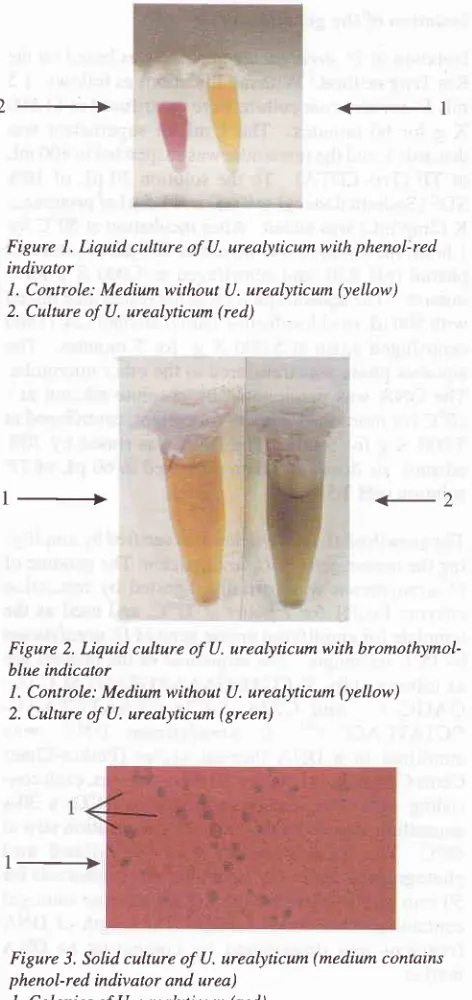
Liquid cuhure of U. urealyticum with phenol:red indivatorl. Controle: Medium
without U. urealyticum (yellow) 2. Culture of U. urealyticum (red)1_+
+2
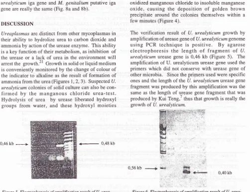
Figure 2. Liquid culture of U. urealyticum with bromothymol-blue indicator
I . Controle: Medium without U. urealyticum (yellow)
2. Culture of U. urealyticum (green)
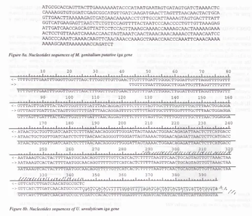
1-+
Figure 3. Solid culture of U. urealyticum (medium contains phenol-red indivator and urea)
l.
Colonies of U. urealyticum (red)Figure 4. Manganous chloride urea-test of U. urealyticum growth of solid culrure
Vol 8, No 4, October - December 1999
urealyticum iga
gene
andM.
genitalium putative
iga
gene are
really
the
same(Fig.
8a and 8b).DISCUSSION
Ureaplasmas
aredistinct from
other mycoplasmas
in
their ability
to
hydrolize
urea
to
carbon
dioxide
andammonia
by
action of
the ureaseenzyme. This
ability
is
akey
function of
their metabolism,
asinhibition of
the
ureaseor
a
lack
of
urea
in
the
environment
will
arrestthe growth.2l
Growth
in
solid
orliquid
medium
is
conveniently monitored
by
the changeof colour of
the
indicator
to
alkaline
asthe result
of
formation of
ammonia
from
the urea(Figures
1,2,3).
Suspected U.urealyticum colonies
of
solid culture can
also becon-firmed
by
the manganous chloride
urea-test.
Hydrolysis of urea by
urease
liberated hydroxyl
groups
from
water,
and these
hydroxyl
moieties
0,46
kb
--|
-
0,48kb
F i gure 5. Electrophores is of amplification re sult of U. urea-lyticum urease gene.
Left: urease gene (0,46 kb); right: marker VIII
The identification of homology of
genes
215oxidized
manganouschloride to
insoluble
manganeseoxide,
causing
the
deposition
of
golden
brown
precipitate
around
the colonies
themselves
within
afew minutes (Figure 4).
The
verification result
of
U. urealyticum growth
by
amplification
of urease geneof
U.urealyticumgenome
using
PCR technique
is positive.
By
agarose
electrophoresis
the length
of
fragment
of
U.urealyticum
urease geneis 0,46 kb (Figure
5).
Theamplification of U. urealyticum
urease gene used theprimers which
did not
conserve
with
urease geneof
other
microbia.
Since theprimers
usedwere specific
ones and the
length
of
the U. Luealyticum
urease genefragment
wasproduced
by this amplification
was
the same asthe
length
of
urease genefragment
that
wasproduced
by Kui Teng,'
thusthat
growth
is
really
thegrowth
of
U.urealyticum.
0,56 kb
[image:5.595.75.577.90.475.2] [image:5.595.77.549.537.725.2]0,40 kb
Figure 6. Ekctrophoresis of amplification result of U. urea-lyticum iga gene.
Left: Marker
II;
right: U. urealyticum iga gene (0,40 kb).0,48 kb
Figure 7. Hybridizntion between U. urealyticum genome and iga gene.
216
Harmiatun etal
Med J IndonesATGCGCACCAGTTACTTGA'\'qÀN\ATACCCATAAT GAATAGT GATAGT GATCTAJNqJ\CTC CAAAAGGTGT GGAT CGAGCGGCATGTT GAT CAAGAT GAACTTAGTTTI\ACAACTACTGCA GTTGAACTTAAÀAAGAGT GAT
GAi\CÀ\\N\CCT
GTTG CCATTru\\\GTAGTGACTTTATT
,
GGT CAT GAAGAGTTAATCT CT GT T C CAGTT TTACTAAT C C CAAC CC CT GT T GT TAAAGAG ATTGATCÀÀCCAGCAGTTATT CCT CCAGTT AÀAGCAÀÀACCAU\i\GC
N\CTAJ\NN\GAi{\
ACTCCTGTTA;\\TCAJqu\\CCÀÀCTAGTN\,\TCAj\CTAiN\CN\i\CA,\jU\CCTru\\CÆ\TCC
Ai\GCCCAJN\TCNfu\'\CAJ\GTTCA'\CA'\;\CCAi{\GCTN\\CCÀ\CCCN\\TTCAJN\CN\N\
[image:6.595.67.579.87.527.2]A'UU\GCA'\TA'q'$;U\i\C CAGAT C T
Figure 8a. Nucleotides sequences of M. genitalium putative iga gene
10 20 30 40 50 60 10 80
....1....1....1.. 1.. 1.. .l I I I I r - r I r I r
< _ TTTTGTTTGAÀTTTGGGTTGGTTTAGCTTTGGTTTGTTGAÀCTTGTT'TTGATTTGGGCTTGGATTGTTTAGGTTTTGTTT
TTTTGTTTGAÀTTTGGGTTGGTTTAGCTTTGGTTTGTTGAÀCTTGTTTTGATTTGGGCTTGGATTGTTTÀGGTTTTGTTT
90
100r10
L20 130 140 150 160< _ GTTTAGTTGÀTTTACTAGTTGGTTTTGATTTAÀCAGGAGTTTTCTTTTTAGTTG AÂCTGGÀGGA
_ > GTTTAGTTGATTTÀCTAGTTGGTTTTGATTTAÀCAGGÀGTTTTCTTTTTAGTTGCTTTTGGTTTTGCTTTAÀCTGGAGGA
GTTTÀGTTGÀTTTACTAGTTGGTTTTGATTTÀÀCÀGGAGTTTTCTTTTTÀGTTGCTTTTGGTTTTGCTTTAÀCTGGAGGA
170
180 190 2002r0
))^
230
240<_ ATAACTGCTGGTTGATCAATCTCTTTAÀCAÀCAGGGGTTGGGATTAGTAÀÀACTGGAACAGAGATTAÀCTCTTCATGACC
->
ATAÀCTGCTGGTTGÀTCAÀTCTCTTTAÀCAACAGGGGTTGGGATTAGTAÀAACTGGAÀCAGAGÀTTAACTCTTCÀTGACC A1'AACTGCl'GGTTGATCAÀTCTCTTTAACAACAGGGGTTGGGATTAGTi\AÀACTGGAACAGAGATTAÀCTCTTCATGACC< _ AATAÀAGTCACTACTTTTAATGGCAÀCAGGTTTTTGTTCATCÀÇTCTTTTTAAGTTCAACTGCAGTAGTTGTTAÀÀCTAA _ > A;\T1U\,\GT\CACTACTTTTAATGGCÀÀCAGGTTTTTGTTCATCACTCTTTTTAÀGTTCAACTGCAGTAGTTGTTAÀÀCTAA
AATAAAGTCACTÀCTTTTAÀTGGCAÀCAGGTTTTTGTTCATCACTCTTTTTAAGTTCÀÀCTGCAGTAGTTGTTAAACTAÀ
330
340
3s0
360
370
380
390/,/,//r//ltt'{(/,ttr,/(,/1,/,t1.t\,,,,1,,,,1,,,,1,,,,t,,,,1,,,,t,,,,1,,,,1,,,,1,,,,t
< _ GTTCA'ICTTGATCAACATGCCGCTC
- >
crrcArcrrcArcAÀcArcccccrçÇ4TçÇ49+ççTTT,
L
...
GTTCATCTTGÀTCAÀCATGCCGCTCGATCCACACCTTTT
/
//
Figure 8b. Nucleotides sequences
ofU.
urealyticum iga geneDesign
of
primers
for amplification of
U. urealyticum
iga
genefragment
basedon nucleotides
sequencesof
M. genitaliumpntative
iga
genewas
successful.
Theprimers are compatible
with
iga
gene
of
U.ur e aly t ic
um.
Agarose electrophoresi sof
amplification
result
showed that thelength of the
U.urealyticum
iga
gene
fragment
was0,40
kb
(Figure
6).There
areseveral
DNA
bands
of hybridization
result
(figure
7). It
means:(l)
The
U.
urealyticum
iga
genefragment
of
PCR
processhybridized with
iga
geneof
U.
urealyticum
genome.
(2)
There
is
homology
be-tween nucleotides
sequences
of
U.
urealyticum
genome
and
M.
genitalium putative
iga
gene.
(3)
Amplification occured
at
the
iga
gene
of
U.
urea-Iyticum genome.
It
was alsoproved by
the sequencingof
iga
gene
of
amplification result, there
is
1007ohomology
with
putative
iga
gene
of M.
genitalium
(Figure
8). SeveralDNA
bandsof hybridization
result
indicates
that there are several sites
of
nucleotides
sequences in the U.
urealyticum
genomehomolog
with
nuclotides
sequencesof
M.
genitalium putative
iga
gene.
It
might
be causedby
anucleotides chain
used asprobe
wastoo short,
so there are samenesswith
theother
genes.
Nevertheless
the
length
of
the
U.urealyticum iga
gene can be estimatedby comparing
it
with
the length
of
the
iga
genes
of
other
microor-ganisms
which
have beenisolated
as: thelength
of
N.Vol 8, No 4, October - December 1999
H.
influenzaeHK368
is 5.1kb.la
Because the genomeof U.
urealyticum
is
smallest between eubacteria's,
probably
the length
of
U.
urealyticumtga
geneis
4,8kb,
(Figure
7),
smaller
thanH.
influenzae
HK368
iga
gene.
CONCLUSION
There
is
1007ohomology between a0,40 kb fragment
of
U.
urealyticumiga
gene anda
0,40kb fragment
of
M. genitaliumputative
iga
gene.Acknowledgment
This
research was supported byURGE Foundation
andChristian
University
of
Indonesia, Jakarta. The authorsare grateful
to
Dr.Ir. F.M.
Mesak
for
his
help in
laboratory
andto Prof.Dr.
H.J. Freislebenfor
hishelp-ful
anddiscussion.
REFERENCES
1. Blanchard A, Hentschel J, Duffy L, Baldus K, Cassell GH.
Detection of Ureaplasma urealyticum by polymerase chain
reaction
in
the urogenital tract of adults, in amniotic fluid,and
in
the respiratorytract
of
newborns.Clin Inf
DisI 993; I 7:S 148-53
2. Brown
TA.
Gene Cloning: an introduction. 3th ed. London,New York, Tokyo: Chapman
&
Hall 19953. Casell GH, Wates KB, Crouse DT, Rudd PT, Canupp KC,
Stagno S, et
al.
Associationof
Ureaplasma urealyticuminfection
of
the lower respiratory tractwith
cronic lungdisease and death in veryJow-birth-weight infants. The
Lan-cet I 988; 7: 240-4
4. Fraser CM, Gocayne JD, White O, Adams MD, Clayton RA,
Fleischmann P.D, et al. The Minimal gene complement
of
Mycoplasma genitalium. Science 1995;270:397 - 403
5. Kapatais-Zoumbos K, Chandler D.K, Barile
MF.
Survey ofimmunoglobulin A protease activity among selected species
of
Ureaplasma and Mycoplasna; Specificity for hostim-munoglobulin A. Int'ect lmmun 1985;3:'704 -9.
6. Koneman EW, Allen
AD,
JandaWN.
Mycoplasma andUreaplasmas.
In:
Koneman
EW,
et al.
Diagnostic
Microbiology.
4th
ed.Philadelphia: JB Lipincot
Co 1992;15:675-702.7. Kui Teng, Muyiao Li, Wanfang Yu, Houyun Li, Dawei Shen,
Dexiang
Liu.
Comparison of PCR with culture for detectionof Ureaplasma urealyticumin cliniCal samples from patients
with urogenital infections J Clin Microbi ol 1994:2'.
The identification of homology of
genes
2178. Lomholt
H,
PoulsenK,
Kilian
M.
Comparativecharac-terization of the iga gene encoding
IgAl
protease inNeis-seria
meningitidis,N.
gononhoeae and
HaemophilusinJluenzae. Mol. Microbiol 1995; 15: 495 - 506.
9. Marmion BP. Mycoplasma: Acholeplasma: Ureaplasmaln:
Collee JG, Duguid JP, Fraser AG, Marmion BP. Practical
Medical
Microbiology
13thed. New
York:
CurchillLivingstone 1989 ; 2: 7 45 -68.
10. Mc Cormack WM, Almeida PC, Bailey PE, Grady EM, Lee
YH.
Sexual activity and vaginal colonization with genitalMycoplasmas, JAMA 1972;221: 1375-'l .
ll.
McCormack WM, Lee YH, Zinner SH. Sexual experienceand urethral colonization with genital Mycoplasmas, Ann Int
Med 1973;78: 696-8.
1 2. Naessens A, Foulun W, Breynaert J, Lauwers S. Postpartum
bacteremia
and placental colonization
with
genitalMycoplasmas and pregnancy outcome. Am J Obstet Gynecol
1989;160:64'l-50.
13. Phillips LE, Goodrich KH, Turner RM, Faro S. Isolation
of
Mycoplasma species and Ureaplasma urealyticum from
obstetrical and gynecological patients by using
commercial-ly
available mediumformulations. J Clin
Microbiol1986;24:371-9.
14. Poulsen K, Brandt J, Hjorth JP, Thogersen HC, Killian, M.
Cloning
and
sequencingof
the immunoglobulin
Al
proteases gene (iga) of Haemophilus influenzae serotipe b.
Infect Immun 1989;57: 3097-105.
15. Poulsen
K,
Jensen JS,Lind
I.
Detectionof
Ureaplasmaurealyticum by PCR
and
biovar determination by lyquidhybridization. J Clin Microbiol 1998;36:321 l-6.
16. Quinn PA, Gillan JE, Markestad T, St John MA, Daneman
A.
Lie KI, et.al.
Intrauterine infection with Ureaplasmaurealyticum as a cause
of
fatal
nenonatal pneumoniae.Pediatric Infect Dis 1985;4: 538-43.
17. Sanchez PJ, Regan
JA.
Ureaplasma urealyticumcoloniza-tion and chronic lung disease in
low
birth weight infants.Pediatric Infect Dis 1977;7:542-6
18. Tjokronegoro
A,
AyuningtyasD,
GanjarI.
PengaruhI-Mycoplasma (Ureaplasma urealyticum) terhadap semen
pria pasangan
infertil.
Indon Med J 1993',3:223-33.19. Waites KB, Duffy LB, Crouse DT, Dworsky ME, Strange
MJ, Nelson KG,
et aI.
Mycoplasmalinfections of
cerebrospinal
fluid in
newbom infants from a communityhospital population. Pediatric Infect Dis J 1990;9:241-5.
20. Wani JH, Gilbert JV, Plaut AG, Weiser
JN.
Identification,cloning, and sequencing of the immunoglobulinA
I
proteasegene of Streptococcus pneumoniae. lnfectlmmun 1 996; 64:
3967-74.
21. Willoughby JJ, Russel WC, Thirkell D, Burdon
MG.
Isola-tion
and
detection
of
urease
genesin
Ureaplasma