Keratin is an insoluble fibrous protein known as major component of feather, hair, wool, and horn. Keratinaceous materials are abundant in nature but their industrial applications are still limited. They showed high stability and resistance to degradative activity of common proteolytic enzymes because of the high degree of cross-linking by disulfide bonds, hydrogen bonds, and hydrophobic interactions (Lin et al. 1992; Cheng et al. 1995; Ignatova et al. 1999). The mechanical stability and resistance to enzymatic degradation depends on tight packing of the protein chains in the α-helix (α-keratin) or β-sheet (β-keratin) structures and linkage of these structures by cystine bridges. The use of keratinolytic enzymes is becoming attractive in biotechnological applications. The enzymes are used for removing hair and feather in the poultry industry, upgrading of feather meal, converting feather into feed protein, and clearing obstructions in the sewage system (Gradisar et al.
2000; Pissuwan and Suntornsuk 2001; Riessen and Antranikian 2001).
Keratinase can be produced by some thermophilic microbes, such as Fervidobacterium penavorans (Friedrich and Antranikian 1996), Streptomyces sp. (Bockle et al. 1995; Letourneau et al. 1998;Bressollier et al. 1999), and Bacillus
sp. (Lin et al. 1992; Takami et al. 1992; Cheng et al. 1995). This paper presents several unique characteristics of chicken feather degrading enzyme excreted by bacteria L-23 isolated from a coastal hot spring of North Sulawesi, Indonesia.
MATERIALS AND METHODS
Bacterial Characterization. The bacteria L-23 in this study was isolated from a costal hot spring in North Sulawesi, Indonesia. Morphology and physiological characteristics of the L-23 isolate were observed using Microbact™ kit 24E (Medvet Australia).
Enzyme Production and Partial Purification. The L-23 isolate was grown in media containing 0.05% NH4Cl, 0.05% NaCl, 0.03% K2HPO4, 0.04% KH2PO4, 0.01% MgCl2
.7H 2O, 0.01% Bacto-yeast exctract (Difco), and 1% feather powder (Lin et al. 1992). A full loop of pure culture was added into 10 ml of the liquid medium. After 10 h of incubation, a seed culture was formed. Ten % (v/v) of the seed culture was transferred into the production medium (same medium). All incubations were performed at 70 °C with shaking at 120 rpm. After 3 days of incubation, the medium was filtered through glass wool in order to remove residual undegraded feathers. The culture medium was then centrifuged at 9 000 x gfor 30 min at 4 °C in order to remove the bacterial cells and other particles. Ammonium sulfate was added up to 80% saturation to the cell free supernatant. After kept overnight at 4 °C, the resulting precipitate was collected by means of centrifugation at 9 000 x g for 15 min and dissolved in a minimal volume of 50 mM phosphate buffer (pH 7), before chromatography on Sephadex G 100.
Enzyme Assay and Measurement of Protein Content.
Assay of enzyme activity was based on the method of Letourneau et al. (1998) with slight modification. Feather powder at 2% was used as substrate. The reaction mixture contained 0.05 ml of the enzyme and 0.2 ml of 2% (w/v) feather powder in 50 mM phosphate buffer (pH 7). The mixture was shaken at 120 rpm for 30 min at 70 °C. A 0.5 ml aliquot of 0.1 M trichloroacetic acid was added prior to centrifugation and the absorbance of supernatant was measured at 578 nm after the addition of Folin reagent. One unit of enzyme activity was defined as the amount of enzyme releasing 1 µmol of tyrosine per minute under the assay conditions. Protein concentration was measured by using Bradford dye reagent with bovine serum albumin fraction V as the standard (Bollag and Edelstein 1991).
Effect of pH. The optimum pH of enzyme was analysed by incubation at various pH’s using 20 mM phosphate buffer (pH 6-8) and Glycine NaOH buffer (pH 9-10). The effects of pH on enzyme stability was assayed by measuring the ISSN 1978-3477
Thermostable Chicken Feather Degrading Enzymes from
L-23 Isolate from Indonesia
ROSITA LINTANG1, MAGGY THENAWIJAYA SUHARTONO2*, JAE KWAN HWANG3, AND YU RYANG PYUN3
1Faculty of Fishery and Marine Science, Universitas Sam Ratulangi, Manado 95115, Indonesia
2Department of Food Technology and Human Nutrition and Research Center of Biotechnology, Institut Pertanian Bogor,
Darmaga Campus, Bogor 16002, Indonesia
3Bioproducts Research Center, College of Engineering, Yonsei University Korea, 134 Shinchon-dong, Seoul 120-749, Korea
The thermostable chicken feather degrading protease enzymes used here was extracted and partially purified from thermophilic bacteria L-23 isolated from a coastal hot spring in North Sulawesi, Indonesia. The L-23 was grown in the selective medium containing 1% chicken feather powder at 70 °C and pH 7. The cell-free culture was precipitated with ammonium sulphate at 80% saturation, followed by heating at 65 °C for 1 h before applied onto Sephadex G-100. The molecular weight of the two enzymes identified were estimated as 47 and 64 kDa. The optimum pH of the mixed enzymes preparation was 7 while the optimum temperature was 65 °C. Zymogram analysis showed that one of the enzymes was still active after being heated at 100 °C for 20 min and was also resistant towards organic solvents and SDS. The activity was enhanced by addition of 1 mM FeCl3.
Key words: Chrysomya bezziana, Saccharomyces cerevisiae, vaccine
_____________________________________________
________________________
*Corresponding author, Phone: +62-251-625965,
Table 1 Characteristics of L-23 isolate
Fig 2 Molecular weight of keratinase L-23. Line 1, marker; Line 2, crude enzyme; Line 3, purified enzymes.
9 4
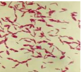
Fig 1 Cells of L-23 isolate in exponential growth phase (1 000x).
Fig 3 Effect of pH on activity of keratinase L-23.
Specific activity (U mg
-1)
residual activity and also through zymogram analysis using gelatin as substrate. For zymogram analysis, enzyme was also incubated at various pH’s before applied onto the gel.
Effect of Temperature. The thermostability was studied by incubating the enzyme at various temperatures (from 70-100 °C) and various time intervals. Evaluation on the thermostability of the enzyme was conducted by measurement of the residual activity and through zymogram analysis.
SDS-PAGE and Zymogram. Preparation for electrophoresis was conducted according to the method of Bollag and Edelstein (1991) with a slight modification as detailed by Riessen and Antranikian (2001). SDS-PAGE was performed in 10% acrylamide. For zymogram analysis, gelatin at 2% was incorporated into the gel. After electrophoresis, the gels were rinsed in 0.25% Triton X-100 for 30 min. The gel was then incubated under optimal assay conditions for 1 h and the protein bands possessing proteolytic (gelatin degrading) were visualized by staining the gels with Coomassie briliant blue. Clear bands were visible at the positions where protein bands showed the enzyme activity. To determine the molecular mass of enzyme protein a low molecular weight marker kit (LMW, Sigma) was used as a standard.
RESULTS
Morphology and Physiological Properties of L-23 Isolate. Isolate L-23 is a gram-negative, motile, rod-shape bacterium that occurs singly, in pairs, or in short chains (Fig 1). Optimal temperature for bacterial growth was 70 °C and optimal pH was 8. No growth occurred at 37 °C. Various biochemical characteristics of the L-23 isolate are presented in Table 1.
Molecular Weight, Optimum pH, and Temperature.
Zymogram analysis was performed to estimate the molecular weight. The molecular mass of two proteases were estimated as 64 and 47 kDa as indicated by the clear bands (Fig 2). The enzyme was active in the range of pH 6 to 10. The optimum pH of enzyme activity was 7. At higher pH values, the enzyme activity remained relatively high loosing only 20% of the maximum activity (Fig 3).
Fig 4 Zymogram showing the effect of pH stability of L-23 enzyme (pH tested: 5, 6, 7 (phosphate), 8, and 9 (Glycine-NaOH).
5 6 7 8 9
Fig 5 Effect of temperature on activity of L-23 enzyme. Temperature incubation (°C)
Relative activity of keratinase (%)
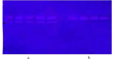
Fig 6 Zymogram showing thermostability at temperature 80 °C for 50 min (a) and 100 °C for 20 min (b).
a b
Table 2 Effect of detergents, inhibitors, and organic solvents
Subtances Relative activity (%)
α-mercaptoethanol 0.1% Thioglikolat 0.1% Fig 8 Effect of metal ions on activity of L-23 enzyme at concentrations 2 mM.
Specific activity of
crude keratinase (U mg
-1)
Fig 7 Effect of heating treatment on activity of keratinase L-23. (u) 70 °C, (n) 80 °C, (p) 90 °C.
Thermostability. To confirm the heat stability of the enzyme it was incubated at 70, 80, and 90 °C for various time intervals and assayed the residual activity using chicken feather substrate as mentioned in the Materials and Methods section. As shown in Fig 7, the enzyme was not stable at temperature of 90 °C despite the fact that when evaluated using zymogram it was stable up to 100 °C for 20 min (Fig 6). Zymogram analysis showed thermostability of the 47 and 64 kd enzymes at temperature 80 °C up to 50 min heating while at temperature 100 °C the 47 kd enzyme was stable for 20 min heating (Fig 6).
Effect of Various Chemicals. Addition of several chloride metal ions, showed an inhibitory effect on the enzyme activity except for FeCl3 (Fig 8). Table 2 showed the effect of other various substances on keratinase activity. The enzyme activity was increased by 1 mM of NaCl and showed high level of stability towards some organic solvents such as DMSO, methanol, and propanol at concentration of 1%. The addition of 1% of Formamide and SDS gave a slightly positive effect on activity. In the presence of Triton X-100 1% and
β-mercaptoethanol 0.1% the enzyme activity was strongly
reduced and completely inhibited by thioglycolate at concentration of 0.1%. None of the proteinase inhibitors, PMSF for serin, EDTA for metallo, and DTT for cysteine-protease tested, gave significant influence on the enzyme activity. Inhibition studies did not lead to a final conclusion regarding their catalytic action and enzyme (protease) classes. However, 0.1% DTT, 0.1% b-mercaptoethanol and 0.1% thioglycolate completely inhibited the enzyme, indicating the importance of cysteine residues in catalytic activity.
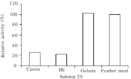
Fig 9 Substrate specificity on various substrates (Casein, Hemoglobin, gelatin, and chicken feather).
100
Casein Hb Gelatin Feather meal
Substrat 2%
Relative activity (%)
120
DISCUSSION
In previous experiments, enzyme from Isolate L-23 was secreted at the late logarithmic growth phase. The amount of enzyme produced depend on substrate concentrations and cultivation condition. Chicken feather substrate at 1% was used because similar to other reports, it is the best substrate concentration for most keratinase production (Lin
et al. 1992; Cheng et al. 1995; Pissuwan and Suntornsuk 2001).
The enzyme excreted by L-23 isolate can be grouped as a gelatinase or keratinase like protease due to the preference for degrading gelatine and chicken feather better over casein or hemoglobin substrate. Our study revealed the presence of two forms of enzyme with molecular weight being estimated as 47 and 64 kD. This is shown through zymogram analysis. The molecular weights of keratinase group vary between 18 kDa (Bressollier et al. 1999) and 135 kDa (Riessen and Antranikian 2001). Most of this group of enzymes show molecular weights around 30 kDa (Lin et al. 1992; Bockle et al. 1995; Cheng et al. 1995; Ignatova et al. 1999; Gradisar et al. 2000).
Most of thermostable keratinases are active in the range of neutral to basal pH. The keratinase from Streptomyces is active over the pH range of 6-11 with maximum activity between pH 7 and 8 (Bockle et al. 1995). The optimum pH of keratinase from Bacillus was 7.5-10.5 (Lin et al. 1992; Cheng
et al. 1995). Our enzyme is active at pH 5-10. The stable conformation at this wide range of pHs is important for the enzyme applications. We used chicken feather powder substrate in the enzyme assay, since as shown in Fig 9 this is a good substrate for the enzyme. For zymogram assay, we used gelatin which is similarly good substrate of the enzymes, but which is also more soluble within the gel compared with chicken feather.
The temperature of 70 °C and above was reported as optimum temperure of keratinase from Fervidobacterium pennivorans (Friedrich and Antranikian 1996), F. islandicum
AW-1 (Nam et al. 2002), Thermoanaerobacter keratinophilus
(Riessen and Antranikian 2001), Bacillus sp. AH-101 (Takami
et al. 1992), Streptomyces sp. SK1-2 (Letourneau et al. 1998). Keratinases which are also active at alkaline pH and showed optimum activity at 60 °C were excreted from Doratomyces microspore (Gradisar et al. 2000), Bacillus licheniformis
(Lin et al. 1992; Cheng et al. 1995), Streptomyces albidoflavus
(Bressollier et al. 1999), and S. pactum (Bockle et al. 1995).
Chicken-feather-degrading enzyme excreted by L-23 isolate is considered highly thermostable, being able to maintain the activity at temperature 80 °C for 30 min when analyzed by spectrophotometric assay using chicken feather substrate. However, our zymogram analysis, using gelatin as the substrate, revealed that the enzyme was highly stable, being able to retain the conformation after heating at 100 °C for 20 min. The difference between these findings may be explained as follows: In the spectrophotometric assay, the enzyme is incubated at the high temperature for the indicated time, and assay with the substrate (chicken feather) at optimum temperature. For zymogram analysis, we also incubated the enzyme at the indicated time and temperature similar to the previous assay, but following incubation, the enzyme was run through gelatine containing gel, and washed with SDS solution before being stained. It is possible that the heated enzyme underwent reversible conformational unfolding, which is still capable of refolding under favorable conditions such as by addition of low SDS solutions. Our study confirmed that 0.1% SDS slightly elevated enzyme activity. This is particularly significant for the smaller form, (the 47 kd molecule). The heavier 64 kd molecule might not be as stable as indicated by our zymogram analysis. The resistance of this enzyme to SDS seems to be related to the rigid structure of protein from extremely thermophilic microorganisms (Friedrich and Antranikian 1996). The stability of enzyme at temperature higher than 75-85 °C might involve several stabilizing factors such as sulfur bridges and ionic interactions because extreme stability of enzyme is unlikely to be maintained at the expense of only hydrophobic interaction as the strength decrease above 70-80 °C (Kim and Kim 1991). The role of these intrinsic factors might explain the stability. Enzymes from thermophilic sources have been found unique for their greater stability are usually caused by additional salt bridges or slightly different amino acid composition (Volkin and Klibanov 1989). The importance of cystine bridges for the catalytic activity of for maintaining stability of L-23 enzyme is suggested by study of the effect of DTT, beta mercaptoetanol, and thyoglycolate which inhibited the enzyme activities. This experiment may imply that the enzyme molecule may be able to maintain its conformation (and thus the activity) and even improved after addition of organic solvents (DMSO, methanol, propanol, and formamide).
In case of the effect of metal ions, the cations added might compete with inner cofactor of the enzyme in binding to the active site. Therefore, it might be concluded that this enzyme did not require additional metal ions for optimum activity. In our experiments, it was only Fe-cation which increased the activity, while other cations tested reduced the activity. Most keratinases required Ca2+ to enhance enzyme activity (Bockle et al. 1995; Bressollier et al. 1999; Ignatova et al. 1999; Riessen and Antranikian 2001; Nam et al. 2002), while for our enzyme only Fe3+ enhanced the assayable activity.
REFERENCES
Bollag DM, Edelstein SJ. 1991. Protein Methods. New York: J Wiley and Sons.
Bressollier P, Letourneau F, Urdaci M, Verneuil B. 1999. Purification and characterization of a keratinolytic serine proteinase from Streptomyces albidoflavus. Appl Environ Microbiol 65:2570-2576.
Cheng SW, Hu HM, Shen SW, Takagi H. 1995. Production and characterization of keratinase of feather-degrading Bacillus licheniformis PWD-1. Biosci Biotech Biochem 59:2239-2243. Friedrich AB, Antranikian G. 1996. Keratin degradation by
Fervidobacterium pennavorans, a novel thermophilic anaerobic species of the order Thermotogales. Appl Environ Microbiol 62:2875-2882.
Gradisar H, Kern S, Friedrich J. 2000. Keratinase of Doratomyces microsporus. Appl Microbiol Biotechnol 53:196-200.
Ignatova Z, Gousterova A, Spassov G, Nedkov P. 1999. Isolation and partial characterization of extracellular keratinase from a wool degrading thermophilic actinomycete strain Thermoactinomyces candidus. Can J Microbiol 45:217-222.
Kim NS, Kim SI. 1991. Some molecular characteristic and improving methods for thermal stability of enzyme. Kor J Appl Microbiol Biotechnol 19:100-108.
Letournaeu F, Soussotte V, Bressollier P, Branland P, Verneuil B. 1998. Keratinolytic activity of Streptomyces sp. S.K1-02: a new isolated strain. Lett Appl Microb 26:77-80.
Lin X, Lee CG, Casale ES, Shih JCH. 1992. Purification and characterization of a keratinase from a feather-degrading Bacillus licheniformis strain. Appl Environ Microbiol 58:3271-3275. Nam GW, Lee DW, Lee HS, Lee NJ, Kim BC, ChoeEA, Hwang JK,
Suhartono MT, Pyun YR. 2002. Native-feather degradation by Fervidobacterium islandicum AW-1, a newly isolated keratinase-producing thermophilic anaerob. Arc Microbiol 178:538-547. Pissuwan D, Suntornsuk W. 2001. Production of Keratinase by
Bacillus sp. FK 28 isolated in Thailand. Kaset start Journal (Natural Science) 34:171-178.
Riessen S, Antranikian G. 2001. Isolation of Thermoanaerobacter keratinophilus sp. Nov, a novel thermophilic, anaerobic bacterium with keratinolytic activity. Extrememophiles 5:399-408. Takami H, Nakamura S, Aono R, Horikoshi K. 1992. Degradation of