THE EFFECTS OF SURFACE CURVATURE ON BIOMECHANICAL BEHAVIOUR OF ARTICULAR CARTILAGE
TAN MEAN YEE
PENUTUP
SEKALUNG BUDI
Fakulti merakamkan tahniah dan terima kasih kepada semua pihak yang terlibat dalam penghasilan Buku Panduan PSM Edisi Kelima
Assoc. Prof. Engr. Dr.Noreffendy Tamaldin Dr. Ruztamreen Jenal
Dr. Abd Rahman Dullah Dr. Mohd Juzaila Abd Latif Prof. Madya. Dr. Azma Putra Dr. Mohd Khairi Bin Mohamed Nor
Prof. Madya Juhari Bin Ab Razak Dr. Abd Rahman Bin Dullah
Ir. Dr. Tan Chee Fai Dr Mohd Ahadlin bin Daud
Encik Nazri Bin Md Daud Encik Ahmad Kamal Bin Mat Yamin
Encik Herdy Rusnandi Encik Fudhail Bin Abdul Munir Encik Mohd Irwan Bin Mohd Azmi
I
SUPERVISOR DECLARATION
“I hereby declare that I have read this thesis and in my opinion this report is sufficient in
terms of scope and quality for the award of the degree of Bachelor of Mechanical
Engineering (Structure & Materials)”
Signature : ………
Supervisor : DR MOHD JUZAILA BIN ABDUL LATIF
THE EFFECTS OF SURFACE CURVATURE ON BIOMECHANICAL BEHAVIOUR OF ARTICULAR CARTILAGE
TAN MEAN YEE
This report was submitted in fulfillment of the requirement for the award of Bachelor of Degree of Mechanical Engineering with Honours (Structure &
Materials)
Faculty of Mechanical Engineering
Universiti Teknikal Malaysia Melaka
III
DECLARATION
“I hereby declare that the work in this report was my own except for summaries and
quotations which have been duly acknowledged.”
Signature : ………
Author : TAN MEAN YEE
IV
ACKNOWLEDGEMENT
In preparing this report, there were many people had involved helping me to
complete the report, so, I would like to express my grateful and thankful to them.
I am very appreciate the help from my final year project supervisor, Dr. Mohd
Juzaila Bin Abdul Latif who has gave a lot of useful idea, suggestions and knowledge to
me. He has spent his precious time in guiding me with his expertise and giving his moral
support.
My sincere appreciation also extends to all my friends and others who have
provided assistance at various occasions. Their views and tips were useful indeed.
Unfortunately, it was not possible to list all of them in this limited space. Once again I
V
ABSTRACT
Osteoarthritis (OA) occurs even more frequently nowaday on the middle aged group,
the senior citizen group and even the youngsters will also affected by this disease.
Osteoarthritis was characterised by the pain of joint and malfunction of the joint, and
then in further stage will be the joint contractures, muscles atrophy and limb deformity
spine. The main causes for Osteoarthritis to occurs will be due to the degeneration of
articular cartilage. Therefore, it is important to understand the behaviour of the cartilage.
In the previous experimental studies, the cartilage are often been assumed to be flat.
However, the actual cartilage surface in human synovial joint will be in curvature shape.
Hence, this report will determine the effect of surface curvature on cartilage behaviour
by using the indentation test and finite element analysis. Finite element model with
several radii of surface curvature will be used to compare with the analysis of model
with flat surface. Based on the comparison result, the percentage of difference by
comparing the smallest cartilage surface of convex 15 mm radius with the flat cartilage
surface for contact pressure is 10 % while for the pore pressure will be 6 %. This shows
that the surface curvature of cartilage does affect the biomechanical behaviour of
VI
ABSTRAK
Pada hari ini, Osteoartritis (OA) bukan sahaja berlaku lebih kerap atas kumpulan
pertengahan umur, kumpulan warga emas, malah remaja juga terjejas oleh penyakit ini.
Osteoartritis boleh dicirikan oleh sakit sendi dan kerosakan sendi, dan pada peringkat
selanjutnya Osteoartritis boleh dicirikan daripada contractures sendi, otot atrofi dan
kecacatan tulang belakang. Punca utama untuk Osteoartritis berlaku ialah disebabkan
kerosakan tulang rawan. Oleh itu, permahaman tentang tulang rawan adalah penting.
Dalam eksperimen sebelum ini, tulang rawan sering diandaikan sebagai permukaan rata.
Namun, permukaan rawan yang sebenar pada sendi sinovia manusia ialah dalam bentuk
lengkung. Oleh itu, laporan ini akan mengkaji tentang kesan kelengkungan permukaan
terhadap ciri-ciri rawan dengan menggunakan ujian indentation dan Finite element
analisis. Model finite element dengan beberapa jejari kelengkungan permukaan akan
digunakan untuk membandingkan dengan analisis model dengan permukaan rata.
Berdasarkan keputusan perbandingan, peratusan perbezaan antara permukaan cembung
rawan terkecil 15 mm jejari dengan permukaan rawan rata untuk tekanan sentuhan
adalah 10% manakala bagi tekanan pore adalah 6%. Ini menunjukkan bahawa
VII
TABLE OF CONTENT
CHAPTER CONTENT PAGE
SUPERVISOR DECLARATION I
DECLARATION III
ACKNOWLEDGEMENT IV
ABSTRACT V
ABSTRAK VI
TABLE OF CONTENT VII
LIST OF FIGURES X
LIST OF TABLES XII
LIST OF ABBREVIATION XIII
CHAPTER 1 INTRODUCTION 1
1.0 Introduction 1
1.1 Problem Statement 2
1.2 Objective 2
1.3 Scope 2
CHAPTER 2 LITERATURE REVIEW 3
2.0 Introduction 3
2.1 Osteoarthritis 3
2.1.1 Causes 4
2.1.2 Diagnosis 5
2.1.3 Treatment 6
2.2 Anatomy of Synovial Joint 6
2.3 Articular Cartilage 7
2.3.1 Structure and Composition 8
VIII
CHAPTER CONTENT PAGE
2.4 Characterisation of Mechanical Properties 10
2.4.1 Investigation Method 10
2.4.2 Indentation Test 11
2.4.3 Finite Element Analysis 11
2.5 Effect on Surface Curvature 12
CHAPTER 3 METHODOLOGY 13
3.0 Introduction 13
3.1 Material and Sample Preparation 13
3.1.1 Phosphate Buffered Saline 13
3.1.2 Sample Preparation 14
3.2 Apparatus 15
3.3 Calibration 16
3.4 Indentation Test 17
3.5 Thickness and Radius Measurement of Cartilage 18
3.5.1 Thickness Measurement of Cartilage 18
3.5.2 Radius Measurement of Cartilage 20
3.6 Computational Method 20
3.6.1 Implementation of Contact Dependent Flow 20
3.6.2 Development of Finite Element Model:
Previous Study 21
3.6.3 Development of Finite Element Model:
Current Study 22
CHAPTER 4 RESULT AND DISCUSSION 24
4.0 Introduction 24
4.1 Radius and Thickness 24
4.2 Validation with Previous Result 25
4.3 The Effect of Surface Curvature on Cartilage
Behaviour in Indentation Test 27
IX
CHAPTER CONTENT PAGE
4.3.2 Pore Pressure 29
4.4 Discussion 30
CHAPTER 5 CONCLUSION 31
5.0 Conclusion 31
5.1 Recommendation 31
X
LIST OF FIGURES
FIGURE TITLE PAGE
2.1 Comparison of Normal Joint and Joint which have
Osteoarthritis
4
2.2 Nomenclature of Synovial Joint 7
2.3 Articular Cartilage 8
2.4 Schematic Diagram of Articular Cartilage 9
3.1 a. Cow Bone that Cut Into smaller pieces, b. Marked Test
Sample
14
3.2 Schematic Diagram of The Test Rig 15
3.3 LVDT and Force Transducer 16
3.4 Specimen Set-up 17
3.5 Calibration Graph 17
3.6 Sample Place in the Test Rig with PBS 18
3.7 Thickness Graph to Determine Thickness of Cartilage 19
3.8 Profile Projector 20
3.9 Fluid Flow at a. 2 s, b. 1000 s 21
XI
FIGURE TITLE PAGE
3.11 FE Model for Current Study a. Fluid Flow Boundary of
Cartilage, b. Convex Cartilage Curvature
23
4.1 Comparison of Stress-Relaxation 26
4.2 Comparison of Creep-Deformation 26
4.3 Contact Pressure at Cartilage Surface for Difference Radius a.
2 s, b. 1000 s
28
4.4 Pore Pressure at Cartilage Surface for Difference Radius a. 2 s,
b. 1000 s
XII
LIST OF TABLES
TABLE TITLE PAGE
3.1 Formulation of Phosphate Buffered Saline 14
3.2 Mechanical Properties According to Previous Study
(Pawaskar, 2006)
22
3.3 Mechanical Properties Used In Current Study(Abd Latif et.
al, 2013)
23
XIII
LIST OF ABBREVIATION
LVDT – Linear variable differential transformer
MRI – Magnetic resonance imaging
OA – Osteoarthritis
PBS - Phosphate Buffered Saline
1
CHAPTER 1
INTRODUCTION
1.0 INTRODUCTION
Osteoarthritis is one of the cartilage diseases which occur due to the abnormal
biomechanics of cartilage. More than 20% of the population in Malaysia may have
Osteoarthritis. Osteoarthritis will causes pain and stiffness, especially in the hip, knee,
and thumb joints. One of the main causes of osteoarthritis is due to the degeneration of
articular cartilage. Articular cartilage is a white dense connective tissue which covers the
bone ends within a diarthrodial joint in human body. The function of articular cartilage is
to provide frictionless bearing in a joint while it uniformly transferring loads on
underlying bone in order to prevent high stress concentrations. Therefore it is essential to
study the behaviour of articular cartilage in order to prevent the degeneration of
2
1.1 PROBLEM STATEMENT
In previous studies, articular cartilage are often assume to be flat surface while study
the biomechanical properties. However, the actual articular cartilage possessed curve
surface in human joint. Therefore, this project will study the effect of cartilage surface
curvature on the biomechanical behavior of articular cartilage.
1.2 OBJECTIVE
The aim of this project is to study the effects of cartilage surface curvature on
biomechanical behavior of articular cartilage.
1.3 SCOPES
The scope of the project includes:
To establish the procedure to determine the thickness of articular cartilage.
To study the effect of the cartilage surface curvature to the contact stress and pore
3
CHAPTER 2
LITERATURE REVIEW
2.0 INTRODUCTION
This chapter will discussed about all the literature review that being study and refer
for this report. In this chapter, the definition for Osteoarthritis, synovial joint, articular
cartilage will be discussed. The method use in previous study in examining articular
cartilage will also be include in this chapter.
2.1 OSTEOARTHRITIS
Osteoarthritis is a condition that affects human’s joints by degenerate the joint
cartilage and the underlying bone. The surfaces within human joints become damaged so
the joint doesn’t move as smoothly as it should. Osteoarthritis occurs most commonly
from middle age and onward. It will cause pain and stiffness at the cartilage surfaces,
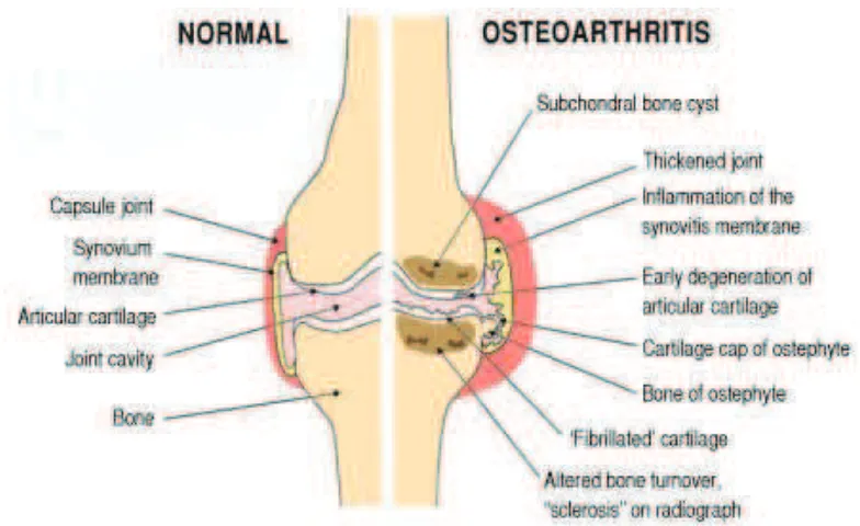
especially in the hip, knee, and thumb joints. The difference of normal joint and joint
4
Figure 2.1 Comparison of Normal Joint and Joint which have Osteoarthritis
As the age of a person increases, the amount of water in the nucleus pulposus
decreases, which reduces the cushioning effect of articular cartilage (Sarzi-Puttini et al,
2005).However, stated that articular cartilage can also be damaged due to several reason
such as osteochondritis dissecans, post traumatic, immobilization. The damage on
articular cartilage also can occur on youngster, especially people in sport and damage
cause by osteochondritis dissecans. By the way, the abnormal bio-mechanics of cartilage
plays an important part in the development of osteoarthritis(Kwan et al, 1986).
2.1.1 CAUSES
The factors that cause osteoarthritis included overweight, joint injury, genetic defect
in joint. However, the degeneration of cartilage will be the general factor to cause
osteoarthritis. The cartilage rub against each other and the friction occur will cause the
5
body will try to repair the damage but sometimes the cartilage can become so thin that it
does not cover the ends of the bones. Therefore the bones rub against each other and
start to wear out. The loss of cartilage, the wearing of bone and the bony spurs can
change the shape of your joint, forcing the bones to be out of their normal position. By
the way, the reduction of the concentration of proteoglycans, breakdown of collagen
network as well as an increase in the water content and softening of the tissue will cause
osteoarthritis(Julkenen, 2008).
2.1.2 DIAGNOSIS
Osteoarthritis is usually diagnosed based on the symptoms and the physical signs
that the doctor finds when examining the joints. The doctor will check for the properties
such as the tenderness of joint, the creaking sounds, excessive fluid, movement and
instability of joint, muscle thinning, etc. Some other methods also can use to diagnose
the osteoarthritis, such as X-rays and Magnetic resonance imaging (MRI) scans.Barry et.
al. (2014) use MRI scans to study the radiographic of knees. Marsh et. al. (2013) use
both MRI scans method and X-rays method to investigate the knee cartilage thickness to
compare their results. X-rays are the most common tools to confirm osteoarthritis.
Although the image of cartilage will not show up on x-rays images, but they will show
the changes of the bone. While MRI scans is rarely to be used on diagnosis of
osteoarthritis but MRI scans can give further information that causes the joint to defect.
MRI scans uses radio waves and strong magnetic field to show the image of bone and
6
2.1.3 TREATMENT
Osteoarthritis has no cure but some medications can be used to help relieve pain.
Physical therapy (PT) or occupational therapy (OT) can be used to help improve strength
and function of the joint. However, when the pain is frequently occur or mobility and
daily activities become difficult, then surgery may be considered.
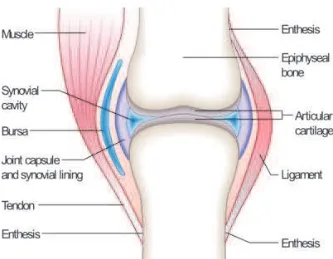
2.2 ANATOMY OF SYNOVIAL JOINT
Synovial joint are the most common joint in mammal’s body. Most of the joint in
human body are synovial joint, such as hinge joint at knee and elbow, condyloid joint at
wrist, saddle joint at thumb, etc. Same as the most of the other joints, synovial joints can
achieve movement at the point of contact of the articulating bones. Synovial joint will
include some component such as articular cartilage, joint capsule, synovial fluid,
ligament, menisci and bursae as shown in Figure 2.2(Shier et. Al., 2008). Joint capsule
is the tubular structure that has two distinct layers; the outer layer is made up of dense
fibrous connective tissue while the inner layer is a shiny vascular membrane called the
synovial membrane. Synovial fluid is a clear viscous fluid secreted by the synovial
membrane for the lubrication of the joint. Ligament is the bundles of tough collagen
fibers that serve to strengthen the joint capsule. Menisci are the disks of fibrous-cartilage
found in some synovial joints that serve as shock absorber. Bursae will be the fluid-filled
7
Figure 2.2 Nomenclature of Synovial Joint
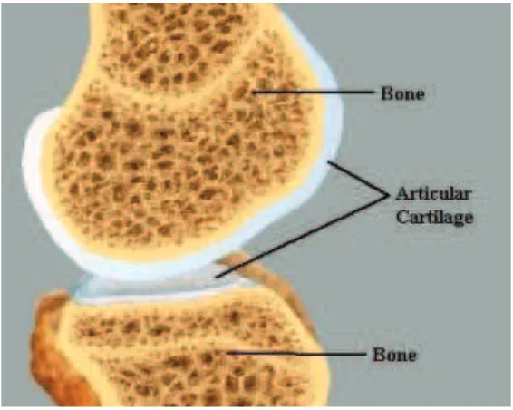
2.3 ARTICULAR CARTILAGE
The main types of cartilage include fibrous cartilage, elastic cartilage and hyaline
cartilage. Fibrous cartilage can be found in such areas as between vertebral discs of the
spinal cord. Elastic cartilage can be found at the outer ear, nose, and larynx. Hyaline
cartilage which also referred to as articular cartilage covers joint surfaces and
additionally may be found in the shoulder, rib cage, knee and hips as well. The general
8
Figure 2.3 Articular Cartilage
2.3.1 STRUCTURE AND COMPOSITION
Articular cartilage is a white dense connective tissue which covers the bone ends
within a diarthrodial joint (Kwan et. Al., 1986). It is a soft, highly hydrated and
permeable tissue. About 80% of articular cartilage is form by water. Articular cartilage
has 3 main structures which is collagen, proteoglycans and interstitial fluid (Julkenen,
2008).
There are 3 layer of collagen in articular cartilage, which is superficial zone, middle
zone and deep zone as shown in Figure 2.4. This 3 layer are being differentiated by the
orientation of the collagen fibrils. The superficial layer is to be described as a tension
resisting diaphragm and is demonstrated by the tendency of articular cartilage to curl
when released from the subchondral bone. In the middle layers, chondrocytes attain a
rounded or spherical shape, proteoglycans content increases and the collagen fibers