Disseminated tuberculosis diagnosed first as leptospirosis in
immunocompetent patient
Keywords: acute onset, disseminated tuberculosis, immunocompetent
pISSN: 0853-1773 • eISSN: 2252-8083 • http://dx.doi.org/10.13181/mji.v24i4.1266 • Med J Indones. 2015;24:252–6 • Received 15 Jul 2015 • Accepted 29 Dec 2015
Correspondence author: Erni J. Nelwan, [email protected]
Copyright @ 2015 Authors. This is an open access article distributed under the terms of the Creative Commons Attribution-NonCommercial 4.0 International License (http://creativecommons.org/licenses/by-nc/4.0/), which permits unrestricted non-commercial use, distribution, and reproduction in any medium, provided the original author and source are properly cited.
Erni J. Nelwan,1 Juferdy Kuniawan,2 Mirna N. Praptini,3 Herdiman T. Pohan1
1 Division of Tropical and Infectious Disease, Department of Internal Medicine, Universitas Indonesia, Jakarta, Indonesia
2 Division of Hepatobilier, Department of Interna Medicine, Universitas Indonesia, Jakarta, Indonesia
3 Department of Internal Medicine, Faculty of Medicine, Universitas Indonesia, Jakarta, Indonesia
Case Report
ABSTRAK
Tuberkulosis diseminata, lebih sering di diagnosis pada pasien dengan status imun yang lemah (immunocompromised). Manifestasi yang tidak khas menyebabkan penegakan diagnosis di klinik sering kali membutuhkan waktu. Studi ini melaporkan kasus tuberkulosis diseminata pada pasien imunokompeten; seorang wanita 42 tahun dengan tuberkulosis paru dan ekstraparu (tuberkulosis hati.) yang berhasil ditegakkan diagnosisnya melalui biopsi hati.
ABSTRACT
Disseminated tuberculosis, mostly seen in immunocompromised patient, can affect several organs.
The diagnosis can be difficult due to various clinical
manifestations. A case of disseminated tuberculosis of lung and liver that develops acutely is rare especially in immunocompetent patient. Here, we present a 42 year old immunocompetent woman with lung and hepatic
Liver involvement in tuberculosis, though common both in pulmonary and extra-pulmonary tuberculosis, is usually clinically silent. Sometimes, signs and symptoms may be prominent in hepatic tuberculosis, and may appear as the initial or sole presenting feature of the disease. However, even in developing countries, liver tuberculosis accompanied by local symptoms is an uncommon entity. A study from South Africa showed that liver tuberculosis accounted for only 1.2% of all cases of tuberculosis diagnosed at a general hospital. Similarly in Indonesia, within the last 10 years, there is only one case of hepatic tuberculosis ever reported from national referral
hospital.1 Lack of familiarity with this condition
was apparently responsible for the diagnosis of
hepatic tuberculosis never been concludedin
the past due to difficulty of diagnosing.2 Since
tuberculosis remains a potentially curable disease, an awareness of diagnosing this disease is very essential.3-5
CASE REPORT
A 42 year old woman was admitted with complaints of fever and yellowish skin since six days before admission. She had been well before. Physical and ultrasonographic examinations of the abdomen revealed hepatomegaly. Leukocyte was low and liver panel showed obstructive jaundice and high liver enzyme [aspartate aminotransferase (AST) at 429U/L and alanine transaminase (ALT)
at 373U/L]. Patient was first diagnosed with
leptospirosis due to contact with rats before and
treated as leptospirosis first with ceftriaxone injections 2 grams qd for five days but there was no
improvement in general condition and fever. The
blood tests for any epidemiologically significant
diseases revealed negative results (tuberculosis polymerase chain reaction, TUBEX test for typhoid fever, malaria antibody, and leptospira antibody). Oral amoxicillin-clavulanic acid was then added to the patient for three more days but patient status was still unchanged and the leukocyte kept declining. Antibiotic was changed to meropenem 1 gram q8h but fever remains. The source of infection was still unknown. Anti-hepatitis A virus HAV), HBsAg, anti-hepatitis C virus
HCV), anti-human immunodeficiency virus
(anti-HIV), gram staining of urine, throat swab, and blood culture were negative. The patient’s CD4
was low (113 cells/μL).
After three days, intravenous (iv) levofloxacin
500 mg qd and iv metronidazole 500 mg q8h were added to patient’s drug regimen but still no improvement. Patient was getting weaker and physical, laboratory, and ultrasonographic examinations of the abdomen revealed hepatomegaly that got bigger during hospitalization and no widening of intra- or extrahepatic ductals, but bilirubin was improved (total of 9.1 mg/dL to 3.7 mg/dL). Due to abdominal pain, patient intake was falling and albumin level also fell. The results of her blood tests showed bicytopenia (Hb 11.3g/dL to 8.4g/dL, and leukocyte 3,900/µL to 2,040/µL). Abdominal computed tomography (CT) scan showed a hepatomegaly and severe fatty liver. Patient was prepared to get liver biopsy and the antibiotics were changed to ertapenem 750 mg qd for seven days, still there was no improvement.
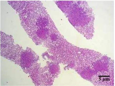
Figure 1. Liver biopsy, acid fast bacilli staining, 100x
Liver biopsy was performed on day 18. Pathologic examination of her liver showed caseous necrosis, granuloma formation, and a positive acid fast bacilli staining (Figures 1 and 2). Patient was diagnosed with milliary tuberculosis of the liver.
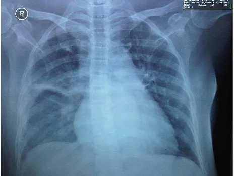
Repeated chest X-ray showed a change from
normal to infiltration at both apices without
any evidence of acid fast bacilli in sputum though repeated testing (Figure 3). Patient was diagnosed as having disseminated tuberculosis. We did not diagnose patient with pneumonia since clinical and laboratory data were not coherent (inconclusive Gram smear result).
Since the liver enzymes were still high (AST 215U/L and ALT 39U/L, bilirubin total of 6.9 mg/dL) and having lactate dehydrogenase (LDH) of 1,871U/L (normal value <215U/L) patient was given rifampicin 450 mg, isoniazide 300 mg, ethambutol 1,000 mg, streptomycin 750 mg as antituberculosis drugs. We gave no pyrazinamide out of concerns that patient also had malignancy besides tuberculosis although the routine malignancy markers came back negative [carcinoembryonic antigen (CEA), Ca 19–9, alpha-fetoprotein (AFP), and Ca 125]. After initiating the antituberculosis treatment for three days, her bilirubin increased and she fell into sepsis with procalcitonin reaching higher than 100ng/mL, so the antituberculosis therapy was changed to
ethambutol, amikacin, moxifloxacin, tygecycline, and fluconazole. She was then getting better and
the antituberculosis regimens were continued
with ethambutol, moxifloxacin, and streptomycin.
Patient was discharged at day-48 with bilirubin
level significantly decreased and clinically much
improved.
DISCUSSION
In all tuberculosis (TB) patients, disseminated TB is responsible for 1–3% cases, while liver tuberculosis is said to be around 1.2% of all
cases or 80–100% of all disseminated TB.3,6,7
Epidemiological data in an endemic area in Iran showed that poor prognosis of disseminated TB often showed blood abnormalities such as low hemoglobin, leucocytes, and thrombocytes.
Disseminated TB mortality rate was significantly
higher in patients with diabetes, injection drug history and blood abnormalities. Old age (median
Figure 3. Chest X-ray day 19. Infiltration at both apices, posi -tive kerley line on the right middle lobe lung
of 52.7 years; 20–76 years) was seen as another factor affecting liver TB survival.6,7
This patient was first came with jaundice and fever.
After narrowing the diagnosis, liver tuberculosis was considered as the primary causation. Liver
tuberculosis was finally diagnosed from liver biopsy
and staining. The positive result of biopsy, as can be seen in this patient, is rare.3,5
Spreading to liver may occur either hematogenously or milliary TB or direct inoculation through the portal venous system from gastrointestinal (GI) lesions. If the spreading was from the lung or the hepatic artery, the liver TB form would be milliary and if it was from portal vein, the form would be focal TB.3,7
Liver tuberculosis has three presentations. The
first and most common presentation is diffuse
hepatic involvement that is usually found together with lung or milliary tuberculosis. This presentation was found in more than half of pulmonary tuberculosis cadaver. The second
presentation is diffuse hepatic infiltration
(granulomatous hepatitis) that happens without any pulmonary involvement. In this presentation, symptoms of liver disease are usually absent. The third and rarest form is a focal/local
tuberculoma or abscess.3 This case belongs to
the second presentation of hepatic tuberculosis
(granulomatous type) with unspecific pulmonary
involvement at the initial that further shows an
Even without caseation or necrosis or positive
acid fast bacilli (AFB), histological finding of liver
granulomas is a gold standard of TB diagnosis in Asia and Africa. The most common symptoms are fever (60–99%), weight loss and abdominal pain, shared almost the same percentages (45– 80%), all were seen in this patient. More than 50% of the patients have hepatomegaly and splenomegaly. Jaundice is not usual for liver tuberculosis and only presents in 20–35% of patients. Biliary compression caused by porta hepatic nodes or pericholangitis or clogging of ruptured TB granuloma into the bile ducts ultimately resulted in jaundice with bilirubin values between 2.3 mg/dL and 5.8 mg/dL. In this patient improvement of bilirubin (total of 9.1 mg/dL to 3.7 mg/dL) was seen after
administration of levofloxacin and metronidazole
which has a potent anti-mycobacterial properties.
Liver function tests are also non-specific but an
elevated alkaline phosphatase level is the only
test that is common and found in 50–87%of
patients. Normal chest X-ray was seen in 75% of patients with isolated hepatic tuberculosis, while only two in 21 patients with local liver tuberculosis
have abnormal chest X-ray finding.3,4,6,7 This case
showed atypical signs of symptoms of hepatic tuberculosis.
Needle biopsy is a proposed method for diagnosis
confirmation. More than 80% of hepatic
tuberculosis case samples will reveal epithelioid granuloma formation; caseous necrosis in 30-83% and positive acid fast bacilli (AFB) staining in less than 60% of cases. There were also some
non-specific features of tuberculosis that can be
found histologically in high percentage of patient with fatty liver appearance, hyperplasia of Kuppfer
cell, focal necrosis and infiltration of inflammatory cell into the liver parenchyma. These unspecific features were also found together with specific
tuberculosis lesions.6,8 Liver biopsy of this patient
confirmed the diagnosis of hepatic tuberculosis
which were shown by caseous necrosis, granuloma formation, and positive AFB staining.
For reasons that stay unanswered, AFB liver tissue results in several studies from India and Philippines have been inconclusive. Negative AFB was found in three series from India, despite the presence of caseous necrosis and epithelioid granulomas. Similarly, even guided percutaneous liver biopsy and laparatomy were successfully
performed in isolated liver TB, diagnosis could still
be missed.8 With the fact of so many challenges in
making definite diagnosis of hepatic tuberculosis,
this patient was successfully diagnosed and treated.
None of common imaging modalities [ultrasound, CT, and magnetic resonance imaging (MRI)] is utilizable because the granulomas are usually too small (2 mm) to be seen by naked eyes, hence imaging is a modality that still needs scrutinizing by analyst. Diffuse hyperechogenicity, with a “bright liver or spleen” pattern could be seen by ultrasound. Multiple hypoechoic and hyperechoic nodules can occasionally be seen in the liver or
spleen. After the disease worsened, calcification
could occur and seen by imaging.3,6,8 This might
explain the bright liver and no tubercle seen in ultrasound while CT scan of the abdomen revealed hepatomegaly and severe fatty liver. Therefore needle biopsy is very important in liver tuberculosis diagnosis.
The CT that was done in this patient was carried out on the concern of possible lymph node malignancy due to high lactate dehydrogenase (LDH). LDH itself can increase in liver disease (LDH-3 that can comprise up to 25% of total LDH) and lung disease (LDH-1 and LDH-2 that comprise up to 27% and 37% of total LDH, respectively), although the maximum level of LDH
in disseminated TB was unknown.9
Other diagnosis to be considered beside hepatic milliary tuberculosis are metastases, lymphoma, sarcoidosis, and fungal infection. This was the reason why this patient was also tested for tumor markers, LDH, liver biopsy, and fungal staining in biopsy. The macronodular form can look similar to metastases, primary malignant tumor, or
pyogenic abscess.6
Due to the nonspecific and wide characteristics of imaging manifestations, definitive diagnosis
for hepatic tuberculosis consecutively starts with ultrasound or CT-guided biopsy (or even open biopsy) with subsequent staining for acid-fast bacilli, culture, and histology examinations. Furthermore, delicate technique of tissue biopsy by multiple passes through all the lesion may be required to yield positive result of histological and microbiological examination, so that the
Disseminated tuberculosis is treated with standard antituberculosis drugs (intensive phase, two months including isoniazide (INH), rifampicin, streptomycin, and pyrazinamide; intermittent phase, seven months including INH and rifampicin)
are required.3-6,10 This can be challenging for some
physicians in giving antituberculosis drugs to people with liver disease, hence liver tuberculosis.
The study of Essop et al11showed that when
both isoniazide and rifampicin are used, it can decrease favorably mortality rates compared to regimen not using rifampicin (0% vs 17–50%). But still we gave no rifampicin to this patient due to high basal liver enzyme and because this may be due to the small number of samples and still good prognosis in several studies. We will follow the patient liver condition. The antituberculosis will be continued for at least one year.
Fluconazole was given to this patient due to prolonged use of antibiotics although the prevalence of candidaemia was just 5%. Intravenous broad spectrum antibiotics (p<0.001), oral broad spectrum antibiotics (p<0.010), and indwelling catheters (p<0.010) were the three important risk
factors of candidemia in TB patients.12
For patients that have obstructive jaundice, a magnetic resonance cholangiopacreatography
(MRCP) order is first to determine problems in
the biliary tract, knowing the site of obstruction to decompress then either by stent insertion during endoscopic retrograde cholangiopancreatography (ERCP) or by percutaneous transhepatic biliary drainage if stent insertion fails. Surgical
decompression should be considered if needed.6
We did not decompress patient due to no visible obstruction in ultrasound or CT and when the bilirubin level rose, the patient condition got worsened. After assessing risk of infection due to MRCP/ERCP, we decided to do no decompression.
Mortality rate of liver tuberculosis was high (15– 42%) and associated with young age, milliary
TB, use of steroid, acquired immunodeficiency
syndrome (AIDS), wasting, chronic end liver disease. About half of the patients with liver tuberculosis mortality in the Philippines study were due to respiratory failure and ruptured esophageal
varices respectively.AIDS is also the main cause of
death in patients with both AIDS and tuberculosis. Report on the drug induced hepatotoxicity is not
found in many studies of hepatic tuberculosis even with the use of rifampicin.6, 7
In conclusion, despite many challenges in diagnosing hepatic TB, the disease can be cured. Liver TB diagnosis can be a problem because there is no easy way to timely diagnose it since
the gold standard is histological finding. Patient’s
symptoms, signs, and imaging manifestations are not typical. This case illustration wants to emphasize the importance of considering the diagnosis of disseminated tuberculosis in prolonged fever even in otherwise healthy adults.
Conflicts of Interest
The authors affirm no conflict of interest in this
study.
REFERENCES
1. Firmansyah I, Jong JB, Noorwati, Nainggolan L. Liver tuberculosis as an etiology in fever of unknown origin. Acta Med Indones. 2005;37(2):91–3.
2. Wu Z, Wang WL, Zhu Y, Cheng JW, Dong J, Li MX, et al. Diagnosis and treatment of hepatic tuberculosis: report
of five cases and review of literature. Int J Clin Exp Med.
2013;6(9):845–50.
3. Purl A, Nayyar AK, Vij JC. Hepatic tuberculosis. Ind J Tub. 1994;41:131–4.
4. Tai W, Kuo C, Lee C, Chuah S, Huang C, Hu T, et al. Liver tuberculosis in Southern Taiwan: 15-years clinical experience. J Intern Med Taiwan. 2008;19:410–7. 5. Andres SC, Tan-Alora A. A case series on disseminated
tuberculosis. Phil J Microbiol Infect Dis. 2001;30(1):29–35. 6. Alvarez S. Hepatobiliary tuberculosis. J Gastroenterol
Hepatol. 1998;13(8):833–9.
7. Hasibi M, Rasoulinejad M, Hosseini SM, Davari P, Sahebian A, Khashayar P. Epidemiological, clinical, laboratory
findings, and outcomes of disseminated tuberculosis in
Tehran, Iran. South Med J. 2008;101(9):910–3.
8. Vanhoenacker FM, De Backer Al, Op de BB, Maes M, Van Altena R, Van Beckevoort D, et al. Imaging of gastrointestinal and abdominal tuberculosis. Eur Radiol. 2004;14(Suppl3):103–15.
9. Sharma PR, Jain S, Bamezai RN, Tiwari PK. Utility of serum LDH isoforms in the assessment of mycobacterium tuberculosis induced pathology in TB patients of Sahariya tribe. Indian J Clin Biochem. 2010;25(1):57–63.
10. Pandit V, Valsalan R, Seshadri S, Bahuleyan S. Disseminated tuberculosis presenting as mesenteric and cerebral abscess in HIV infection: case report. Braz J Infect Dis. 2009;13(5):383–6.
11. Essop AR, Posen JA, Hodkinson JH, Segal I. Tuberculous hepatitis: a clinical review of 96 cases. Q J Med. 1984;53(212):465–77.