www.elsevier.com/locate/ibmb
Transcriptional activation of the
Drosophila
ecdysone receptor by
insect and plant ecdysteroids
Keith D. Baker
a, James T. Warren
b, Carl S. Thummel
c, Lawrence I. Gilbert
b, David
J. Mangelsdorf
a,*aHoward Hughes Medical Institute, Department of Pharmacology, University of Texas Southwestern Medical Center at Dallas, Dallas, TX
75390-9050, USA
bDepartment of Biology, Campus Box #3280 Coker Hall, University of North Carolina at Chapel Hill, Chapel Hill, NC 27599-3280, USA cHoward Hughes Medical Institute, Department of Human Genetics, University of Utah, 50 North 2030 East Room 5100, Salt Lake City, UT
84112-5331, USA
Received 15 December 1999; received in revised form 10 February 2000; accepted 22 March 2000
Abstract
A number of insect ecdysteroids, plant ecdysteroids and juvenoids were assayed for their ability to activateDrosophilanuclear receptors in transfected tissue culture cells. Discrete modifications to 20-hydroxyecdysone, the apparent natural ligand for the ecdysone receptor (EcR), conferred dramatic changes on the transcriptional activity of this receptor, suggesting that other biologically relevant EcR ligands may exist. Conversely, none of the compounds tested had a significant effect on the activity of threeDrosophila
orphan nuclear receptors: DHR38, DHR78 or DHR96. Taken together, these results demonstrate the selectivity of EcR for a series of natural and synthetic ecdysone agonists and suggest that as yet untested compounds may be responsible for activating DHR38, DHR78 and DHR96. 2000 Elsevier Science Ltd. All rights reserved.
Keywords:Ecdysone receptor; Ultraspiracle; Orphan nuclear receptor;Drosophila; Ecdysteroids
1. Introduction
Pulses of ecdysteroids function as temporal cues that control the insect life cycle, triggering a wide range of developmental responses including molting and meta-morphosis (Riddiford, 1993). Release of protho-racicotropic hormone (PTTH) from the central nervous system signals the synthesis and subsequent release of ecdysone from the ring gland (Henrich et al., 1987). This precursor is then modified by peripheral tissues into a wide range of ecdysteroids, including the biologically active hormone 20-hydroxyecdysone (20E) (Gilbert et al., 1996). 20E exerts its effects on development through a heterodimeric complex of two nuclear receptor super-family members, ecdysone receptor (EcR) and Ultraspir-acle (USP) which, in turn, trigger cascades of gene
* Corresponding author. Tel.:+1-214-648-6349; fax: +1-214-648-5419.
E-mail address: [email protected] (D.J. Mangelsdorf).
0965-1748/00/$ - see front matter2000 Elsevier Science Ltd. All rights reserved. PII: S 0 9 6 5 - 1 7 4 8 ( 0 0 ) 0 0 0 7 5 - 8
expression (for reviews, see Ashburner, 1974; Richards, 1997; Thummel, 1996). USP is the insect homolog of vertebrate RXR and, like its mammalian counterpart, functions as a heterodimer partner for the EcR ecdysone receptor, which determines the specificity of hormone recognition (Yao et al. 1992, 1993).
A second systemic hormonal signal has been ident-ified in insects, juvenile hormone (JH), which acts to maintain the larval state until appropriate maturation has taken place (Riddiford, 1996). Although several lines of evidence suggest that JH acts through a nuclear receptor (Harmon et al., 1995; Jones and Sharp, 1997; Ismail et al., 1998), a specific JH receptor has not yet been ident-ified.
known about their transcriptional and mechanistic action (Fisk and Thummel, 1995). DHR38 is the insect homo-log of the vertebrate NGFI-B nuclear receptor, and is required for proper development of the adult cuticle (Kozlova et al., 1998). Like its vertebrate homolog, DHR38 can heterodimerize with the RXR homolog USP, suggesting that it may define a unique hormone signaling pathway (Sutherland et al., 1995). Molecular and genetic studies of DHR78 have demonstrated that it can bind to a subset of target sites recognized by the EcR/USP heterodimer (Fisk and Thummel, 1995), and that it exerts an essential function during the third instar (Fisk and Thummel, 1998). DHR96 can also bind a subset of EcR/USP response elements, but its biological functions remain undefined (Fisk and Thummel, 1995).
A wide spectrum of ecdysteroids and juvenoids has been identified in the insect hemolymph, raising the possibility that at least some of these compounds may function as ligands for orphan nuclear receptors (Gilbert et al., 1996). In addition, given the specificity of nuclear receptors, it is important to establish a rank order of potency for ligands among receptors that function at the same place and time. The purpose of this study is to establish transcriptional response profiles for the ecdy-sone receptor using naturally occurring insect and plant ecdysteroids that have characteristics typical of nuclear receptor ligands (e.g., low molecular weight, lipophilic, etc.). Additionally, we have examined the effects of these and other candidate ligands on DHR38, DHR78 and DHR96, in an attempt to further elucidate their con-tribution to developmental signaling.
2. Materials and methods
2.1. Hormones
9-Cis-retinoic acid was purchased from Sigma. 22a-Hydroxycholesterol was purchased from Steraloids. 26-Hydroxyecdysone was isolated from eggs (0–12 h old) as described previously (Warren et al., 1986), while 3-dehydroecdysteroids were chemically oxidized from the corresponding parent compounds. 3-(α)-Epiecdysteroids were synthesized by chemical reduction of the corre-sponding 3-dehydroecdysteroids and purified from the by-products, i.e., from 20E and makisterone A (Spindler et al., 1977; Dinan and Rees, 1978). Inokosterone, pona-sterone and 22-isoecdysone were gifts of Professor Koji Nakanishi, while 2-deoxyecdysone, 7-dehydro-25-hyd-roxycholesterol and 7-dehydro-25-hyd7-dehydro-25-hyd-roxycholesterol- 7-dehydro-25-hydroxycholesterol-5,8-peroxide were gifts of Professor Rene´ Lafont.α -5,6-Epoxy-7-dehydrocholesterol, α-5,6-epoxycholesterol and β-5,6-epoxycholesterol were synthesized as described previously (Warren et al., 1995). RH5489 and RH5992 were a gift of the Rohm and Haas Company. All other ecdysteroids were obtained from commercial
suppliers. Stock solutions of compounds were dissolved in 1:1 solution of ethanol/dimethyl sulfoxide (DMSO).
2.2. Cell culture
SL2 Schneider cells, an embryonic Drosophila cell line, were grown in Schneider’s Drosophila medium (GIBCO) supplemented with 13.5% super-stripped fetal bovine serum (Gemini) and 1% antibiotic–antimycotic (GIBCO) in atmospheric conditions at 24°C.
2.3. Plasmids
pA5C–EcR was a gift from Dr William A. Segraves. Chimeric Gal4–orphan receptor expression plasmids were created by fusing the yeast Gal4 DNA binding domain (DBD) to the ligand binding domain (LBD) of the respective receptor using the Drosophila pA5C expression vector (Koelle et al., 1991). pA5C– GalDHR38 was constructed as follows: the LBD of DHR38 was amplified by polymerase chain reaction (PCR) using pBluescript–DHR38 as a template (Fisk and Thummel, 1995) and T7 and DHR38.GALLBD–RI (59 CCTGGTCGTAGAATTCGTCAAGGAAGTG 39) pri-mers. The DHR38.GALLBD–RI primer contains an Eco RI site. The resulting PCR product was digested with Eco RI and Eco RV, and inserted into pCMX–Gal4 (Willy et al., 1995). This construct encodes a protein with the Gal4 DBD fused in-frame to the DHR38 LBD beginning at amino acid 288. GalDHR38 was then excised from the pCMX vector by digestion with Hind III and Nhe I, blunted with Klenow DNA polymerase and inserted into the Eco RV site of pA5C. pA5C– GalDHR78 was constructed as follows: the LBD of DHR78 was amplified by PCR using pBluescript– DHR78 as a template (Fisk and Thummel, 1995) and
DHR78.GALLBD–RI (59 AAAGAATTCCGAAGT
GATTCTGTGCAC 39) and DHR78.REV–BAM (59
AAAGGATCCGCCTACAGTCCACTAGTGTTG 39)
primers. The resulting PCR product was digested with Eco RI and Bam HI enzymes, and inserted into pCMX– Gal4. The plasmid encodes a protein that contains the Gal4 DBD fused in-frame to the LBD of DHR78 at amino acid 118. GalDHR78 was excised from pCMX with Hind III (blunted) and Bam HI, and inserted into pA5C between the Eco RV and Bam HI sites. PACKN– GalDHR96 was constructed as follows: the LBD of DHR96 was amplified by PCR using pBluescript– DHR96 as template (Fisk and Thummel, 1995) and
DHR96.GALLBD–BAM (59 AACGGGATCCAGA
GTTGAAAACATTATGTCC 39) and DHR96.REV–
NHE (59 AAAGCTAGCATCGGTTGTCTAGTGATT
DHR96 at amino acid 74. GalDHR96 was excised from pCMX with Hind III (blunted) and Nhe I, and inserted into the PACKN vector between the Xho I (blunted) and Nhe I sites.
The ecdysone response reporter plasmids pADH– hspEcRE–LUC and pADH–UAS–LUC were derived from the pAdh vector (Koelle et al., 1991). These plas-mids contain either the hsp27 ecdysone responsive element (EcRE) or the yeast Gal4 upstream activating sequence (UAS) inserted just upstream of the minimal
Drosophila Adh promoter, which drives expression of the firefly luciferase gene. The following oligonucleot-ides were synthesized for generation of pADH– hspEcRE–LUC and pADH–UAS–LUC: 59 AGCTTCA GGTCATTGACCTGAG 39, 59 AGCTCTCAGGTC
AATGACCTGA 39, 59 AGCTCGGAGTACTG
TCCTCCG 39 and 59 AGCTCGGAGGACAG
TACTCCG 39. Three tandem copies of these oligonucle-otides, which have compatible Hind III overhangs, were inserted at the Hind III site of pADH.
2.4. Transfection
100µl/well of SL2 cells at a density of 8×10⁵cells/ml
were plated in 96-well opaque plates (Costar), covered with Parafilm, and allowed to grow overnight. After |17 h of growth, 20µl of transfection mix was added per well. Transfection mix was prepared using the stan-dard calcium phosphate method buffered with 1X 4-(2-Hydroxyethyl)-1-Piperazineethanesulfonic Acid (HEPES) pH 7.4 and contained 25 ng receptor, 15 ng reporter, 5 ng internal control plasmid (A5C–βGal, which drives high-level expression of the E. coli β-galactosidase protein) and 100 ng carrier DNA (pGEM) for each well that was transfected. Cells were dosed with 20µl of the indicated compounds in media 6 h post-transfection and harvested 17 h after dosing with hormone. The medium in each well was replaced by 50µl per well of luciferase lysis buffer [3 mM tricine pH 7.8, 0.8 mM magnesium acetate, 0.02 mM ethylenediamine-N,N,N9,N9-tetraacetic acid (EDTA), 0.15 mM adenosine triphosphate (ATP), 100 mM 2-mercaptoethanol, 1% Triton X100, 0.5 mM Coenzyme A (Sigma) and 0.5 mM d-luciferin, sodium salt (Molecular Probes)] and the plates were incubated at room temperature under aluminum foil for 30 s. Light units were then read with an AML-34 luminometer (Torcon). Cell lysates were transferred to a clear 96-well plate (Costar) and 100µl/well of 2-Nitrophenyl-β -D-gal-actopyranoside (ONPG) buffer (60 mM Na2HPO4,
40 mM NaH2PO4) containing 2 mg/ml ONPG was
added. After color development at 37°C, the reaction was terminated with the addition of 50µl/well 1 M sodium carbonate. β-Galactosidase activity was meas-ured on a Dynatech MR5000 plate reader (test filter 410 nm, reference filter 630 nm). The relative light units
(RLU) reported in Figs. 1 and 2 were calculated as: (light units/OD 420)×reaction time.
3. Results and discussion
Although the identification of the EcR/USP heterod-imer as the functional ecdysone receptor is well docu-mented (Koelle, 1992; Yao et al., 1993), the structure/activity relationship of ecdysone receptor agon-ists has not yet been fully characterized. To better under-stand the ligand selectivity of the EcR/USP heterodimer, a directed series of insect and plant ecdysteroids was assayed for their ability to induce ecdysone-receptor-mediated transcription of a luciferase reporter gene in transfected tissue culture cells. This cotransfection assay has been widely used to characterize ligands for many vertebrate nuclear receptors; in addition, this assay has been crucial to the identification of novel ligands for ver-tebrate orphan receptors (for examples, see Kliewer et al., 1998; Janowski et al., 1996; Janowski et al., 1999; Makishima et al., 1999). To implement our studies, a reporter gene (pADH–hspEcRE–LUC) was cotrans-fected with an EcR expression plasmid (pA5C–EcR) into
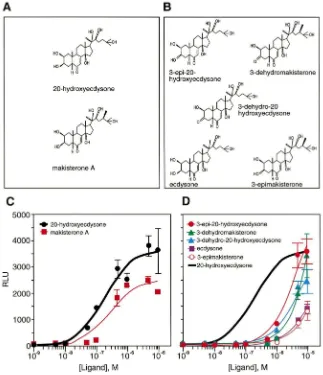
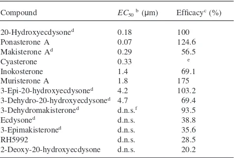
Drosophila SL2 cells, which express significant quan-tities of endogenous USP (Yao et al., 1992). The reporter plasmid carries three tandem copies of thehsp27 ecdy-sone response element (EcRE), which functions as a strong binding site for the EcR/USP heterodimer (Yao et al., 1993). Insect ecdysteroids with modifications at the C3, C20 or C24 position [Fig. 1(A) and (B)] all had varying effects on ecdysone-receptor-mediated acti-vation of the luciferase reporter gene [Fig. 1(C) and (D), Table 1].
Of the insect ecdysteroids, 20E was the most potent and among the most efficacious (EC50=0.18 mM),
fol-lowed by makisterone A (EC50=0.29 mM) (Fig. 1, Table
1). In contrast to previous reports that used insect bioas-says to monitor activity (Weirich et al., 1989; Somme´-Martin et al., 1990; Bergamasco and Horn, 1980; Nigg et al., 1974; Kaplanis et al., 1979), we found that 3-epi-20-hydroxyecdysone has a high efficacy, although it is a relatively poor activator at lower concentrations [Fig. 1(D), Table 1]. This resulted in an EC50 for
3-epi-20-hydroxyecdysone (4.2 mM) that is an order of magnitude greater than theEC50obtained for either 20E or
Fig. 1. Dose–response curves for activation of EcR by insect ecdysteroids. Shown are the structures and dose–response curves ofDrosophila
ecdysteroids that have high (A and C) or low (B and D) potency for activating EcR. The dose–response curve for 20-hydroxyecdysone generated in (C) is provided in (D) as a bold solid line for comparative purposes. Compounds were assayed for their ability to activate transcription of an ecdysone-inducible luciferase reporter gene in SL2 cells. Transcriptional activation is represented as relative light units (RLU), and is plotted relative to the concentration of the compound tested. Individual points in (C) and (D) are the average±standard deviation from three independent experiments.
Somme´-Martin et al., 1988; Cherbas and Cherbas, 1970), has been shown to have a potency indistinguishable from 20E in Manduca (Hiruma et al., 1997) and was also observed to have high activity in Drosophila larval fat body (Somme´-Martin et al., 1990), but is a relatively poor activator of EcR/USP when tested in the Droso-phila cotransfection assay [Fig. 1(D)].
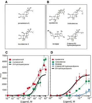
Dose–response curves for the Drosophila ecdysone receptor were also generated with phytoecdysteroids (Fig. 2). 20E deoxygenated at the C25 position [ponas-terone A; see Fig. 2(A)] proved to be the most potent activator of EcR among all compounds tested. Ponas-terone A was clearly distinguishable from 20E, having a lower EC50 (significant activation was observed with
a concentration as low as 1 nM) and a higher efficacy [Fig. 2(C), Table 1]. This finding is consistent with a previous report by Cherbas et al. (1980) which showed that ponasterone A is eight times more effective than
20E at initiating theDrosophila Kc-H cell morphologi-cal response. Hydroxylation at the C5 and C11 positions yields muristerone A [Fig. 2(A)], another phytoecdys-teroid with potent ecdysphytoecdys-teroid activity (Yao et al., 1993). Consistent with previous reports (Yao et al., 1993), mur-isterone A initiated the highest levels of EcR/USP-mediated transcription in the cotransfection assay, and had anEC50comparable to that of the natural ligand 20E
non-Table 1
Compounds that activateathe ecdysone receptor
Compound EC50b(µm) Efficacyc(%)
20-Hydroxyecdysoned 0.18 100
Ponasterone A 0.07 124.6
Makisterone Ad 0.29 56.5
Cyasterone 0.33 e
Inokosterone 1.4 69.1
Muristerone A 1.8 175
3-Epi-20-hydroxyecdysoned 4.2 103.2 3-Dehydro-20-hydroxyecdysoned 4.7 69.4 3-Dehydromakisteroned d.n.s.f 93.5
Ecdysoned d.n.s. 38.8
3-Epimakisteroned d.n.s. 35.6
RH5992 d.n.s. 28.5
2-Deoxy-20-hydroxyecdysone d.n.s. 20.2
aCompounds tested but not active were: 26-hydroxyecdysone; 3-dehydroecdysone; 22-isoecdysone; 14,trideoxyecdysone; 22,25-dideoxy-5α-(H)-ecdysone; 5β-(H)-cholestane-6-one-2β,3β-diol; 22α-hydroxycholesterol; 5α,6α-iminocholesterol; 5β,6β-iminocholesterol; 5β,6β-iminocholesterol-N-acetate; 5α,6α-epoxycholesterol; 5β,6β-epoxycholesterol; dehydrocholesterol; 5α,6α-epoxy-7-dehydrocholesterol-N3-OH adduct; 7-dehydro-25-hydroxycholesterol; 7-dehydro-5α,8α-peroxide-25-hydroxycholesterol; 7α-hydroxycholes-terol; 7β-hydroxycholes7α-hydroxycholes-terol; 5-cholestene-3β-ol-7-one; 5α-choles-7-ene-3β-ol; cholesta-4-ene-3-one; cholesta-5-ene-3-one; 5-pregnene-3β,20β-diol; 5-pregnene-3β,20α-diol; RH5849; 9-cis-retinoic acid; JH0; JHI; JHII; JHIII; JHIII bisepoxide; JH acid; hyrdoprene; methop-rene; fenoxycarb; farnesoic acid; and methoprene acid.
b EC50=effective concentration for 50% maximal activation. cEfficacy=maximal activation at 10µM relative to 20-hydroxyec-dysone.
d Naturally occurringDrosophilaecdysteroid. eToxic at 10µM.
f d.n.s.=does not saturate.
steroidal compound RH5992 [Fig. 2(B)], which has been reported to have an ecdysone-receptor-dependent LC50
(lethal dose concentration where 50% of cells die) of 600 nM in C18+Drosophilaimaginal disk cells (Cottam and Milner, 1997), had only modest affects on EcR/USP-mediated transcription [Fig. 2(D), Table 1]. It should be noted that, although SL2 cells were used in the experi-ments reported here, no significant metabolism of 20E has been seen in Kc cells, a relatedDrosophilacell line (L. Cherbas and P. Cherbas, personal communication).
Table 1 lists 37 other compounds (ecdysteroids, chol-esterol derivatives, pregnanes, a retinoid and juvenoids) that were tested in the ecdysone receptor transcriptional assay, all of which had no significant activity (data not shown). Together with the EcR agonists, these com-pounds were also tested for their ability to activate Gal4 chimeras (Harmon et al., 1995) in which the Gal4 DNA binding domain is fused to the ligand binding domains of the DHR38, DHR78 and DHR96 orphan receptors. However, in each case, the measured level of transcrip-tional activity for these receptors did not exceed those that were treated with vehicle alone (data not shown). The observation that natural and synthetic juvenoids
failed to activate the EcR/USP heterodimer and the orphan receptors tested suggests that USP is not a juven-ile hormone receptor in the context of a homodimer or a heterodimer with EcR or DHR38. This is despite the fact that USP is the insect homolog to the mammalian receptor RXR, and RXR can be activated by retinoids and juvenile hormone analogues (Heyman et al., 1992; Harmon et al., 1995), compounds that have isoprenyl chains similar to those found in juvenoids. The possi-bility still remains, however, that USP may serve a role in juvenoid-mediated signaling when paired with an unidentified partner.
This report represents the first comprehensive study of the effects of insect and plant ecdysteroids on the tran-scriptional activity of the EcR/USP heterodimer in Dro-sophila cells. Our transcriptional data correspond rela-tively well with a previous study by Mao et al. (1995). This group defined the binding affinities of ecdysteroids in the ixodid tick and found the relative affinities to be: ponasterone A.muristerone A.makisterone A.20E. ecdysone. Our data are also in agreement with Hiruma et al. (1997), who showed in vivo activity for the induc-tion of mRNA inManduca sextafor 20E and 3-dehydro-20-hydroxyecdysone, but failed to see biological activity for 3-dehydroecdysone and 26-hydroxyecdysone. As previously noted, a study performed by Cherbas et al. (1980) detailed the effectiveness of different ecdystero-ids in the extension of processes in Drosophila Kc-H cells. Again, their qualitative assignments of cell mor-phology correspond well with the quantitative measure-ments obtained in this report for the induction of an ecdysone-receptor-inducible reporter.
The observation that the natural ecdysteroid makis-terone A is able to generate a half-maximal transcrip-tional response in a range approaching that of 20E raises some interesting questions regarding the true in vivo ligand for the ecdysone receptor in Drosophila. Indeed, makisterone A may play a role in ecdysone receptor sig-naling throughout development. The titers of individual ecdysteroids supplied by the ring gland change drasti-cally and, as demonstrated by Pak and Gilbert (1987), the most abundant free ecdysteroids at pupariation in
Drosophila are makisterone A and 20E. Redfern (1986) noted that an increase in dietary campesterol resulted in the increase of 20-deoxymakisterone A relative to ecdy-sone, which are converted to makisterone A and 20E, respectively. That makisterone A may serve the role of 20E in insect signaling is not unprecedented. For example, makisterone A is thought to be the major ecdy-steroid in the last larval instar of the honeybee (Rachinsky et al., 1990).
Fig. 2. Dose–response curves for activation of EcR by phytoecdysteroids and a synthetic analog. Shown are the structures and dose–response curves of phytoecdysteroids that have high potency (A and C) or phytoecdysteroids and a synthetic analog that have low potency (B and D) for activating EcR. The highest concentration of cyasterone in (D) is toxic and is therefore not shown. The dose–response curve for 20-hydroxyecdysone generated in Fig. 1(C) is provided as a solid bold line in (C) and (D) for comparative purposes. Compounds were assayed for their ability to activate transcription of an ecdysone-inducible luciferase reporter gene in SL2 cells, as in Fig. 1. Transcriptional activation is represented as relative light units (RLU), and is plotted relative to the concentration of the compound tested. Individual points in (C) and (D) are the average±standard deviation from three independent experiments.
transcriptional activation, provide important future direc-tions for understanding the hormonal regulation of insect development.
Acknowledgements
We thank Drs K. Nakanishi, R. Lafont, and Rohm and Haas for some of the hormones used in this study and W.A. Segraves for the pA5C–EcR expression construct. D.J.M. and C.S.T. are investigators of the Howard Hughes Medical Institute (HHMI). This work was funded by HHMI (D.J.M. and C.S.T.), by grants from the Robert A. Welch Foundation and Human Frontier Science Program (D.J.M.), and grants from NIH (DK30118) and NSF (IBN 9603710) (L.I.G.).
References
Ashburner, M., 1974. Sequential gene activation by ecdysone in poly-tene chromosomes ofDrosophila melanogaster. II. The effects of inhibitors of protein synthesis. Dev. Biol. 39, 141–157.
Bergamasco, R., Horn, D.H.S., 1980. The biological activities of ecdy-steroids and ecdysteroid analogues. In: Hoffman, J.A. (Ed.), Pro-gress in Ecdysone Research. Elsevier/North-Holland Biomedical Press, Amsterdam, pp. 299–324.
Cherbas, L., Cherbas, P., 1970. Distribution and metabolism of alpha-ecdysone in pupae of the silkwormAntheraea polyphemus. Biol. Bull. 138, 115–128.
Cherbas, L., Yonge, C.D., Cherbas, P., Williams, C.M., 1980. The morphological response of Kc-H cells to ecdysteroids: hormonal specificity. Roux’s Arch. Dev. Biol. 189, 1–15.
Cottam, D.M., Milner, M.J., 1997. The effects of several ecdysteroids and ecdysteroid agonists on two Drosophila imaginal disc cell lines. Cell. Mol. Life Sci. 53, 600–603.
Fisk, G.J., Thummel, C.S., 1995. Isolation, regulation, and DNA-bind-ing properties of threeDrosophilanuclear hormone receptor super-family members. Proc. Natl. Acad. Sci., USA 92, 10604–10608. Fisk, G.J., Thummel, C.S., 1998. The DHR78 nuclear receptor is
required for ecdysteroid signaling during the onset ofDrosophila
metamorphosis. Cell 93, 543–555.
Gilbert, L.I., Rybczynski, R., Tobe, S.S., 1996. Endocrine cascade in insect metamorphosis. In: Gilbert, L.I., Tata, J.R., Atkinson, B.G. (Eds.), Metamorphosis: Postembryonic Reprogramming of Gene Expression in Amphibian and Insect Cells. Academic Press, New York, pp. 59–107.
Grau, V., Lafont, R., 1994. Metabolism of ecdysone and 20-hydrox-yecdysone in adult Drosophila. Insect Biochem. Mol. Biol. 24, 49–58.
Harmon, M.A., Boehm, M.F., Heyman, R.A., Mangelsdorf, D.J., 1995. Activation of mammalian retinoid X receptors by the insect growth regulator methoprene. Proc. Natl. Acad. Sci., USA 92, 6157–6160. Heyman, R.A., Mangelsdorf, D.J., Dyck, J.A., Stein, R.B., Eichele, G., Evans, R.M., Thaller, C., 1992. 9-Cis retinoic acid is a high affinity ligand for the retinoid X receptor. Cell 68, 397–406.
Henrich, V.C., Pak, M.D., Gilbert, L.I., 1987. Neural factors that stimulate ecdysteroid synthesis by the larval ring gland of Droso-phila melanogaster. J. Comp. Physiol. 157, 543–549.
Hiruma, K., Bo¨cking, D., Lafont, R., Riddiford, L.M., 1997. Action of different ecdysteroids on the regulation of mRNAs for the ecdysone receptor, MHR3, dopa decarboxylase, and a larval cuticle protein in the larval epidermis of the tobacco hornworm,Manduca sexta. Gen. Comp. Endocrinol. 107, 84–97.
Ismail, S.M., Satyanarayana, K., Bradfield, J.Y., Dahm, K.H., Bhaska-ran, G., 1998. Juvenile hormone acid: evidence for a hormonal function in induction of vitellogenin in larvae ofManduca sexta. Arch. Insect Biochem. Physiol. 37, 305–314.
Janowski, B.A., Willy, P.J., Devi, T.R., Falk, J.R., Mangelsdorf, D.J., 1996. An oxysterol signalling pathway mediated by the nuclear receptor LXRa. Nature 383, 728–731.
Janowski, B.A., Grogan, M.J., Jones, S.A., Wisely, G.B., Kliewer, S.A., Corey, E.J., Mangelsdorf, D.J., 1999. Structural requirements of ligands for the oxysterol liver receptors LXRa and LXRb. Proc. Natl. Acad. Sci., USA 96, 266–271.
Jones, G., Sharp, P.A., 1997. Ultraspiracle: an invertebrate nuclear receptor for juvenile hormones. Proc. Natl. Acad. Sci., USA 94, 13499–13503.
Kaplanis, J.N., Thompson, M.J., Dutky, S.R., Robbins, W.E., 1979. The ecdysteroids from the tobacco hornworm during pupal–adult development five days after peak titer of molting hormone activity. Steroids 34, 333–345.
Kliewer, S.A., Moore, J.T., Wade, L., Staudinger, J.L., Watson, M.A., Jones, S.A., McKee, D.D., Oliver, B.B., Willson, T.M., Zettersto¨m, R.H., Perlman, T., Lehmann, J.M., 1998. An orphan nuclear recep-tor activated by pregnanes defines a novel steroid signaling path-way. Cell 92, 73–82.
Koelle, M.R., Talbot, W.S., Segraves, W.A., Bender, M.T., Cherbas, P., Hogness, D.S., 1991. The Drosophila EcR gene encodes an ecdysone receptor, a new member of the steroid receptor super-family. Cell 67, 59–77.
Koelle, M.R., 1992. Molecular analysis of theDrosophila ecdysone receptor complex. Ph.D. thesis, Stanford University, Stanford, CA. Kozlova, T., Pokholkova, G.V., Tzertzinis, G., Sutherland, J.D., Zhi-mulev, I.F., Kafatos, F.C., 1998. Drosophila hormone receptor 38 functions in metamorphosis: a role in adult cuticle formation. Gen-etics 149, 1465–1475.
Makishima, M., Okamoto, A.Y., Repa, J.J., Tu, H., Learned, M., Luk, A., Hull, M.V., Lustig, K.D., Mangelsdorf, D.J., Shan, B., 1999. Identification of a nuclear receptor for bile acids. Science 284, 1362–1365.
Mao, H., McBlain, W.A., Kaufman, W.R., 1995. Some properties of
the ecdysteroid receptor in the salivary gland of the ixodid tick,
Amblyomma hebraeum. Gen. Comp. Endocrinol. 99, 340–348. Nigg, H.N., Svoboda, J.A., Thompson, M.J., Kaplanis, J.N., Dutky,
S.R., Robbins, W.E., 1974. Ecdysone metabolism: ecdysone dehydrogenase–isomerase. Lipids 9, 971–974.
Pak, M.D., Gilbert, L.I., 1987. A developmental analysis of ecdystero-ids during the metamorphosis of Drosophila melanogaster. J. Liquid Chromatogr. 10, 2591–2611.
Rachinsky, A., Strambi, C., Strambi, A., Hartfelder, K., 1990. Caste and metamorphosis titers of juvenile hormone and ecdysteroids in last instar honeybee larvae. Gen. Comp. Endocrinol. 79, 31–38. Redfern, CH.P.F., 1986. Changes in patterns of ecdysteroid secretion
by the ring gland ofDrosophilain relation to the sterol composition of the diet. Experientia 42, 307–309.
Richards, G., 1997. The ecdysone regulatory cascades inDrosophila. Adv. Dev. Biol. 5, 81–135.
Riddiford, L.M., 1993. Hormones and Drosophila development. In: Bate, M., Martinez-Arias, A. (Eds.), The Development of Droso-phila melanogaster, vol. 2. Cold Spring Harbor Laboratory Press, Cold Spring Harbor, pp. 899–939.
Riddiford, L.M., 1996. Juvenile hormone: the status of its “status quo” action. Arch. Insect Biochem. Physiol. 32, 271–286.
Smith, S.L., Mitchell, M.J., 1986. Ecdysone 20-monooxygenase sys-tems in a larval and an adult dipteran. Insect Biochem. 16, 49–55. Somme´-Martin, G., Colardeau, J., Lafont, R., 1988. Conversion of ecdysone and 20-hydroxyecdysone into 3-dehydroecdysteroids is a major pathway in third instar Drosophila melanogaster larvae. Insect Biochem. 18, 729–734.
Somme´-Martin, G., Colardeau, J., Beydon, P., Blais, C., Lepesant, J.A., Lafont, R., 1990. P1 gene expression in Drosophila larval fat body: induction by various ecdysteroids. Arch. Insect Biochem. Physiol. 15, 43–56.
Spindler, K.-D., Koolman, J., Mosora, F., Emmerich, H., 1977. Cata-lytical oxidation of ecdysteroids to 3-dehydro products and their biological activities. J. Insect Physiol. 23, 441–444.
Sutherland, J.D., Kozlova, T., Tzertzinis, G., Kafatos, F.C., 1995. Dro-sophilahormone receptor 38: a second partner forDrosophilaUSP suggests an unexpected role for nuclear receptors of the nerve growth factor-induced protein B type. Proc. Natl. Acad. Sci., USA 92, 7966–7970.
Thummel, C.S., 1995. From embryogenesis to metamorphosis: the regulation and function ofDrosophilanuclear receptor superfamily members. Cell 83, 871–877.
Thummel, C.S., 1996. Flies on steroids —Drosophilametamorphosis and the mechanisms of steroid hormone action. Trends Genet. 12, 306–310.
Warren, J.T., Steiner, B., Dorn, A., Pak, M., Gilbert, L.I., 1986. Metab-olism of ecdysteroids during the embryogenesis ofManduca sexta. J. Liquid Chromatogr. 9, 1759–1782.
Warren, J.T., Rybczynski, R., Gilbert, L.I., 1995. Stereospecific, mech-anism based, suicide inhibition of a cytochrome P450 involved in ecdysteroid biosynthesis in the prothoracic glands of Manduca sexta. Insect Biochem. Mol. Biol. 25, 679–695.
Weirich, G.F., Thompson, M.J., Svoboda, J.A., 1989. Ecdysone oxi-dase and 3-oxoecdysteroid reductases inManduca sextamidgut: kinetic parameters. Arch. Insect Biochem. Physiol. 12, 201–218. Willy, P.J., Umesono, K., Ong, E.S., Evans, R.M., Heyman, R.A.,
Mangelsdorf, D.J., 1995. LXR, a nuclear receptor that defines a distinct retinoid response pathway. Genes Dev. 9, 1033–1045. Yao, T.-P., Segraves, W.A., Oro, A.E., McKeown, M., Evans, R.M.,
1992.Drosophila ultraspiraclemodulates ecdysone receptor func-tion via heterodimer formafunc-tion. Cell 71, 63–72.