CHAPTER IV
RESEARCH METHOD
IV.1. Scope of study, place and time of the research
Scope of the study was included Pathobiology, histopathology.
IV.2. Place and time of the study
The study was conducted in Animal Laboratory at Gadjah Mada
University and the liver tissues examination were done at the Pathology Anatomy
Department of Diponegoro University Semarang between September–November
2011.
IV.3. Research design
The study design sign was experimental study, namely post test only
control group design. The samples were randomly divided into 3 intervention
groups and 1 group for control .
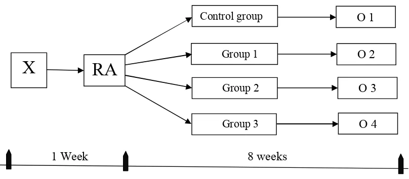
1 Week 8 weeks
Figure 6 . The scheme of the study
O 1 Control group
X
O 2Group 1
RA
Group 2 O 3
Notes :
X = Adaptation time.
RA = Random Allocation into 4 treatment groups.
Control Group = Each rat were received ethanol (12 g/kg of b.w per day) by using
the intragastric feeding model for 8 weeks
Group 1 (G1) = Each rat were received extract of Nigella sativa seeds in a dose
(0.5g/kg of b.w per day) along with ethanol (12 g/kg of b.w per
day) by using the intragastric feeding model for 8 weeks.
Group 2 (G2) = Each rat were received extract of Nigella sativa seeds in a dose
(1g/kg of b.w per day) along with ethanol (12 g/kg of b.w per
day) by using the intragastric feeding model for 8 weeks.
Group 3 (G3) = Each rat were received extract of Nigella sativa seeds in a dose
(1.5 g/kg of b.w per day) along with ethanol (12 g/kg of b.w per
day) by using the intragastric feeding model for 8 weeks.
Every group gets (1) additional rat for replacing rat which die during the study
period.
O1 = Output from the control group.
O2 = Output from the group 1.
O3 = Output from the group 2.
O4 = Output from the group 3.
The doses of Nigella sativa seeds extract were devided according of
experimental groups to three doses in two times per day by using the intragastric
treated with Nigella sativa seeds extracts as follows : - Group 1, Rats were
received 1ml of Nigella sativa extract given in two doses daily (0.5ml in the
morning and 0.5ml in the afternoon), equivalent to 0.5 g/kg b.w. Group 2, Rats
were received 1ml of Nigella sativa extract given in two doses daily (0.5ml in the
morning and 0.5ml in the afternoon), equivalent to 1 g/kg b.w. Group 3, Rats were
received 1ml of Nigella sativa extract given in two doses daily (0.5ml in the
morning and 0.5ml in the afternoon), equivalent to 1.5 g/kg b.w. This research
used doses gradual of Nigella sativa extract (0.5, 1, 1.5 g\kg b.w) according of
previous studies were used doses gradual. 16
The dose of ethanol was 4 ml of 40% ethanol given as two doses daily
(2ml in the morning and 2 ml in the afternoon), equivalent to 12 g/kg b.w as an
aqueous solution, using the intragastric feeding model (sonde tube) for 8 weeks.
This research used a fixed dose (12 g\kg b.w) that is the optimal dose of ethanol
on rats. It is based on preliminary experiment that shows this dose did not kill the
rats according of previous studies were used doses gradually, which was
considered an optimal dose. All rats both of Control group, group 1, group 2 and
group 3 were received 4 ml of 40% ethanol given in two doses daily (2 ml in the
morning and 2 ml in the afternoon) by using the intragastric feeding model (sonde
tube) for 8 weeks.11,12
IV.4. Population of The study
The population of the study was healthy male Wistar rats inbred strain of
Animal Laboratory at Gajah Mada University. Samples were taken from such rats
IV.4.1. inclusion criteria
Wistar rat (male), in healthy condition (move actively), measuring the
temperature of the rats and 12-16 weeks old and weights is between (250-300 g).
IV.4.2 exclusion criteria
The Wistar rats were showed behavioral changes (e.g., Low activity or
lazy)
IV.5. Sample size
The sample size of the study was determined according to the WHO
requirement for traditional medical study. According to WHO guideline minimal
5 rats are necessary in each group. In this study, according to Federer's formula,
the sample size in each group were 6 of Wistar rats. The total animals were 24 of
Wistar rats.
Federer's formula = (T-1) (N-1) >15
Abbreviation: T = Number of the group
N = Number of the rats
= (3N) – (3 ) >15 = (3N) < 15+3 = (N) < 6
By using sampling techniques (simple random sampling), samples as
many as 6 tails. Determination of the treatment of the sample was
randomly by using a random table.
IV.6. Research variable
IV.6.1. Independent variable
IV.6.2. Dependent variable
The dependent variable was hepatic tissue changes (hepatic steatosis state,
hepatic inflammation state, Mallory body state).
IV.7. Operational definition
IV.7.1. Hepatic steatosis state
The hepatic steatosis state is accumulation of either small or large fat
droplets in hepatocytes. It was diagnosed by evaluating the range of hepatic
steatosis in hepatocytes stained with HE staining with light microscope by using
NASH activity scoring system. (The NASH activity scoring system was attached
at appendix 2).
The scale is ordinal.
IV.7.2. Hepatic inflammation state
The hepatic inflammation state is infiltration of inflammatory cells that
consists of white blood cells. It was diagnosed by counting the number of white
blood cells of hepatic tissue stained with HE staining under light microscope by
using the Knodell scoring system. (The Knodell scoring system was attached at
appendix 2).
The scale is ordinal.
IV.7.3. Mallory bodies
Mallory bodies are large irregular masses which occur in the cytoplasm of
damaged liver cells. It was diagnosed by using Mallory bodies present or none
The scale is nominal.
IV.7.4. Nigella sativa seeds
Three doses of Nigella sativa seeds extractwere (0.5 g/kg of b.w, 1 g /kg
of b.w, 1.5 g /kg of b.w daily) given daily by intragastrically for 8 weeks.16
IV.8. Research tools and material
IV.8.1. Research material
A. Ethanol.
B. Nigella sativa seeds extract.
C. Wistar rats.
D. Food and drink for Wistar rats.
E. Hematoxylin Eosin staining.
F. Paraffin wax.
G. Formalin.
H. xyline.
I. Albumin.
IV.8.2. Research tools
A. Glass slides and cover slides.
B. Light Microscope for examination of tissues.
C. Hot plate or Water bath.
D. Wash bottle.
E. Racks for slides.
G. Microtome.
H. Hot plate or water bath.
I. Syringe.
J. Sonde tube.
IV.9. Research procedure
IV.9.1. Preparation of extract of the thymoquinone-rich fraction (TQRF) of
Nigella sativa seeds :-
Nigella sativa seeds were cleaned and dried in an oven at 40°C until a
constant weight was attained. Identification of Nigella sativa seeds were
conducted and the extraction have done by using ethyl acetate by maceration
procedure. Nigella sativa extracts at three doses, 0.5, 1 and 1.5 g/kg of b.w. All
doses were given to rats by intragastrically for 8 weeks.16 Nigella sativa seeds
extract were prepared in the Pharmacology Department of Gadjah Mada
University.
IV.9.2. Preparation of ethanol.
Rats received 12 g/kg b.w of 40 % ethanol. Liver cells damage were
induced in rats by administering 4 ml of 40 % ethanol, equivalent to 12g/kg of
body weight as an aqueous solution by using sonde tube for 8 weeks.12
IV.9.3. Histology specimen preparation
After the total duration of the experiment 8 weeks, at the end of it, the rats
from all groups were terminated. Initially the rats were subjected to whole-body
anesthesia. The procedure used for termination of rats pay attention to the
principles stated in the Helsinki Declaration of 1975 and the National Guidelines
for Health Research Ethics, and then were operated to take its liver tissue. The
liver tissues were removed and stored immediately in 10% formalin to preparation
of histology specimen for histopathological examination. The liver tissues were
fixed for at least 48 hours in 10% formalin. The specimens were dehydrated in
ascending grade solutions of alcohol and cleared in xylene and then impregnating
and embedding in paraffin wax for 2 hours. Sections were cut at 5 µm by using
the rotary microtome and then stained with hematoxylin and eosin for
examination using a light microscope.12 (Procedure for preparing of liver
histology slides was attached at appendix 1)
IV.10. Data collection and analysis
IV.10.1. Data collection
The study was conducted in Animal Laboratory at Gajah Mada University.
After terminated the rats, the liver tissue of the rats were taken to do
histopathology examinations. The slides for this examination were made in the
Pathology Anatomy Laboratory of Kariadi hospital Semarang and the tissue
examination at the Pathology Anatomy Department of Diponegoro University
Semarang.
IV.10.2. Statistical analysis
At first, the data collected from examination of hepatic tissue changes in 5
Kappa Statistic (K) was used to assess inter- pathologists agreement for the
reliability and validity to each diagnostic score. All of the grouped data from the
examination of hepatic tissue changes among all groups were descriptively
analyzed by calculating the standard deviation and median. The number of liver
hepatocyte changes of each group showed in box-plot graph. For categorical
variable (Mallory bodies) the results were presented in the form of a crosstabs and
analyzed using the Kendall's tau-b test. Hepatic tissue changes data (inflammation
and steatosis) were analyzed using nonparametric tests through Kruskal-Wallis
test followed by Mann Whitney test. Assisted statistical analysis with SPSS. The
level of statistical significance was set at P < 0.05.
IV.11. Ethics
Protocol of the study has been submitted to Ethical Committee of Medical
and Health Research Medical Faculty Diponegoro University and then ethical
clearance was obtained. All animals have been care according to Helsinki’s
Declaration, as well as, all invasive treatment have been conducted under
anesthesia.
IV.12. Preparations followed for the study.
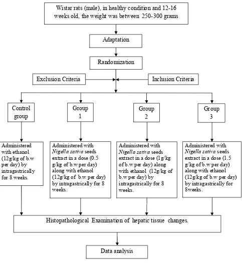
The study was conducted in Animal Laboratory at Gajah Mada University.
Twenty four healthy, male, adult Wistar rats 12-16 weeks old and the weight was
between 250 - 300 grams. They were procured from Animal Laboratory at Gajah
All animals were cared on Animal Research Care Facility of Faculty
Medicine Gajah Mada University. Adaptation time was done in one week to
prepare the rats, that was weighing, examining the health status, and help the rats
to adapt to new environments. They were housed in metal cages under controlled
conditions, and raised by balancing feeding at room temperature and constant
wetness degree according to the alteration of day and night. The rats were divided
into four groups of six rats each. All groups of rats were received a normal diet of
standard pellets. Liver cell damage was induced in rats of all Groups by
administering 4 ml of 40% ethanol (2 ml in the morning and 2 ml in the
afternoon) by intragastrically (sonde tube) for 8 weeks.11,12 Through of this
period the Wistar rats were treated as follows for 8 weeks :-
The first group was an experimental control group of rats, which were received 4
ml of 40% ethanol given in two doses daily (2 ml in the morning and 2 ml in the
afternoon), equivalent to 12 g/kg b.w as an aqueous solution, using the intragastric
feeding model (sonde tube). This research used a fixed dose (12 g\kg b.w) that is
the optimal dose of ethanol on rats.11,12 Group 1, Rats were received two doses of
Nigella sativa seeds extract and ethanol at two times in the morning and in the
afternoon. One dose was consisted of 0.5 ml of Nigella sativa seeds extract,
along with 2 ml of 40% ethanol after two hours from receipt of Nigella sativa
seeds extract by intragastrically.11,12,16 Group 2, Rats were received two doses of
Nigella sativa seeds extract and ethanol at two times in the morning and in the
afternoon. One dose was consisted of 0.5 ml of Nigella sativa seeds extract,
seeds extract by an intragastrically.11,12,16 Group 3, Rats were received two doses
of Nigella sativa seeds extract and ethanol at two times in the morning and in the
afternoon. One dose was consisted of 0.5 ml of Nigella sativa seeds extract, along
with 2 ml of 40% ethanol after two hours from receipt of Nigella sativa seeds
extract by an intragastric tube .11, 12, 16
The total of duration of the experiment was 8 weeks, at the end of it, the
rats from all groups were terminated, which was according to the principles stated
in the Helsinki Declaration of 1975 and the National Guidelines for Health
Research Ethics. Initially the rats were subjected to whole-body perfusion using
normal saline and 10% formalin under light ether anesthesia, and then were
operated to take its liver tissue. The liver was removed and stored immediately in
10% formalin to preparation of histology specimen for histopathological
examination using a light microscope by using magnification of 400x in 5 fields
IV.13. FLOW CHART OF STUDY
Figure 7. Flow Chart Of Study Wistar rats (male), in healthy condition and 12-16 weeks old, the weight was between 250-300 grams
Adaptation
Histopathological Examination of hepatic tissue changes.