CHAPTER II LITERATURE REVIEW
II.1. ETHANOL INDUCED HEPATIC TISSUE CHANGES II.1.1. Ethanol
Ethanol is a commonly used substance among people all over the world with its intoxicating effects and potential for abuse. The main component of all alcoholic drinks is ethanol (ethyl alcohol). Ethanol is one of direct hepatotoxins agents, ethanol ingested in high doses impairs tissues by a variety of mechanisms,17 oxidative stress plays surely a crucial role. Most ethanol is broken down (i.e., metabolized) in the liver through a series of chemical reactions, known as oxidation reactions, which involve hydrogen and oxygen atoms. In the predominant biological pathway for ethanol metabolism, called the alcohol dehydrogenase pathway (ADH), a second pathway of ethanol metabolism, the microsomal ethanol-oxidizing system (MEOS), is activated by long-term heavy ethanol consumption.18
II.1.2. Definition of liver injury induced by ethanol
chronic hepatitis with hepatic fibrosis or cirrhosis. These latter stages may also be associated with a number of histologic changes (which have varying degrees of specificity for ALD), including the presence of Mallory bodies.19
II.1.3. Ethanol metabolism
Most tissues of the body, including the skeletal muscles, contain the necessary enzymes for oxidative or nonoxidative metabolism of ethanol. However, the major site of ethanol metabolism is the liver. Within the liver, there are three enzyme systems for metabolism of ethanol, These enzymes are: -Cytosolic alcohol dehydrogenase (ADH) uses nicotinamide adenine dinucleotide (NAD) as an oxidizing agent.
1. The microsomal ethanol-oxidizing system (MEOS) uses nicotinamide adenine dinucleotide phosphate (NADPH) and molecular oxygen. The central enzyme of MEOS is cytochrome P-450 2E1 (CYP2E1). Ethanol up regulates CYP2E1, and the proportion of alcohol metabolized via this pathway increases with the severity and duration of alcohol use.
2. Peroxisomal catalase uses hydrogen peroxide as an oxidizing agent.20
II.1.4. Factors that influence the liver injury II.1.4.1. Genetic factors
inconsistent results, as have studies of genetic polymorphisms of collagen, ADH, ALDH, and CYP2E1. The genetic factor that most clearly affects susceptibility is sex. For a given level of ethanol intake, women are more susceptible than men to developing alcoholic liver disease.21
II.1.4.2. Malnutrition
In the past, nutritional deficiencies were assumed to play a major role in the development of liver injury. This view changed after key studies, that demonstrated that alcohol ingestion could lead to steatohepatitis and cirrhosis in the presence of a nutritionally complete diet. However, subsequent studies have suggested that enteral or parenteral nutritional supplementation in patients with alcoholic hepatitis may improve survival.22
II.1.5. Diagnosis of liver injury induced by ethanol II.1.5.1. Clinical symptoms
II.1.5.2. Laboratory diagnosis of liver injury induced by ethanol II.1.5.2.a. Laboratory tests
Laboratory tests show increases in serum aspartate aminotransferase (AST), Gamma glutamyl transferase (GGT), while the increase in alanine aminotransferase (ALT) is less pronounced. Other markers include hypoalbuminemia, prolonged prothrombin time (PT), increase in mean corpuscular volume (MCV) of erythrocytes.24
II.1.5.2.b. Liver biopsy
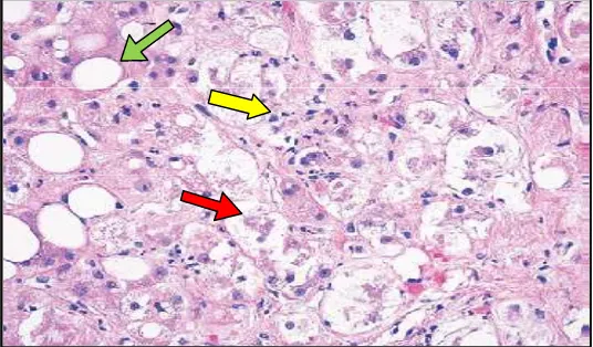
bodies called Mallory bodies (red arrow), which are often surrounded by neutrophils (yellow arrow) (hematoxylin and eosin).19,26 (Figure 1).
Figure 1. Histopathological features in a liver-biopsy specimen in AH. Adapted from : - Michael R. Lucey M R, Mathurin P and Morgan T R .26
II.1.6. Pathogenesis of liver injury induced by ethanol : II.1.6.1. Mechanism of ethanol-induced hepatic steatosis
triglycerides, fatty acids and the TCA and may thereby increase lipogenesis (Figure 2C1).26,27
S-adenosylmethionine (SAMe) is the primary methyl donor and a precursor to glutathione. Oxidative stress activation via long term alcohol consumption, through the activity of cytochrome P-450 2E1, that alters the intracellular balance between levels of S-adenosylmethionine (SAMe) and S-adenosylhomocysteine (SAH), resulting in a decrease in hepatic methionine levels, decrease in the activity of methionine adenosyl transferase, the enzyme
which converts methionine to SAMe 28 and a decrease in the ratio of S-adenosylmethionine to S-adenosylhomocysteine. A decrease in these agents
may contribute to alcohol induced liver injury, since S-adenosylhomocysteine exacerbates TNF- hepatotoxicity, whereas S-adenosylmethionine diminishes it. Reduction in the enzymes that convert ((S-adenosylhomocysteine (SAH) to methionine , then convert methionine to S-adenosylmethionine (SAMe) )), lead to increase in homocysteine. This causes a decrease in glutathione synthesis which results in impaired clearance of oxidative species such as 4-hydroxynonenal (a marker of lipid peroxidation) and mitochondrial injury caused by a decrease in glutathione transport from the cytosol into the mitochondria. Increases in concentration of S-adenosylhomocysteine(SAH), stressing the endoplasmic reticulum (ER stress), that lead to release of sterol regulatory element-binding protein 1c (SREBP-1c) from ER stress (Figure 2C1).26,29
AMP-activated protein kinase (AMPK) activates hepatic fatty acid synthesis by activating hepatic sterol regulatory element-binding protein (SREBP-1c) transcription.30 These mechanisms lead to SREBP-1c initiates the transcription of genes involved in triglyceride and fatty-acid synthesis. Also ethanol has been shown promotes lipid metabolism by inhibiting peroxisome-proliferator–activated receptor (PPAR- ). Decrease in the binding of peroxisome-proliferator–
activated receptor (PPAR- ) to DNA reduces the expression of genes involved in fatty acid oxidation (Figure 2C1). 26,27
II.1.6.2. Liver injury due to ethanol metabolites
Hepatocytes convert ethanol to acetaldehyde through three mechanisms: Alcohol dehydrogenase (ADH), cytochrome P-450 isoenzyme-1 (CYP2E1), and catalase. 31 Lipid-peroxidation products can combine with acetaldehyde and with proteins to produce neoantigens, which can stimulate an autoimmune response. Against the background of steatosis and existing liver damage, heavy and continued drinking in some patients causes alcohol hepatitis. Two main mechanisms are involved: inflammation and oxidative stress.32
II.1.6.2. a. Mechanism of ethanol-induced hepatic inflammation
with toll-like receptor (TLR4) receptors.33 TLR4 adapter pathway activates NF-in Kupffer cells, that is translocate to the nucleus NF-in response to stress signals and binds to the promoter region of pro-inflammatory, thereby activating multiple cytokine genes.26,34,35 Kupffer cells are pivotal for the development of hepatic inflammation induced by ethanol.36 The ongoing ethanol-induced LPS absorption may provide a continuing stimulus to Kupffer cells that perpetuates hepatic inflammation induced by ethanol( figure 2B).26, 37
Ethanol is broken down (i.e., metabolized) in the liver cells by two enzymes, alcohol dehydrogenase (ALD) and, particularly after chronic ethanol consumption, cytochrome P450 2E1 (CYP2E1). Both enzymes convert ethanol to acetaldehyde, a toxic substance. Some of the acetaldehyde interacts with proteins in the cells such as malondialdehyde, which results from lipid peroxidation, interact, through a covalent binding, with the reactive lysine residues of proteins, formation malondialdehyde-acetaldehyde proteins adducts (MAA) which called adducts, located on the membranes of hepatocytes. MAA proteins adducts capable to produce neoantigens, which can activate certain immune cells to produce various cytokines, including interleukins such as (IL–1, IL–2 ), interferon gamma (IFN– ), and tumor necrosis factor alpha (TNF– ), that in turn attacks healthy liver cells, resulting in tissue damage (figure 2C2).26,38,39
accumulate primarily in cell structures called mitochondria (figure 2B1). 26,40 ROS normally are eliminated from the cells by compounds known as antioxidants, particularly a small molecule called glutathione (GSH). Alcohol, however, depletes the cell’s GSH stores, thereby further exacerbating ROS accumulation in the mitochondria. This process leads to the release of cytochrome c from the mitochondria, which then activates enzymes called caspases and promotes production of IL–8 in the cell ( figure 2C3).26, 37 ROS can also activate ERK1/2, p38 MAPK kinases, and NF- B which stimulates TNF- , complementing the LPS-induced pathways of TNF- production and creating a vicious cycle of inflammation and oxidative stress( figure 2B1).26, 41
II.1.6.3. Mechanism of ethanol-induced Mallory bodies
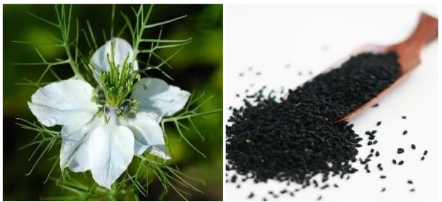
II.2. NIGELLA SATIVA SEEDS II.2.1. Description
Nigella sativa is bisexual and forms a fruit capsule which consists of many
white trigonal seeds. Once the fruit capsule has matured, it opens up and the seeds contained within are exposed to the air, becoming black in color (black seeds).
Nigella sativa and its black seed are known by other names, varying between
places.43 (fig. 3)
Figure 3 : Black Seed Flower , black Seed Nigella seeds: the miraculous Black
Seed. Adapted from:
http://www.healthinfo21.com/2010/11/nigella-seeds-miraculous black-seed.html.44
Nigella sativa Scientific classification:- Kingdom: Plantae, Division:
Magnoliophyta, Class: Magnoliopsida, Order: Ranunculales, Family: Ranunculaceae, Genus: Nigella, Species, Nigella sativa. The scientific name is a derivative of Latin Niger (black). Original black cumin seed is Carum bulbocastanum. The botanical name is Nigella sativa, while In English, Nigella
sativa seed is variously called fennel flower, nutmeg flower, Roman coriander,
kalonji (Hindi/Urdu) or mangrail (Hindi), ketzakh (Hebrew), chernushka (Russian), çörek otu (Turkish), siyah daneh (Persian), karim jeerakam (Malayalam or in Sinhala).45
II.2.2. The chemical composition of Nigella sativa seeds
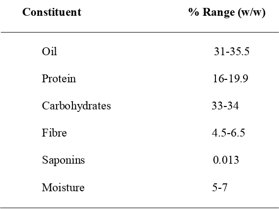
The general chemical composition of the seeds is depicted in Table 2. Table 2 : The general chemical composition of Nigella sativa seeds Constituent % Range (w/w). Adapted from: Ismail M Y M.46
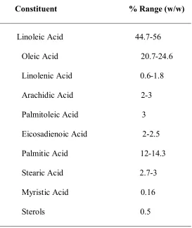
II.2.3a. The chemical composition of Nigella sativa oil
The chemical analysis of Nigella sativa total oil revealed the presence of both a fixed oil and a volatile oil. The major component was the fixed oil whereas the volatile oil ranged from 0.4- 0.7% of the seeds’ weight. The fixed oil chemical composition is outlined in Table 3.
Constituent % Range (w/w)
Table 3 : The chemical composition of Nigella sativa fixed oil .( Adapted from : - Ismail M Y M. ) .46
Generally, there were no significant variations in the chemical composition of the fixed oils of seeds grown in Egypt, Sudan, Ethiopia, India, Turkey and Syria. However, Al-Jassir noted that the seeds grown in Qassim, Saudi Arabia, contained, in addition to the fatty acids depicted in Table 2, two more acids which were lignoceric acid about (1%) and myristoleic acid (0.18%) without the presence of eicosadienoic acid. Specific chemical analyses of the volatile oil started by Mahfouz and El-Dakhakhny and Canonica et al. These studies were complemented by most recent ones which revealed various pharmacologically active constituents that included Thymoquinone that may
Constituent % Range (w/w)
attain up to 27.8% of the volatile oil (w/w), Carvacrol phenol which is also known as isothymol (5.8-11.6% (w/w)), p-cymene in the range of (15.5-31.7% (w/w)), –pinene in the range of (9.3% (w/w)), –terpineol in the range (2-6.6% (w/w), longifolene (1-8% (w/w)), t-anethole (p-Propenyl anisole)benzene in range (0.25-2.3%(w/w)) and the reduction product of thymoquinonethymohydroquinone together with some esters about 16% .
II.2.3b. Non-oily components of Nigella sativa seeds 1. Minerals
Analysis of Nigella sativa seeds, ash revealed the presence of 0.5-1% calcium, 0.6% phosphorus, 0.6% potassium and 0.1% sodium of the total seeds weight .
2. Saponins
The major saponin in the defatted seeds of Nigella sativa is the glycoside -hederin or Helixin or melanthin which on acid hydrolysis releases its sugar rhamnose / arabinose and gives the aglycone hederagenin (or melanthigenin) or caulosapogenin.
3. Alkaloids
Three types of alkaloids were isolated from the defatted seeds of Nigella
sativa. These were identified as the indazole nigelicine, the isoquinoline
II.2.4.The therapeutic potential of Nigella sativa seeds II.2.4.1. Anti inflammation of Nigella sativa seeds
Inflammation has been known to produce proinflammatory cytokines and diverse reactive oxygen species (ROS) and reactive nitrogen species (RNS) creating pre-disposition to various patho-physiological disorders.47 The amelioration of the Experimental Allergic Encephalitis by TQ was examined, who showed potent effects, which were thought to occur via induction of glutathione.48
TQ was found to decrease NF-K activation, thereby NF-K is a molecular target of TQ, where previous studies showed the effects of TQ on the inhibition of activation of NF-K in an experimental autoimmune encephalomyelitis in the rat model of multiple sclerosis. TQ has been shown inhibit of activation of NF-K in an experimental autoimmune encephalomyelitis. TQ was able to increase the red blood cell glutathione, and inhibit the activation of NF-K in the brain and spinal cord. These results clearly provide some early indication that many of the biological activities of TQ could in part be due to inactivation of NF-K and its downstream genes. Collectively, these results suggest that NF-K is a molecular target of Thymoquinone.49, 50, 51
II.2.4.2. Antioxidant activity of Nigella sativa seeds
hyperhomocysteinemia (Hhcy) and its associated state of oxidative stress. Pre-treatment of rats with an oral dose of 100 mg/kg of TQ for 30 min and for one week provided complete protection against induced HHcy after methionine load (100 mg/kg). Treatment with the different doses of Thymoquinone (TQ) produced significant reductions in hepatic superoxide dismutase (SOD), and catalase (CAT) and glutathione peroxidase (GSH-Px) activities although neither produced any change in GST activity nor influenced reduced glutathione content in any of the tissues studied. These results revealed that Thymoquinone (TQ) and dihydrothymoquinone (DHTQ) acted not only as superoxide anion scavengers but as general free radical scavengers.59
The effects of TQ on carbon tetrachloride (CCl4)- induced hepatotoxicity have been investigated in male Swiss albino mice. Oral administration of TQ in a single dose (100 mg/Kg) resulted in a significant protection against the hepatotoxic effects of CCl4. The result suggests that the protective action.The effects of TQ against CCl4-induced hepatotoxicity may be mediated through the combined antioxidant properties of TQ and its metabolite DHTQ.60,50,51
II.2.5. Side effects of Nigella sativa seeds
Nigella sativa seeds are a safe and effective herb that can be used by
should not take Nigella sativa seeds if their wombs are sensitive unless otherwise instructed by a medical professional (Many Muslim women take it while pregnant and no harm has been found).61, 62
II.3. Animal models of ethanol-induced liver damage
Animals have been administered ethanol by various methods in attempts to try to develop liver lesions resembling those seen in human ALD. The animals continued to gain weight with ethanol concentrations up to 50%, showed increased ethanol metabolism, and sustained elevated blood ethanol levels, but liver damage occurred at most in only a minority of the animals. To overcome the failure of most animals otherwise to consume higher amounts of ethanol voluntarily, it has been administered in a nutritionally adequate liquid diet that provided a maximum of 35-40 % of total calories from alcohol. This situation resembles that of many alcoholics, who often receive more than 50 % of their total energy as ethanol. In rats this has been achieved by administration of ethanol as a component of the liquid diet, either orally or by forcing intragastric infusion.63
II.3.1. Intragastric ethanol feeding model