58 CHAPTER IV RESEARCH METHODS
IV.1. Scope of the study
Scope of study was pathology anatomy ,theraby , histopathology and specially in the field of pathobiology.
IV.2. Place and time of the study
The study was conducted in Animal Laboratory at Gajah Mada University and the tissue examination was done at Pathology Laboratory of RSUP Dr. Kariyadi Semarang between Agust-October 2011.
Group A exposed to cigareete smoke for 10 weeks
Group B exposed to cigareete smoke for
13weeks
Group C exposed to cigareete smoke + oral adminestration of 80 mg/kg B.W.daily of curcuma
longa extract for 10 weeks
Group D exposed to cigareete smoke + oral adminestration of 80 mg/kg B.W.daily of
curcuma longa extract for 13 weeks
59 IV.3. Subjects of the study
Subjects of the study were Sprague Dawley albino rats inbred strain. The rats were obtained from Animal Laboratory at Gajah Mada University. Criteria of samples as follow:
IV.3.1. Inclusion Criteria
- Male
- Age 4 weeks, body weight was between 40-65g.
- Healthy characterized by active moving and no sign anatomic anomaly by inspection
IV.3.2. Exclusion Criteria
- Rats had diarrhea during the study, the activity looks abnormal , sneezing and
semi-dormant
-Rats were death during experiment
IV.3.3. Sampling methods
Rats were allocated into 4 experiment group by simple random sampling using computer generated random number.
IV.4. Sample Size
60
in each group. In this study according to Fredreic test the sample in each group
were 6 SD rats. The total animals were 24 rats.
Fredreic test =
Abbreviation:
T= number of the group
N= number of the rats = (4-1 ) (N-1) >15
= ( 3) (N-1) >15 = ( 3N) – (3 ) >15 = (3N) < 15+3 = (N) < 6
By using sampling techniques (simple random sampling) sampled as many as 6 tails
IV.5. Study variables
IV.5.1. Independent variable
Cigarette smoke and curcuma administration status: - Cigarette smoke exposure for 10 weeks
- Cigarette smoke exposure for 13 weeks
- Cigarette smoke exposure followed by oral administration of Curcuma L extract 80 mg/kg B.W. daily for 10 weeks
61
- Cigarette smoke exposure followed by oral administration of Curcuma L extract 80 mg/kg B.W. daily for 13 weeks
IV.5.2. Dependent variables
a. Liver cells change ( odeama cell ,karyiohexis ,karyiolysis ) b. Liver tissue TNF- expression score
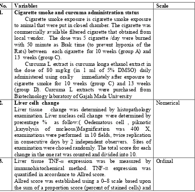
Table (2) Dependent variables definition
No. Variables Scale
1. Cigarette smoke and curcuma administration status Cigarette smoke exposure is cigarette smoke exposure to animal that were put in closed chamber. The cigarette was commercially available filtered cigarette that obtained from local vendor. The dose was 5 cigarette /day were burned with 50 minute as Brak time (to prevent hypoxia of the Rats) between each cigarette for 10 weeks (group A) and 13 weeks (group C).
Curcuma L extract is curcuma longa ethanol extract in the dose of 80 mg/kg (in 1 ml of 5% DMSO) daily administered using orally immediately after exposure to cigarette smoke for 10 weeks (group C) and 13 weeks (group D). Curcuma L extracts were purchased from Biotechnology laboratory of Gajah Mada University
2. Liver cells change
Liver tissue change was determined by histopathology examination. Liver nucleas cell change were determined by precentage % as follow:( Oedematous cell , piknotic ,karyolysis of nucleous)Magnification was 400 X, examinations were performed in 10 fields, twice replication in consecutive days by 2 independent observers. Sites of examination were chosed randomly. The total score for each change in the one rat was counted and divided into 10.
Nomerical
3. Liver tissue TNF- expression was be measured by immunohistochemical method. TNF- expression was quantified in accordance to Allred score.
Allred score was established using a 0–8 scale based upon the sum of a proportion score (percent of stained cells) and
62
No. Variables Scale
intensity score (weak, intermediate, and strong). The possible values of Allred score are: 0 – Allred 0*; 1 – Allred 2, 3, 4; 2 – Allred 5, 6; 3 – Allred 7, 8 (*Allred score 1 is
B. Curcuma longa extract in dose 80 mg/kg B.W.daily C.Sprague Dawley albino rats
D.Closed chamber designed for cigarette smoke exposure E.Water
F. General pellet food for rats
G.TNF- immunohistochemical staining kit (RND system, USA) H.Hematoxyllin (HE) eosin staining kit
63 IV.7.2 RESEARCH Material
A. Glass slides.
B. Light Microscope for examination of tissues. C. Hot plate or Water bath .
D. Wash bottle. E. Racks for slides.
F. Marker of marking of slides . G. Microtome .
H. Hot plate or water bath .
IV.7.3. Animal care
64 IV.7.3 Data collection method
All animal were injected by ketamine injection as ( anaesthesia ) through tail vein,Rats liver tissue were taken for histopathology and immunohistochemistry examination. The slide for those examination was made at Pathology Laboratory of RSUP Dr. Kariyadi Semarang. Rats in group (A) was exposed to the cigarettes Smoke in dose 5 cigarettes with 50 minute intervals as preak time (to prevent hypoxia of the Rats in the cage ) between each cigarettes smoke exposure for 10 weeks. Group (B) was exposed to the cigarette smoke in dose 5 cigarettes with 50 minute intervals as preak time between each cigarettes smoke exposure for 13 weeks. Group (C) was exposed to the cigarette smoke in dose 5 cigarettes with 50 minute intervals as preak time between each cigarettes smoke exposure and followed by oral administration of Curcuma L in dose 80 mg/kg B.W.daily for the 10 weeks. Group (D) was exposed to the cigarette smoke in dose 5 cigarettes with 50 minute intervals as preak time between each cigarettes smoke exposure and followed by oral administration of Curcuma L in dose 80 mg/day B.W. daily for the 13 weeks.
IV.8. RESEARCH PROCEDURE
IV.8.1. Preparation of Curcuma L extract
65
dried under reduced pressure at a room temperature not exceeding C. were Rats received 80mg/kg B.W. daily of curcuma longa rhizoma extract orally by feeding ,
IV.8.2 Preparation of Marlboro ciggarete smoking
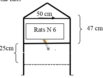
A Marlboro cigarettes were brought from a local shop were Nicotine with level is 1.5 mg on the baket , each group has been giving a total number of 5 cigarettes with Dividing time up to 50 minutes between each cigarette as preak time to prevent hypoxia of the Rats
50 cm
47 cm
25cm . ...
Fig 10. Design of the cage size of passive smoking
IV.8.3. Histology Specimen Preparation and staining (Liver cells change)
After the total duration of the experiment 10 weeks for (A&C),and 13 weeks for (B&D) at the end of it, the animals subjected to whole-body perfusion using normal saline and buffered formalin under light ether anesthesia. way to termination pay attention to the principles stated in Helsinki Declaration of 1975 and the National Guidelines for Health Research Ethics(PNEPK ),The liver removed
66
and stored immediately in buffered formalin for histopathologyy examination. The tissue fixed for at least 48 hours in buffered formalin. The specimens were dehydrated in ascending grades solutions of alcohol and cleared in xylene and then impregnating and embedding in paraffin wax for 2 hours . Sections cuted at (Three pieces ) with the rotary microtome and then stained with hematoxylin and eosin for examination by light microscopy. 11
Were the liver cells change is Liver Odeama Cell & Karyopiknotic or/and karyorhexis and Karyolysis
Odeamatous cell
hepatocytes exhibited cloudy swelling with pale cytoplasm and poorly delineated and displaced nuclei . Cellular swelling might be accompanied by leakage of lysosomal hydrolytic enzymes that lead to cytoplasmic degeneration and macromolecular crowding
Karyopyknosis
pyknotic nuclei is clumping and condensation of the chromatin materials in the periphery of the nuclei together with irregularity nuclear membranes Karyopyknosis ia an irreversible condensation of chromatin in the nucleus of a cell undergoing necrosis or apoptosis
Karyolysis
67
IV.7.3. Liver tissue TNF- expression immunohistochemistry procedure - Animals were sacrifice by overdose of ketamine injection.
- Liver was remove by clean surgery procedure.
- Liver tissue were fixed in formalin and embed in paraffin blocks according to standard procedures,were each slide got three pieces of the tissue section - Object glass slides were cleaned with 95% ethanol and treated with subbing
solution and air dry, or by using pre-treated slides.
- Tissue sections were cut 4–6 micron thick and applied to slides. Tisue were deparaffinize in xylenes using three changes for 5 minutes each. Hydrate sections gradually through graded alcohols: wash in 100% ethanol twice for 10 minutes each, then 95% ethanol twice for 10 minutes each. Wash in deionized H2O for 1 minute with stirring. Aspirate excess liquid from slides. - Antigen unmasking was performed at this point. Certain antigenic
determinants are masked by formalin fixation and paraffin embedding and may be exposed by Pepsin: Incubate sections for 10–20 minutes in 0.1% pepsin in 0.01 N HCl at room temperature. Slides were washed several times in deionized H2O. Aspirate excess liquid from slides.
68
- TNF- quantified in accordance to Allred
score by two independent pathologists and compared across histological categories using Kappa test.
Were TNF- is;
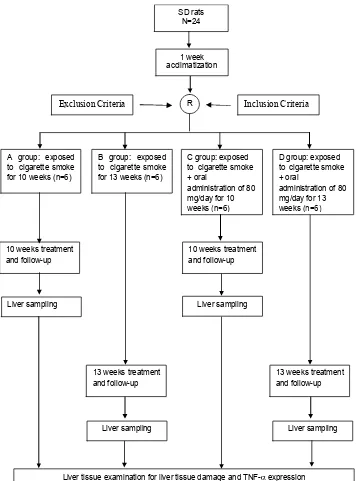
69
Liver tissue examination for liver tissue damage and TNF- expression
Fig. 11. Flow chart of the study
70 IV.9. Data analysis
Before analysis data was checked for correct and completeness. Data were coded, tabulated and entry in to computer.At first ,the data collected from examination of hepatic tissue changes in 5 fields for each slide were evaluated statistically by calculating median , then Kappa Statistic(K) was used to assess inter- pathologists agreement for the reliability and validity to each diagnostic scores . Number of liver hepatocyte changes of each group showed in box-plot graph
Data analysis was include descriptive analysis and test of hypothesis. Data of liver cell change and TNF- expression score were expressed as mean and standard deviation (SD). Data normality distribution was checked by Saphiro-Wilk test. Saphiro-Wilk test showed liver cell change were normality distribute and TNF- expression score were abnormally distributed. The difference of liver cells change score were analyzed by using independent t-test. P value < 0.05 is considered significant.and TNF- expression score between A group and C group, B group and Dgroup were analyzed by using Mann-Whitney Test. P value < 0.05 is considered significant.Statistical analysis were performed by computer program.
IV. 10. Ethics
71 CHAPTER V
RESULTS
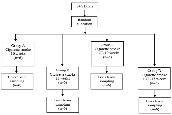
This study used 24 SD rats as study objects. Rats were randomly allocated by simple random sampling into 4 groups: Group A, rats were exposed to smoke of 5 cigarettes for 10 weeks (n=6); group B, rats were exposed to smoke of 5 cigarettes for 13 weeks (n=6); group C, rats were exposed to smoke 5 cigarettes and followed by oral administration of 80 mg/kg B.W.daily for 10 weeks; group D, rats were exposed to smoke 5 cigarettes and followed by oral administration of 80 mg/kg B.W. daily for 13 weeks. During study period no rats were sick or death. At final experiment all rats were used for data analysis. The number of rats during study period as showed on figure 12.
24 SD rats
72 4.1. LIVER CELLS CHANGES
Histopathology feature of liver tissue on study groups at the end of experiment were shown on figures below. Tissue damage were observed from H&E staining examination by Olympus PX51 light microscope with 400x magnification in 10 fields from randomized choosing. The examiner counted the number of liver cells changes (i.e, odeama cell, karyopiknotic, and karyolysis). The results were expressed in
percentage (%) of abnormal cells per all cells counted on those fields. From total 24 rats, 24 livers of the rats were made into tissue slide
Were the Rats distrubuted as group as ,Group A as control group was exposed to passive cigaratte smoke for 10 weeks after that was terminated and liver of it was taken,Group B as control group was exposed to passive cigaratte smoke for 13 weeks after that was terminated and liver of it was taken, Group C as treated group was exposed to passive cigaratte smoke for 10 weeks followed by curcuma longa rhizoma
extract after that was terminated and liver of it was taken, Group D as treated group was
exposed to passive cigaratte smoke for 13 weeks followed by curcuma longa rhizoma extract after that was terminated and liver of it was taken,
73
B
C
D
A
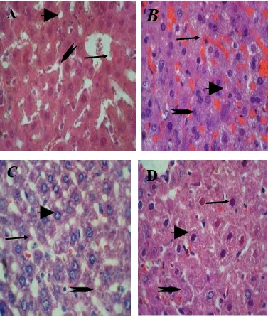
Figure 13. HE staining examination of group (A,B,C,D) (400X magnification) Histopathology feature of liver cells changes of SD rats after SD rats after 10
weeks (A)13 weeks (B )cigarette smoke exposure only,10 weeks cigarette smoke exposure + Curcuma L (C). 13 weeks cigarette smoke exposure +
74
Figure 13 showed large number of liver cells with liver cells change such as odeama, karyolysis and karyopicnotic on SD rats that exposed to cigarettes smoke for 13 weeks, in other hand, on SD rats that exposed to cigarette smoke with Curcuma L
administration for 10 weeks , the number of cells with liver cells changes were smaller , Liver cell change score on study groups were showed on tabel 3.
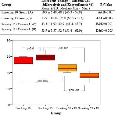
Table 3. Liver cells change on study groups (n=24)
Group
Smoking 10 Smoking 13 Smoking 10 + CL Smoking 13 + CL
Group
Figure 14 Box plot diagram of liver cells change score on study groups. P value were obtain from Boferroni post hoc test.
p=0.0
p=0.003
p<0.001
75
Table 3 showed the mean of liver cells change score on SD rats on study groups. The highest score was in group (B) 13 weeks cigarettes smoke exposure group, followed by group (D)13 weeks cigarettes smoke exposure group+ Curcuma , group (A) with 10 weeks smoke exposure and the lowest was group
(c)
10 weeks cigarette smoke exposure + Curcuma L. The difference of Liver cells change score between groups were statistically significant (p=0.01). Figure 14, show the comparison of liver cells change score of each study group. Multiple comparison by Bonferroni post hoc test showed liver cells change score of 10 weeks exposure cigarette smoke + Curcuma L was significantly lower than 10 weeks exposure of cigarette some only (p=0.003). Furthermore, liver cells change score of 13 weeks exposure cigarette smoke + Curcuma L was significantly lower than 13 weeks exposure of cigarette some only (p<0.003). On Figure also was showed liver cells change score of 13 weeks cigarette smoke exposed group was higher than 10 weeks exposure, however the different was statistically significant (p=0.01). In contrary, the liver cells change score of 13 weeks cigarettes smoke exposure + Cucruma L was significantly lower than 10 weeks cigarette smoke exposure + Curcuma L (p=0.005). These results may also can be interpreted as longer administration of Curcuma L may give bigger liver cells change score reduction.76 V.2. Liver cells TNF- expression
Expression of TNF- observed by immunohistochemistry examination using
77
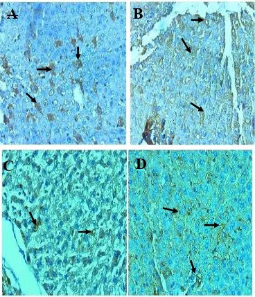
A
B
C
D
Figure 15. Morphology of TNF- expressions of liver tissue of SD after 10 for group A cigarette smoke exposure ,13 weeks for group B cigarette smoke
exposure ,10 weeks for group C cigarette smoke exposure + Curcuma L. 13 weeks for group D cigarette smoke exposure + Curcuma L using 400X
78
Table 4 showed the highest TNF- expression of liver cells was found on 13 weeks cigarettes smoke exposure group, followed by 10 weeks cigarettes smoke exposure, 10 weeks cigarettes smoke exposure + Curcuma L and the lowest was 13 weeks cigarettes smoke exposure + Curcuma L.
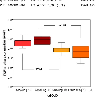
Table 4. Liver cells TNF- expression score in study groups (n=24)
Group TNF- expression score
Sig
Mean ± SD; Median (Min - Max) P/Value
Smoking 10 Group (A) 2,0 ± 0,63; 2,00 (1 - 3) A&C=0,5 Smoking 13 Group (B) 2,25 ± 0,75; 2,25 (1 – 3) C&A=0,5
Smoking 10 + Curcuma L (C) 1,83 ± 0,40; 2,00 (1 - 2) B&D=0.04 Smoking 13 + Curcuma L (D) 1,8 ± 0,75; 2,00 (1- 3 ) D&B=0.04
Figure 16. Box plot diagram of liver tissue TNF- expression score on study groups. P value were obtain Mann-Whitney Test post hoc test.
Smoking 10 Smoking 13 Smoking 10 + CLSmoking 13 + CL
Group 0,0
0,5 1,0 1,5 2,0 2,5 3,0 3,5
P=0.04
79
Figure 16, showed liver tissue TNF- expression of group (C) 10 weeks cigarettes smoke exposure + Curcuma L group was lower than group (A)10 weeks cigarettes smoke exposure only group, however, the difference was not significant (p=0.5). The liver tissue TNF- expression of Group (D) 13 weeks cigarettes smoke exposure + Curcuma L was significantly lower than Group (B) 13 weeks cigarettes smoke exposure only (p=0.004). Figure 22 also showed liver tissue TNF- of 13 weeks cigarettes smoke exposure group was higher than 10 weeks exposure, however, the difference was more as median (2,25). This findings suggest with increasing period exposure of cigarettes smoke that cause increase of liver tissue TNF- , longer exposure time was supra-maximal exposure time . Longer administration of Curcuma L may give bigger reduction of liver tissue TNF- expression.
Base on those findings the hypothesis that administration of Curcuma L have potency to reduce up-regulation of liver tissue TNF- expression due to cigarette smoke exposure for 13 weeks has been proven. On the other side the administration of the curcuma L dident have potency to reduce up-regulation of liver tissue TNF- expression due to cigarette smoke exposure for 10 weeks may give suggest that with increasing time using of curcuma L inhance the regenirative role aganist the expusore effect of passive cigarette smoke
80
more potency reduction than regenirative role on the TNF- expression may be because the curcuma longa extract act as antioxidant much more than act as anti-inflammatory production and also may be because curcuma longa extract can be regenirative role much more against early stage of liver damage like liver cell change than than serious liver cell damage like TNF- expression as inflamation procces inside liver cell at the same period of the curcuma longa administration
V.Reliability of measurement:
` The current study calculated reliability coefficient of scores by measuring agreement between two observers, using Kappa coefficient test, which examines the agreement between two raters for a sureness whether there is a concordance in reading the data, the table below shows the result of Kappa test for Liver cells
.
sAccording to table(Appendix 3) there is agreement between both of reading , because the values of chi square (16.7 & 0.22) were not significant in two variables (tliver cells change