! ""# $% & ' () $
*)&+# , -. *#! $ / - + ). + - + - ! $ 01233! $ $
-- " $ $4 )+ - . &5 " ""# $% & 6784. & * .
( +- *+
The study was aimed to investigate the growth inhibiting effect of ,
(Lour.) Spreng hexane extract (PAN) in combination with doxorubicin againts HeLa cell
Lines, to observe the apoptotic induction and immunocytochemistry of HeLa cell Lines on cyclin D1, Bcl 2, and Cox 2 after treatment of PAN. The percentage viability of the cell were
carried out by using MTT [3 (4,5 dimethylthiazole 2 yl) 2,5 diphenyl tetrazolium bromide]
assay. The effect of apoptosis was observed by flowcytometry assay in single dose of PAN. The expression of cyclin D1, Bcl2 and COX 2 proteins were investigated on HeLa cell lines by using immunocytochemistry. The result showed that PAN showed strong synergistic effect with doxorubicin againts HeLa cells based on Combination Index analysis. No significant apoptotic induction of PAN in HeLa cell lines, but the extract caused necrosis.
The immunocytochemical study showed suppression of cyclin D1, Bcl2 and COX 2
expression on HeLa cell lines. The results concluded that PAN could be a potential co chemoterapeutic agent with doxorubicin on cervix cancer cells but need to be explored further by its combination on specific molecular target.
9 #: - 5 , (Lour.) Spreng., combination, HeLa cell lines
INTRODUCTION
The latest world report provides clear evidence that global cancer rates could increase to 15 million by 2020 and is expected to grow to 21.4 million new cancer cases and 13.2 million
cancer deaths by 20301. Cancer is one of the most life threathening diseases with more than
100 different types. Due to lack of effective drugs, expensive cost of chemotherapeutic agents
and their side effects, cancer can be a cause of death2. Conventional cancer therapies,
including surgery and chemotherapy have a limited but important role in the overall treatment
of most solid tumors 3. Chemotherapy drugs are not selective in killing cells because they
affect the synthesis of nucleic acids and protein, so that normal body cells also die4.
Doxorubicin as chemoterapeutic agent causes serious problems such as drug resistance and toxic effect on normal tissue especially on heart. The manifestations are heart failure,
treatment using combined therapies or combined agents are considered more promising for
higher efficacy, and become alternative, resulting in a better survival6.
Plant have been source of medicine for thousands of years and phytochemicals continue to
play an essential role in medicine2. One of the medicinal plant is
(Lour.) Spreng. The previous studies had showed that the hexane, ethylacetate and ethanol
extracts of , (Lour.) Spreng. had antioxidant activities. The
hexane and ethylacetate extracts exhibited strong cytotoxic effect on T47D breast cancer
cells with IC50 value of 44.716 µg/mL and 37.61 µg/mL, respectively7. Antioxidant activity is
usually correlated with cancer prevention. Thus, the extract has potential effect as a
chemoprevention. The cytotoxic effect of hexane, ethylacetate and ethanol extracts were
also examined on the HeLa cell lines. The study showed that the three extracts had cytotoxic
effect on HeLa cell with IC50 values 76.322 µg/mL,143.291 µg/mL, and 88.997 µg/mL,
respectively8. The aims of this research are to investigate cytotoxic activity of PAN
doxorubicin combination, to analyze apoptotic induction and the proteins expression of HeLa cell lines after single treatment of PAN.
Plant material
Fresh leaves of (Lour.) Spreng. was collected from Pematang
Siantar, Simalungun regency, Sumatera Utara province, Indonesia.
(Lour.) Spreng. was identified in Research Centre for Biology, Indonesian Institute of Science, Bogor, and the voucher specimen was deposited in herbarium.
Preparation of Hexane extract (PAN)
The air dried and powdered leaves of (Lour.) Spreng. (1 kg) were
repeatedly extracted by cold maceration with hexane (3x3 d, 7.5 L). The powder were dried in the air and extracted with ethylacetate (3x3 d, 7.5 L) at room temperature on a shake. The filtrate was collected, and then evaporated under reduced pressure by rotary evaporator (Heidolph VV 200) to obtain a viscous extract and the concentrated extract was dried by freeze dryer (Edwards).
chemicals: hexane and ethylacetate were purchased from Merck (Darmstadt, Germany), DMSO (Sigma Aldrich Chemie GmBH Germany), [3 (4,5 dimethylthiazol 2 yl) 2,5 diphenyl tetrazolium bromide] (MTT) (Sigma Chemical, St. Louis, MO), RPMI media and
Phosphate Buffer Saline (FBS) 10% v/v (Gibco, Grand Island, NY, USA), Doxorubicin
(Ebewe).
Cytotoxicity assay
Cytotoxicity was determined by the MTT assay.
Briefly, HeLa cells were plated at 104 cells/well in a 96 well plate. After incubation for 24 h
different concentration and incubated for 24 h. MTT solution was added to each well and
further incubated for 4h at 370C, optical density was read with an ELISA reader at 595 nm8.
Flowcytometry assay Apoptosis assay
HeLa cells (5x10⁵ cells/well) were seeded into 6 well plate and incubated for 24 h. After that, the cells were treated with PAN, and then incubated for 24 h. Both floating and adherent cells were collected in conical tube using tripsin 0.025%. The cells were washed thrice with cold PBS and centrifuged 2500 rpm for 5 min. The supernatant was separated, while the sediment was collected and fixed in cold 70% ethanol in PBS at 20oC for 2 h. The cells were washed thrice with cold PBS and centrifuged in 2500 rpm for 5 min. The supernatant was separated,
while the sediment was collected and fixed in cold 70% ethanol in PBS at 20oC for 2 h. The
cells were washed thrice with cold PBS then suspended then centrifuged 3000 rpm for 3 min
and Annexin V kit added to sediment and suspended and incubated at 37oC for 30 min. The
samples were analyzed using FACScan flowcytometer9.
Immunocytochemistry
HeLa cells (5x10⁴ cells/well) were seeded on coverslips in 24 well plate and incubated for 24 h. After that, the cells were treated with PAN, and then incubated for 24 h. After incubation, the cells were washed with PBS and then fixed with cold methanol at 4oC for 10 min. After that, the cells were washed with PBS and blocked in hydrogen peroxide blocking solution for 10 min at room temperature, incubated using primary antibody Bcl 2, cyclin D1, and COX 2 for 1 h, then washed thrice with PBS, then incubated with secondary antibody for 10 min. The cells were washed with PBS, then incubated in 3,3 diaminobenzidin (DAB) solution for 10 min, and washed with aquadest. Afterward, the cells were counterstained with Mayer Haematoxylin for 5 min, and the coverslips were taken and washed with aquadest, and then immersed with xylol and ethanol 70%. Protein expression observed by light microscope (Nikon YS100). Cells that express a particular protein will provide the brown colour, while the cells that does not give a specific protein will provide blue colour6.
The aims of the study were to investigate the efficacy of PAN as a co chemotherapy on doxorubicin treatment, to analyze apoptotic induction and proteins expression of HeLa cell lines after treatment of the extract. PAN, doxorubicin and their combination were investigated for their cytotoxicity effect on HeLa cell lines. Cell viability was determined by MTT method after incubation for 24 h, and the effect of combination was analyzed by
Combination Index analysis. The (CI) analysis is one of the most popular
method for evaluating drug interactions in combination cancer chemotherapy10. The method
is used to categorize the effect of the combination which is synergistic, additive, or antagonis. In every treatment (PAN and their combination) was showed the inhibition of cells growth.
The IC50 value of PAN 76.322 µg/mL8 and doxorubicin 1.8 µg/mL, and the combination was
showed higher inhibitory effect if compare with single treatment. The optimum combination
of doxorubicin (0.6 µg/mL) categorized with strong synergistic effect (CI < 0.1). These effects supposed to be related to apoptotic induction and expression of some proteins.
In this study, the apoptotic induction and expression of proteins were done at PAN single treatment, because we wanted to know its effect alone. The effect of its combination with doxorubicin need further study. Evaluation of apoptotic induction was performed by using flowcytometry assay with Annexin V. As shown in Figure 1, the cells in the upper and lower right quadrants represent late apoptotic/necrotic and early apoptotic cells, respectively. The percentage of cells treatment by PAN, in early apoptotic was 0.20%, in late apoptotic/ early necrotic 11.04% and in late necrotic 79.03%.
!
Figure 1. Apoptotic analysis of PAN on HeLa cell lines.
(a) control cells; (b) PAN 1/5 IC50 (15.26 µg/mL)
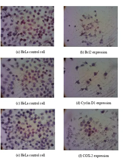
The expression of apoptosis regulator protein Bcl2, the protein that play important role
in cell cycle, Cyclin D1, and expression of COX 2 which contribute to the development of human cancer, were observed in HeLa cells by using PAN treatment. COX 2 derived prostaglandin E2 induces angiogenesis of tumor development by increasing of angiogenic
factors, or decreased expression of anti angiogenic factors, or a combination of both event11.
(a) HeLa control cell (b) Bcl2 expression
(c) HeLa control cell (d) Cyclin D1 expression
(e) HeLa control cell (f) COX 2 expression
Figure 2. Expression of Bcl2, cyclin D1, and COX 2 after treatment of PAN 1/5 IC50 (15.26
µg/mL) observed by immunocytochemistry assay
The observation of apoptosis regulator protein Bcl2 was conducted in HeLa cells after treatment of PAN. Immunocytochemistry assay with Bcl2 antibody showed the expression of Bcl2 was decreased by PAN, therefore it is strengthen the apoptosis mechanism of PAN. One
of secondary metabolite in is ursolic acid12. Ursolic acid inhibit
EGFR/MAPK and suppress Bcl2 expression on colon cancer cell by activation caspase 3 and
need further study. As seen in Figure 2, the untreated cells (control) showed high intensity for Bcl2, cyclin D1 and COX 2. A single treatment of PAN was decreased on Bcl2, cyclin D1 and COX 2 expression. Inhibition of cyclin D1 protein expression strengthen the mechanism
of modulating cell cycle especially in inhibition of cell cycle on G0 G1 phase. Cyclin D1 play
important role in G0 G1 phase with established complex with CDK 4 or CDK 6 to controlled
G1 to S phase transition14. However, we need to be explored more detail about the molecular
mechanism of apoptosis induction, cell cycle modulation, and antiangiogenic regulation of
PAN. Based on the results, it can be concluded that combination of hexane extract of
(Lour.) Spreng. leaves and doxorubicin sinergistically inhibit the
HeLa cell lines. Based on the immunocytochemistry assays, the hexane extract of
can be used in development of chemopreventive agent on cervical cancer.
9
Authors would like to thank to DP2M DIKTI (Directorate of Higher Education) Ministry of Education and Culture, Indonesia, through “Hibah Fundamental” Research Grant 2015 for financial support in the study.
1. Gupta M, Dahiya J, Marhawa RK, Dureja H. Therapies in Cancer Treatment: an Overview. International Journal of Pharmacy and Pharmaceutical Sciences 2015; 7(4):1 9
2. Mahadev R, Ramakrishnaiah H, Khrisna V, Deepalakshmi AP, Kumar NN. Cytotoxic
Activity of Methanolic Extracts of D.Don. International Journal of
Pharmacy and Pharmaceutical Sciences 2015; 7(2):106 108
3. Sukardiman, Suharjono, Balqianur T. The Role of Ethylacetate Fraction of
and Doxorubicin Toward The Increase of Apoptosis and Decrease of VEGF Protein Expression of Mice Fibrosarcoma Cells. International Journal of Pharmacy and Pharmaceutical Sciences 2015; 7(4):347 350
4. Macdonald F, Ford CHJ, Casson AG. Molecular Biology of Cancer. Garland Scientific Publisher, London. 2004. 209 266.
5. Yeh ETH. Cardiotoxicity Induced by Chemotherapy and Antibody Therapy. 2006; 57:485 498
6. Satria D, Furqan M, Hadisahputra S, Rosidah. Combinational Effects of Ethylacetate
Extract of Lour. And Doxorubicin on T47D Breast Cancer Cells.
International Journal of Pharmacy and Pharmaceutical Sciences 2015; 7(7):73 76
7. Hasibuan PA, Rosidah, Ilyas S, Nasution MP. Antioxidant and Cytotoxic Activities of , (Lour.) Spreng. Extracts. International Journal of Pharmacy Teaching & Practices 2013;4(3):755 758
8. Rosidah, Hasibuan PA. Cytotoxic Effect of n Hexane, Ethylacetate, and Ethanol Extracts
of , (Lour.) Spreng. on HeLa and Vero Cells Lines. International
9. Hostanka K, Nisslein T, Freudenstein J, Reichling J, Saller R. Evaluation of Cell Death Caused by Triterpene Glycosides and Phenolic Substances form
extract in Human MCF 7 Breast Cancer Cells. Biol Pharm 2004; 27(12):1975 1970 10. Zhao L, Wientjes MG, Au JLS. Evaluation of Combination Chemotherapy: Integration
of Nonlinear Regression, Curve Shift, Isobologram, and Combination Index Analyses. 2004;10:7994 8004
11. Fosslien E. Review: Molecular Pathology of Cyclooxygenase 2 in Cancer induced Angiogenesis. Annals of Clinical & Laboratory Science, 2001;31(4):325 341
12. Kaliappan ND, and Viswanathan PK. Pharmacognostical Studies on The Leaves of (Lour) Spreng. International Journal of Green Pharmacy, 2008;8(3): 182 184.
13. Shan J, Xuan Y, Zheng S, Dong Q, Zhang, S. Ursolic Acid Inhibits Proliferation and Induces Apoptosis of HT 29 Colon Cancer Cells by Inhibiting the EGFR/MAPK
Pathway. ! " #, 2009;10(9): 668 674.