Biosaintifika
Journal of Biology & Biology Educationhttp://journal.unnes.ac.id/nju/index.php/biosaintifika
Evaluation of Chromosomal Aberrations and Micronuclei in Medical
Workers Chronically Exposed to Low Dose Ionizing Radiation
Yanti Lusiyanti, Iin Kurnia, Viria Agesti Suvifan, Sardini, Sofiati Purnami, Nastiti
Rahajeng
DOI: 10.15294/biosaintifika.v9i3.12382
Center for Radiation Safety Technology and Metrology, National Nuclear Energy Agency (BATAN) Indonesia
Abstract
Medical workers representing the group is the most consistently are exposed to low doses of ionizing radiation, prolonged low-level ionizing radiation can induce chro-mosomal aberrations (CAs). This study would evaluate the cytogenetic effect using the CAs based on dicentric, and cytokinesis-blocked micronucleus (CBMN) assay on hospital workers. The exposed group dividedto Interventional and Diagnostic groups then compared to non exposed group. The accumulated absorbed doses cal-culated for the radiation workers were below 5mSv. Blood samples were obtained from 29 samples of medical workers , and 15 samples of control. The Study showed that the frequency of dicentric chromosomes both in exposed and control were not found. In case of micronuclei, the mean frequencies were observed in exposed group that was (19 ±6.22) and (16.25 ± 6.04) respectively and the control group was (10.4±7.79). Frequency MN/1000 cell in the lymphocytes both in the two exposed group was relatively higher compared to control group. However the MN frequen-cies in all sample group was still in normal range . In this study chronic low radia-tion dose exposure in the hospital had no significant effect on chromosome aberra-tion nor micronuclei. The benefit of the study is to enrich the potential usefulness of cytogenetic assay providing safety index in medical surveillance programs. The results suggest that education and retraining of staff concerning radiation safety guidelines need to be done to maintain the safety aspects of radiation.
How to Cite
Lusiyanti, Y., Kurnia, I., Suvifan, V. A., Sardini, S., Purnami, S., & Rahajeng, N. (2017). Evaluation of Chromosomal Aberrations and Micronuclei in Medical Workers Chronically Exposed to Low Dose Ionizing Radiation. Biosaintifika: Jour-nal of Biology & Biology Education, 9(3), 585-591.
© 2017 Universitas Negeri Semarang
History Article
Received 13 August 2017 Approved 10 November 2017 Published 31 December 2017
Keywords
Chromosome aberrations; Human lymphocytes; Ion-izing radiation; Occupational exposure
Correspondence Author:
Jl. Lebak Bulus Raya 49, Jakarta 12070 E-mail: [email protected]
The results of several follow-up studies have been published by Bouraoui et al. (2013)age and smo-king habits was used. The clastogenic/aneugenic effect of IR was evaluated using the CBMN assay in combination with fluorescence in situ hybridi-zation with human pan-centromeric DNA in all the exposed subjects and controls. Results: The study showed a significant increase of the micro-nucleus (MN and was reporting on a significant increase of the micronucleus frequency in the lymphocytes of the exposed workers compared to the control group. On the other hand, the pro-liferation of markers that can be used to evaluate general toxicity caused by radiation exposure is the Mitotic Index (MI) and Nuclear Division In-dex (NDI) used to define cell cycles progression of the lymphocytes after mitogenic stimulation in culture cells. Another cell proliferation used at the tissue level is the use of AgNORs staining techniques used to evaluate cell morphology and kinetics of biopsy results. Kurnia et al. (2012) re-ported that the AgNOR staining technique was effective to assess the proliferation rate in tissue biopsy of cancer patients. At present, the increa-sing application of radiation for medical purposes indicates the need to conduct cytogenetic analy-sis in order to evaluate the level of chronic ex-posure. Although the baseline frequency of CAs from the normal population has a wide range of variation which might reflect interlaboratory dif-ferences in culture conditions or characteristics of the sample (Zakeri & Hirobe, 2010)36 nuclear medicine physicians and 33 conventional radio-logists were included in this study, along with 35 healthy age- and sex-matched individuals as the control group. We used conventional metaphase chromosome aberration (CA. The purpose of the study was to assess occupationally induced chro-mosomal damage in medical workers who expo-sed to low levels of ionizing radiation using the cytogenetic endpoints of CAs and MN and also evaluate MI and NDI index . The results of this study are expected to emphasize the importance of biomonitoring aspects of health in implemen-ting safety programs of radiation exposure.
METHODS
Subjects and Sampling
The sample group of hospital workers con-sisted of 29 exposed staff and according to the job classification of 13 as radiographer staff in the diagnostic division and 16 Interventional staff. The mean duration of occupational exposure to radiation respectively16.41 ± 9.94 years (from 4 to 34 years) and the control staff consisted of 14
INTRODUCTION
Hospital staffs are an occupational group exposed to different agents suspected to indu-ce genetic damage, such as ionizing radiation (x- and gamma- rays), radionuclides, cytostatic drugs, and anaesthetic gases, in which all of them have been investigated for their cytogenetic effect (Kasuba et al., 2008). Occupational radiation ex-posure is normally less than a few cGy per year. The International Commission on Radiological Protection (ICRP) has set the following limits on exposure to ionizing radiation: the general public shall not be exposed to more than 1 mSv per year (over and above natural background) and occupa-tional exposure shall not exceed 20 mSv per year. These limits exclude exposure due to background and medical radiation. The limit for normal occu-pational exposure is 0.05 Sv/ year. Monitoring of personnel occupationally exposed to ionizing ra-diation consists of regular film dosimetric control and periodic health examination (UNSCEAR, 2000).
unexposed individuals. Peripheral blood samples were collected by venipuncture using heparini-zed vacutainer tubes (BD Vacutainer systems). The study was approved by the Ethics Commit-tee of the National Institute of Health Research and Development, the Indonesian Ministry of Health, number LB.02.01/5.2.KE.051/ 2015. In-formed consent was obtained from all staff. A de-tailed questionnaire was used to get information on age and occupation.
Dicentric Assay and Mitotic Index Evaluation (MI)
Blood cultures were set up according to the IAEA standard procedures with minor mo-difications (IAEA 2011) as described in previous research (Lusiyanti et al., 2013) one ml of whole blood samples were cultured for 48 hours in the incubator at 37°C containing 5% CO2. The cultu-re medium consisted of 7.5 ml of Roswell Park Memorial Institute (RPMI) 1640 supplemented with 20% heat-inactivated fetal bovine serum (FBS), 1% streptomycin/penicillin, 2.5% ml of phytohemagglutinin (PHA). Colchicine was ad-ded to the culture medium for the last 3 hours at a final concentration of 0.05 µg/ml. The cells then were centrifuged for 10 minutes at 1500 rpm and resuspended in 10 ml of 0.075 M KCl (pre-war-med to 37°C) for 25 minutes. The cells then were centrifuged again for 10 minutes at 1500 rpm and re-suspended in 2 ml of fresh fixative that was a mixture of methanol and acetic acid (3:1). The fixation step was repeated four times until white sediment was obtained. Fixed cells were dropped onto clean, wet slides, dried and stained with 4% Giemsa solution (pH 6.8) for 10 minutes. Mitotic Index then was calculated based on the protocol in the IAEA publication as shown in Formula (1). Typically, 500 cells were counted for a full mitotic index analysis for each sample (Figure 1).
Mitotic Index = (#metaphases) x 100 x 500 (#metaphases+blast)
The cytokinesis-block micronucleus assay and Nuclear Division Index Evaluation
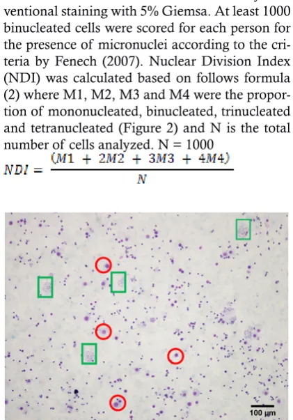
Micronuclei were prepared in cytokinesis-blocked cells using cytochalasin B following the method suggested by Fenech (2007) with minor modification as described in previous research (Lusiyanti et al., 2016) . The cultures were incuba-ted for 72 h. After 44 h of incubation cytochalasin B (Sigma) was added to the cultures at a concent-ration of 6 µg/ml to block cytokinesis. The cells were collected by centrifugation, and treated with a mild hypotonic solution containing 0.075M
KCl for 3 min. After centrifugation and remo-val of the supernatant, the cells were fixed with a fresh mixture of methanol/acetic acid (3:1). Centrifugation and resuspension were carried out three times and the cells were then dropped on to clean slides for detection of micronuclei by con-ventional staining with 5% Giemsa. At least 1000 binucleated cells were scored for each person for the presence of micronuclei according to the cri-teria by Fenech (2007). Nuclear Division Index (NDI) was calculated based on follows formula (2) where M1, M2, M3 and M4 were the propor-tion of mononucleated, binucleated, trinucleated and tetranucleated (Figure 2) and N is the total number of cells analyzed. N = 1000
Figure 1. View of a typical lymphocyte culture slide. Red circles are nuclei counted as blasts, white circles are nuclei which are not counted, boxes are metaphase spreads (200x magnifica-tion).
of 400X).
Statistical Analysis
Unpaired t-test was used to compare the MI and NDI, and also the frequency of unstable CAs and micronuclei between exposed workers and control samples using SPSS 22.0 statistical software. The influence of micronuclei and ex-posure duration of employment were tested using the medical software.
RESULTS AND DISCUSSION
Cytogenetic analysis by mean CAs and MN in cytokinesis block (CBMN) in human periphe-ral blood lymphocytes is an important technique for risk assessment in occupational exposure. In this study, radiation workers chronically exposed to ionizing radiation have evaluated the frequen-cies of CAs, and MN. On the other hand in this study,tests of cell proliferating markers were done by using Mitotic Index (MI) and Nuclear Divisi-on Index (NDI). The mean MI and NDI amDivisi-ong exposed group compared to control group were shown in Table 1.
MI and NDI are markers for estimating the general toxicity. The proportion of metapha-se cells and binucleated cells may be umetapha-sed as a biomarker of the lymphocytes mitogen response, immune functions and cytostatic effects of vario-us studied agents included the low radiation dose exposure (Eroğlu, 2011, Alaloc & Eroğlu,2011). Table 1 showed that the MI of the exposed staff in diagnostic and interventional is slightly higher compared to control but statistically it is not sig-nificantly different. It means that chronic low radiation dose exposure in exposed staff has no significant effect on lymphocytes proliferation. Furthermore, larger sample number should be used in further research to ensure the effect of chronic low radiation dose exposure on lympho-cytes proliferation. In this study a total of 11000 metaphases or on average about 250 cells (exposed
group and control) were analyzed and the result showed that observation chromosome aberration such as dicentric and centric rings chromosome was not detected in all exposed and control staff. Similar results had been reported in another rese-arch concerning with the induction of chromo-some based on dicentric type, reporting the ab-sence of centric ring and dicentric chromosome. In fact at this level of exposure a very high num-ber of scored metaphases was required in order to detect the presence of dicentric (Cardoso & Peitl, 2001). Cigarran et al. (2001) also reported that no significant increase in chromosomal abnormali-ties in their study population as there was a wide variation in the individual frequencies. According to Terzoudi & Pantelias (2006) the cytogenetic response to ionizing radiation is intrinsic for each individual so that radiosensitivity between indivi-duals may be an effect due to differences in DNA repair capacity, which can be explained by spe-cific mutations or polymorphism in DNA repair genes or alternatively may be linked to cell cycle and feedback control mechanism. Thieren et al.
(2002) suggested that exposure to low radiation doses might induce inter-individual differences in susceptibility and the activation of DNA repair and the transcription of early response genes and stimulate DNA repair enzymes.
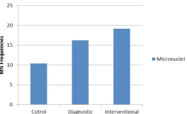
In case of micronuclei the observations of micronucleus in binucleate cells (BNC ) frequen-cies were carried out in each of 1000 binucleate cells per sample. The mean frequencies and distri-bution of MN frequencies in between medical ra-diation workers group (Diagnostic and interven-tional) compared to control staff were shown in Table 2 and Figure 3.
The frequencies of MN in the exposed group (diagnostic and interventional) varied bet-ween each exposed in range of 6-24/1000 BNC and 11-27/1000BNC for diagnostic and interven-tional staff respectively, whereas in control staff in range of 1-23/1000 BNC (Figure 3).
Table 1. The mean MI and NDI in radiation workers group compared to control group in hospital staff.
Group Number of
sub-jects
Age (yr) x¯ ± SD
(%)
Duration of employment (yr) x¯ ± SD (%)
Mean MI ± SD (%)
Mean NDI ± SD
Diagnostic 13 42.31 ± 11.76 15.92 ±9.68 10.05. ± 2.07 1.32 ± 0.14 Interventional 16 47.88±11.42 17.13 ± 10.74 10.82 ± 1.98 1.31 ± 0.13 Control 15 42.13 ± 11.43 - 9.16 ± 2.69 1.39 ± 0.18 Total 44
Figure 3. The mean frequencies of Micronuclei in lymphocytes from exposed group in medical workers, compared to control group.
According to the IAEA manual (2011), the MN frequencies in both exposed group and cont-rol group were still in normal range ( 1-36/1000 BNC) ( International Atomic Energy Agency, 2011) particularly when there are difficulties in interpreting the data, in cases where there is rea-son to believe that perrea-sons not wearing dosimeters have been exposed to radiation, in cases of claims for compensation for radiation injuries that are not supported by unequivocal dosimetric eviden-ce, or in cases of exposure over an individual\ u2019s working lifetime. The IAEA has main-tained a long standing involvement in biological dosimetry commencing in 1978. This associati-on has been through a sequence of coordinated research programmes (CRPs. In this study the existence of the variation in MN between indi-viduals, according to Joseph et al. (2004), that a high individual variability exists as a consequen-ce of intrinsic genomic instability in peripheral lymphocytes, which may be affected by the qua-lity and the intensity of exposure in modulating the cellular’s response to radiation injury. The lack of an increase in micronuclei frequency in the exposed population might indicate that the chronic doses received by the exposed subjects are either too low to be detected or the damage caused might have been repaired during the long span of time. (Cigarran et al,2001). Association based on the mean frequencies in both exposed group compared to control is shown in Figure 4.
Figure 4. The mean frequencies of micronuclei in control subject compare to Diagnostic workers (A) and Interventional workers in Hospital (B).
B
In this study the mean frequencies of MN for the diagnostic and interventional groups were respectively 15.77±6.51 and 19.20 ± 6.42. Whe-reas for the control staff was (10.47±6.33), and showed significant differences (P=0.67). The higher frequencies of MN were found for radia-tion workers in Intervenradia-tional staff. This was caused probably by the increase of chromosome damage that can be due to the accumulation of DNA damage by chronic exposure from gamma /or X-rays along the years of employment (Car-doso & Peitl, 2001). Similar results had been re-ported in another research concerning to enhan-cement of MN frequencies using CBMN method where this was significantly higher in the exposed group than the control group (Zakeri & Hirobe, 2010)36 nuclear medicine physicians and 33 con-ventional radiologists were included in this study, along with 35 healthy age- and sex-matched in-dividuals as the control group. We used conven-tional metaphase chromosome aberration (CA. Related to job classification between two exposed group the mean frequencies on MN are shownin Figure 5.
MN
12 14 16 18 20 22 24
Diagnostic Intervention
p>0.05 ns
n=13 mean=15.77
n=16 mean=19.20
Figure 5. The mean frequencies of micronuclei in Diagnostic workers compare to Interventional workers in the hospital.
The higher mean frequencies were found in interventional staff compare to diagnostic staff. This finding is in agreement with other stu-dies reporting from another researcher that was found no significant, increase in MN frequency in medical radiation workers (Maffei et al, 2002). The interventional group working in a high-vo-lume catheterization laboratory had higher levels of MN, when compared with diagnostic. Years of radiological practice were correlated with the MN value in the exposed group, which suggests the presence of cumulative damage (Andreassi
as length of employment, and in some cases, is not related to continuous exposure to ionizing ra-diation during the years
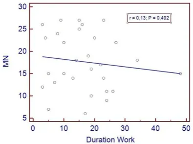
Figure 6. The mean frequencies of micronuclei in medical radiation workers based on working duration
In the present work according to work du-ration of employment the mean frequencies of MN were no significant correlation was observed between exposing group compared to the control group as shown in Fig. 6, (Andreassi et al., 2005) reported the similar result that was no significant correlation between occupational radiation doses during the past year and MN frequency. (Zake-ri & Hirobe, 2010) also reported that no obvious trend of increased chromosomal damages as a function of either duration of employment, ex-posed dose, sex or age. Rapolo et al. (2012) also reported that length of work does not affect the extent of genetic damage induced by chronic ex-posure to ionizing radiation, but an increased fre-quency of binucleated and mono-nucleated cells with micronuclei was observed, based on the ac-cumulated radiation dose.
Based on the research result it can be pointed out that although at low-level radiation exposure, unstable aberrations (such as dicentric and rings) could not be detected, the analysis of MN in cytokinesis-blocked binucleated cells were the most sensitive biomarker to evaluated early clastogenic effect induced by low chronic radia-tion exposure, even though the absorbed doses calculated for the exposed individuals were be-low the permissible limit. In order to monitoring, detection of early genotoxic effects may allow for the adoption of preventive biological control measures, such as reduction of hours of occupa-tional exposure.Because of the sensitivity of the CBMN assay for low-dose detection, in the future research needs to be done by combining the assay with fluorescence in situ hybridization
centro-mere staining technique. Referring to Vral et al.
(2011) the development of a combined automa-ted MN-centromere scoring procedure remains a challenge for the future, as it will allow systema-tic biomonitoring of radiation workers exposed to low-dose radiation.
CONCLUSIONS
In conclusion chronic low radiation dose exposure in medical workers was not significantly affect on lymphocytes proliferation. Further rese-arch using larger sample number should be con-ducted to ensure the effect of chronic low radia-tion dose exposure on lymphocytes proliferaradia-tion. Micronuclei in cytokinesis-blocked binucleated cells demonstrated to be the most sensitive bio-logical marker to determine the cellular respon-se to low levels of irradiation. It is important to develop procedures that allow the occupationally exposed hospital staff to be biomonitored and protected from ionizing radiation.
ACKNOWLEDGMENTS
The authors gratefully acknowledge to Center for Technology of Radiation Safety and Metrology (PTKMR). This research was sup-ported by grants from the National Nuclear Ener-gy Agency of Indonesia (Badan Tenaga Nuklir Nasional) as a BATAN Annual Research Project in 2015.
REFERENCE
Agency, International Atomic Energy Agency. (2011). Cytogenetic Dosimetry : Applications in Pre-paredness for and Response to Radiation Emergencies. Manual Series 247.
Alakoç, C. & Eroğlu, H. E. (2011). Determining Mitot-ic Index in Peripheral Lymphocytes of Welders Exposed to Metal Arc Welding Fumes. Turkish Journal of Biology, 35(3), 325-330.
Andreassi, M. Cioppa, A., & Botto, N. (2005). Somatic DNA Damage in Interventional Cardiologists: A Case-Control Study. FASEB Journal, 17(3), 1–17.
Boffetta, P., van der Hel, O., Norppa, H., Fabianova, E., Fucic, A., Gundy, S., ... & Kelecsenyi, Z. (2006). Chromosomal aberrations and cancer risk: results of a cohort study from Central Eu-rope. American journal of epidemiology, 165(1), 36-43.
625–31.
Bouraoui, S., Mougou, S., Drira, A., Tabka, F., Bouali, N., Mrizek, N., ... & Saad, A. (2013). A cyto-genetic approach to the effects of low levels of ionizing radiation (IR) on the exposed Tunisian hospital workers. International journal of occupa-tional medicine and environmental health, 26(1), 144-154.
Cardoso, R. S., Takahashi‐Hyodo, S., Peitl, P., Ghilar-di‐Neto, T., & Sakamoto‐Hojo, E. T. (2001). Evaluation of chromosomal aberrations, mi-cronuclei, and sister chromatid exchanges in hospital workers chronically exposed to ion-izing radiation. Teratogenesis, carcinogenesis, and mutagenesis, 21(6), 431-439.
Cigarrán, S., Barquinero, J. F., Barrios, L., Ribas, M., Egozcue, J., & Caballin, M. R. (2001). Cytoge-netic analysis by fluorescence in situ hybridiza-tion (FISH) in hospital workers occupahybridiza-tionally exposed to low levels of ionizing radiation. Ra-diation research, 155(3), 417-423.
Dias, F. L., Antunes, L. M., Rezende, P. A., Carvalho, F. E., Silva, C. M., Matheus, J. M., ... & Bala-rin, M. A. (2007). Cytogenetic analysis in lym-phocytes from workers occupationally exposed to low levels of ionizing radiation. Environmen-tal toxicology and pharmacology, 23(2), 228-233. Eroğlu, H. E. (2011). The Cytogenetic Effects of Black
Tea and Green Tea on Cultured Human Lym-phocytes. Brazilian Archives Of Biology And Tech-nology, 54(6), 1159–65.
Fenech, M. (2007). Cytokinesis-block micronucleus cytome assay. Nature protocols, 2(5), 1084-1104. Joseph, L. J., Patwardhan, U. N., & Samuel, A. M.
(2004). Frequency of micronuclei in peripheral blood lymphocytes from subjects occupation-ally exposed to low levels of ionizing radiation.
Mutation Research/Genetic Toxicology and Envi-ronmental Mutagenesis, 564(1), 83-88.
Kašuba, V., Rozgaj, R., & Jazbec, A. (2008). Chromo-some aberrations in peripheral blood lympho-cytes of Croatian hospital staff occupationally exposed to low levels of ionising radiation. Ar-chives of Industrial Hygiene and Toxicology, 59(4), 251-259.
Kurnia, I., Bintari, S. H., & Khaisuntaha, M. (2012). Tingkat Keganasan Kanker Serviks Pasien Pra-Radiasi Melalui Pemeriksaan AgNORs, MIB-1 dan Cas-3. Biosaintifika: Journal of Biology & Bi-ology Education, 4(2), 53-61.
Lusiyanti, Y., Alatas, Z., Syaifudin, M., & Purnami, S. (2016). Establishment of a dose-response curve for X-ray-induced micronuclei in human lym-phocytes. Genome integrity, 7(49), 1–4.
Lusiyanti, Y., Alatas, Z., Lubis, M., Suvifan, V. A., Ramadhani, D., & Purnami, S. (2013). Dose-response curve of chromosome aberrations in human lymphocytes induced by gamma-rays.
Atom Indonesia, 39(3), 124-128.
Maffei, F., Angelini, S., Forti, G. C., Lodi, V., Violante, F. S., Mattioli, S., & Hrelia, P. (2002). Micronu-clei frequencies in hospital workers occupation-ally exposed to low levels of ionizing radiation: influence of smoking status and other factors.
Mutagenesis, 17(5), 405-409.
Terzoudi, G. I., & Pantelias, G. E. (2006). Cytogenetic methods for biodosimetry and risk individu-alisation after exposure to ionising radiation.
Radiation protection dosimetry, 122(1-4), 513-520. Thierens, H., Vral, A., Barbe, M., Meijlaers, M., Baey-ens, A., & Ridder, L. D. (2002). Chromosomal radiosensitivity study of temporary nuclear workers and the support of the adaptive re-sponse induced by occupational exposure. In-ternational journal of radiation biology, 78(12), 1117-1126.
UNSCEAR (United Nations Scientific Committee on the Effects of Atomic Radiation). Sources and Effects of Ionizing Radiation. Report to the General Assembly, (2000); Volume II: Effects. New York: United Nations.
Vral, A., Fenech, M., & Thierens, H. (2011). The mi-cronucleus assay as a biological dosimeter of in vivo ionising radiation exposure. Mutagenesis, 26(1), 11-17.
Zakeri, F., & Hirobe, T. (2010). A cytogenetic approach to the effects of low levels of ionizing radia-tions on occupationally exposed individuals.