Serial Case: Colorectal Malignancy in Young Age
Lia Sasmithae*, Bogi Pratomo**
*Faculty of Medicine, Universitas Brawijaya/Dr. Saiful Anwar General Hospital, Malang **Division of Gastoentero-hepatology, Department of Internal Medicine, Faculty of Medicine
Universitas Brawijaya/Dr. Saiful Anwar General Hospital, Malang
Corresponding author:
Bogi Pratomo. Division of Gastroentero-hepatology, Department of Internal Medicine, Dr. Saiful Anwar General Hospital. Jl Jaksa Agung Suprapto No.2 Malang Indonesia. Phone: +62-341-362101; facsimile: +62-341-369384. E-mail: [email protected]
ABSTRACT
Colorectal cancer was the third most common cancer found worldwide. In 2002, colorectal cancer was the second most common cancer in men, while it ranked third among women. Based on Indonesian Ministry of Health data, its prevalence was 1.8 per 100,000 population. We report four cases of colorectal cancer in this case series, and all cases was occured among person aged 28-32 years old. Age was the main relevant risk factors for colorectal cancer in most population. Only 3% of colorectal cancer found in individual aged less than 40 years old. This case series also aimed to show that risk factors was various and changing by the time, but its determinant factors could not be explained yet.
Keywords: colonoscopy, colorectal cancer, young age
ABSTRAK
Kanker kolorektal menduduki peringkat ketiga jenis kanker yang paling sering terjadi di dunia. Pada tahun 2002 kanker kolorektal menduduki peringkat kedua pada kasus kanker yang terdapat pada pria, sedangkan pada wanita kanker kolorektal menduduki peringkat ketiga dari semua kasus kanker.Data dari Depkes didapati angka 1,8 per 100.0000 penduduk. Dilaporkan 4 buah kasus keganasan kolorektal,dari keempat serial kasus yang disajikan rata-rata usia penderita keganasan kolorektal adalah usia muda sekitar 28-32tahun. Usia merupakan faktor paling relevan yang mempengaruhi risiko kanker kolorektal pada sebagian besar populasi. 3% dari kanker kolorektal muncul pada orang dengan usia dibawah 40 tahun. Serial kasus ini ingin menunjukkan bahwa mulai terjadi perubahan faktor risiko terjadinya kanker kolorektal dan sampai saat ini juga belum diketahui secara pasti penyebab terjadinya hal tersebut.
Kata kunci: kanker kolorektal, usia muda, kolonoskopi
INTRODUCTION
Colorectal cancer was the third most common cancer found worldwide. Globally, colorectal cancer was occured in 9.5% and 9.3% of men and women, respectively, from all cancer patients.1 Europe was the region with the most colorectal cancer incidence. In 2004, there were 2,886,800 incidence and 1,711,000 death in cancer patients, where colorectal cancer was ranked the 2nd most.2 In Indonesia, colorectal cancer
has a high incidence and mortality.3 In 2002, colorectal cancer was the 2nd most common cause of cancer in men and 3rd most cause in women.4 Many reports showed an increase incidence of colorectal cancer, while Indonesian Ministry of Health reported a 1.8 per 100,000 incidence in Indonesian population.5
developing countries.6 There were also difference among Western Countries and Indonesia in colorectal cancer incidence. In Indonesia, incidence was similar in male and female, higher in younger age, and about 75% of them was a rectosigmoid colon cancer; meanwhile in Western Countries, colorectal cancer was found mostly in male, older age, and only 50% have a rectosigmoid colon cancer.3 Symptoms of right colon cancer is abdominal fullness, symptomatic anemia, and bleeding, while a left colon cancer present an altered defecation pattern, bleeding, constipation, and obstruction.2
Histological findings were important as it would much influence the treatment and prognosis.7 The most
common histological findings were adenocarcinoma (90-95%), mucinous adenocarcinoma (17%), signet ring cell carcinoma (2-4%) and sarcoma (0,1-3%).7,8 Data also showed that mortality among colorectal cancer was about 50%.1 This happened because colorectal cancer was asymptomatic at the early stage, so that patient came to physician in late stage and further curative approach was not optimal.
CASE ILLUSTRATION
Case 1
Patient came with chief complaint of bloody stool, soft consistency, once in a day, since two month ago. Patient also complaining pain in lower abdominal during defecation. There were no nausea and vomiting. Patient also felt fatigue and looks pallor. Four months ago, patient have similar symptoms so patient been hospitalized and have a blood transfusion. In physical examination, patients look mildly ill, GCS E4M6V5, good nutrition status, body weight 72 kg, body height 167 cm, blood pressure 120/80 mmHg, temperature 36,6oC, with both conjunctiva is pale, and pain in left abdominal iliac fossa. A digital rectal examination showed a strong anal sphincter tonus, smooth mucosa, and no palpable mass.
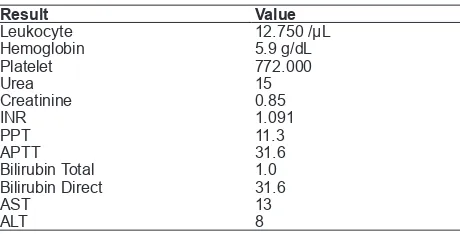
Table 1. Laboratory data
Result Value
INR: international normalized ratio; PPT: plasma protrombine time APTT:
activated partial thromboplastine time; ALT: alanine aminotransferase; AST:
aspartate aminotransferase
Pathology examination from biopsy specimen showed macroscopically a small white tissue, with macroscopic result of malignant epithelial cell in colon mucosa tissue forming an acinar. Pathologist conclusion is invasive adenocarcinoma with moderate differentiation.
Case 2
Patient complaining intermitten bloody stool since 5 month ago. There were also altered defecation pattern, soft stool, without any abdominal pain, but patient feels fatigue after defecating. Patient also complaining epigastric pain since one week before admission. Nausea and vomiting happened after eating food. Patient had a 5 kg-weight loss since 5 months. No history of herbal consumption, alcohol, nor NSAID. Patient’s mother was having ovarian cancer. Physical examination showed a mildly ill, GCS E4M6V5, good nutritional status, body weight 47 kg, body height 156 cm, blood pressure 120/80 mmHg, temperature 36,5oC, no pallor conjunctiva, and no abdominal pain. Digital rectal examination showed a good anal sphincter tonus, smooth mucosa, and no palpable mass.
Table 2. Laboratory examination
AST: aspartate aminotransferase; ALT: alanine aminotransferase; CEA: carcinoembryonic antigen
Figure 1. Endoscopy examination showed a lobulated mass,
fragile, tend to bleed, obstructing + 60% of colon lumen.
Endoscopist conclusion was an internal hemorrhoid with
Pathology examination from biopsy specimen showed a macroscopic small white tissue and microscopic a mucosal rectosigmoid tissue with group of malignant epithelial cell forming an acinar. Pathologist conclusion is an invasive adenocarcinoma with moderate differentiation.
Case 3
Patient with chief complain of intermittent bloody stool since 5 months before admission without any abdominal pain. There were also alteration in defecation pattern, with small stool like a ‘goat stool’. Patient also complaining a weight loss although dietary consumption was normal as usual. There were no history of herbal consumption nor NSAID before.
Physical examination showed a mildly ill, GCS
E
4M
6V
5,good nutritional status, body weight 62
kg, body height 157 cm, blood pressure 110/80
mmHg, temperature 36,2
oC, no pallor or icteric
conjunctiva, no abdominal pain and mass. Digital
rectal examination showed a good anal sphincter
tonus, smooth mucosa, and a lobulated palpable
mass with blood on handscoen after digital
removal.
Table 3. Laboratory examination result Result Value
Leukocyte 8.290 /μL
Hemoglobin 13.7 g/dL
Platelet 516.000/uL
Case 4
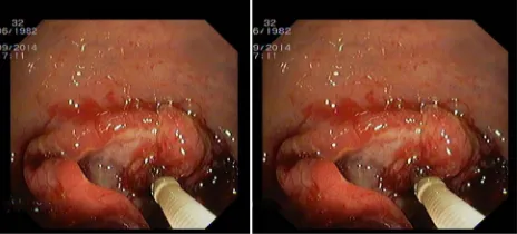
Patient complaining bloody stool since one month before admission with fresh blood during or after defecation. Stool was hard and shape like a ‘goat stool’. Patient also having difficulty during bowel movement because of hard stool. Patient felt an uncomplete defecation with a burning sensation in anus area. Patient also complaining a weight lost about 5 kg since a month. Patient was regularly consuming herbal (jamu) when feeling myalgia and sometimes consuming NSAID. No history of malignancy among family. Physical examination showed a mildly ill, GCS E4M6V5, good nutritional status, body weight 63 kg, body height 165 cm, blood pressure 120/80 mmHg, temperature 36,6o C, no pallor conjungtiva, positive bowel sound, palpable liver about 8 cm, timphanic percussion in Traube’s space, and a palpable mass in left upper quadrant. Digital rectal examination showed a strong anal sphincter tonus, smooth mucosa, and a palpable lobulated mass with pain and tend to bled. Endoscopy examination showed an internal hemorrhoid, anular mass, fragile, tend to bled in rectum, suspect of malignancy. Later, biopsy was done and sent to pathologist.
Figure 2. Endoscopy examination showed a large mass + 30 cm
from anus, lobulated, fragile, tend to bleed, obstructing + 90% of colon lumen, so that further endoscopic examination was
not feasible. Endoscopist conclude a suspect of rectosigmoid
cancer.
Figure 3. Endoscopy examination showed a suspect of rectal carcinoma. A further pathology examination was planned.
Figure 4. Endoscopy examination showed an internal hemorrhoid, anular mass, fragile, tend to bled in rectum, suspect of malignancy
DISCUSSION
Cancer incidence in Indonesia was approximately 100 per 100,000 population. But, only 3,2% of cancer patient got a treatment in hospital. Nowadays, colorectal cancer was one of the most common cancer in Indonesia. Data from 13 cancer centers in Indonesia showed that colorectal cancer was among the five most cancer in both male and female.9,10
findings were similar in both male and female, younger age, with 75% among it was a rectosigmoid cancer. Otherwise, in most Western Countries, colorectal cancer was found higher in male, older age, and rectosigmoid colon proportion was 50%.3,11
From four cases above, the mean age of patient is 25-35 years old. The main symptoms are alteration in defecation pattern, bloody stool, uncomplete defecation, weight loss, and fatigue. All four patients undergo colonoscopy and a mass was found with characteristic of fragile and tend to bled. Only one patient having a malignancy history among family, and two patients have a history of smoking for more than 20 years. Based on literature, smoking was an independent risk factors for colorectal cancer.11-15 The risk to develop colorectal cancer was increasing in older people, mainly in both male and female aged 50 years old or more,1,5 while only 3% of colorectal cancer found in patient aged less than 40 years old.2
Fifty five percent of colorectal cancer was aged > 65 years old,16 with incidence of 19 and 337 per 100,000 population for people aged less than and more than 65 years old, respectively.2 This serial case present four colorectal patients aged less than 65 years old. There were no exact risk factor to develop colorectal cancer in this patients.
Further approach in all patient after colonoscopy was to examine a biopsy tissue taken during endoscopy. This was aimed to identify the cell type and planning further approach based on the result. Exact cancer stage is depending on histopathologic examination result and confirmed by surgical staging. Differ to other malignancy, tumor size was less correlated to colorectal cancer prognosis. Factors that affect prognosis in colorectal cancer is the depth of tumor penetration to colon wall, involvement of regional lymph node, and metastasis status. Several staging systems have been developed in last decade, but cancer staging system by Dukes in 1982 was still widely used because of its simplicity and acceptability. On the other hand, this system did not explain several important information for prognostication, such as vascular invasion, and histology and DNA differentiation of tumor cell. In Dukes Stage A, > 90% patients survive in 5 years. In Dukes Stage B, its survival rate was decrease to 60-80%. An involvement of regional lymph node (Dukes Stage C) reduce its survival rate to 20-50%, and if metastasis occur (Dukes Stage D), its survival was only < 5% in 5 years. Yet, TNM staging system combined to Dukes system that modified by Astler-Coller was recommended.12-17
Tumor located in transverse and descenden colon have a worse prognosis compared to ascenden and rectosigmoid colon. Patient with obstruction or bowel perforation have a worse prognosis than patient without it.2 In first case, mass was found in transverse colon, so that this patient has a worse prognosis based on literature.
In this case series, all four patients were planned to undergo surgery. Surgery was a curative approach in colorectal cancer. This surgery was done by wide excision but try to preserve colon function maximally.17-18 Patients would be consulted to digestive surgeon after agreed by patient and family. Counseling and education was done to patient about the procedure and further treatment approach after surgery. After surgical resection, patients were planned to treat with adjuvant chemotherapy. Chemotherapy followed by tumor extirpation theoretically was increase the effectivity of chemotherapeutic agents. Chemotherapy was effective if given in a small number of tumor cell. Otherwise, there were several studies reporting a chemotherapy-resistant colorectal cancer case. Therefore, it is a challenging for physician to found risk factors of colorectal cancer development in order to both prevent and cure it in the future.
REFERENCES
1. Depkes. 2006. Gaya hidup penyebab kolorektal, [serial online] [cited 2015 March 13]. Available from: URL: http:// www.depkes.go.id/index.php?option=news&task=viewarticl e &sid=2058&Itemid=2
2. Casciato DA. Manual of Clinical Oncology 5th ed. Lippincott
Willi ams & Wilkins: USA 2004.p.201
3. Syamsuhidajat R, Jong Wim D. Buku ajar Ilmu Bedah. 2nd ed.
EGC: Jakarta 2004.p.661-3.
4. WHO. 2006. The Impact of Cancer, [serial online] [cited 2015
March 27]. Available from: URL:http://www.surveillance/ infobase/web/InfoBasePolicyMaker/reports/ReporterFull View.aspx?id=5.
5. Depkes. 2006. Deteksi dini kanker usus besar [serial online] [cited 2006 December 3]. Available from: URL: http://www. depkes.go.id/download.php?file=download/kanker.pdf 6. Hansen J. Common Cancers In The Elderly. Drug Aging
1998;13:467-78.
7. Stewart SL, Wike JM, Kato I, Lewis DR, Michaud F. A population based study of colorectal cancer histology in United States 1998-2001. American Cancer Society Cancer 2006;107:5.
8. Fahlevi R. 2002. Karsinoma Rekti. [serial online] [cited 2015 March 27] https://usebrains.wordpress.com/2008/09/14/ kanker-kolorektal/
[serial online] 2003 [cited 2015 March 13]. Available from: URL: http://www.who.or.id/ind/products/ow6/sub2/display. asp?id=1
10. Soeripto. Gastro-intestinal cancer in Indonesia. Asian Pacific Journal of Cancer Prevention 2003;4:289-96.
11. Kastomo DR, Soemardi A. Tindakan bedah pada keganasan kolorektal stadium lanjut. Majalah Kedokteran Indonesia 2005;55:499-500.
12. Silalahi J. Antioksidan dalam diet dan karsinogenesis. Cermin Dunia Kedokteran 2006;153:40.
13. Schwartz SI. Schwartz’s Principles of Surgery. 8th Ed. United
States of America: The McGraw-Hill Companies 2005.p.420-2.
14. Michels KB. Prospective study of fruit and vegetable consumption and incidence of colon and rectal cancers. J Natl Cancer Inst 2001;93:879.
15. Geovannuci E. An updated review of the epidemiological evidence that cigarette smoking increases risk of colorectal cancer 2001;10:725-3.
16. Devita VT, Hellman S, Rosenberg SA. Cancer Principles & Practice of Oncology 6th ed. Lippincott Williams & Wilkins.
USA 2001.p. 2191–210.
17. Beaumont hospitals. 2006. Colorectal Cancer, [serial online] [cited 2006 September 21]. Available from: URL: http://www.hospitals.com/pls/ portal30/site. Web pkg. page?xpageid=P07164.