A quantitative competitive PCR assay for the covalently
closed circular form of the duck hepatitis B virus
William R. Addison
a,*, Winnie W.S. Wong
b, Karl P. Fischer
a,
D. Lorne J. Tyrrell
a,baDepartment of Medical Microbiology and Immunology,and Glaxo Wellcome-Heritage Research Institute,622HMRC,
Uni6ersity of Alberta,Edmonton,AB.,Canada T6G2S2
bDepartment of Medicine,622HMRC,Uni6ersity of Alberta,Edmonton,AB.,Canada T6G2S2
Received 1 April 2000; accepted 17 July 2000
Abstract
A crucial step in the establishment and maintenance of a hepadnavirus infection is the formation of a pool of covalently closed circular viral genomes in the nucleus. Changes in the size of this pool occur when an infection is established, when acute infections are resolved, and when chronic infections are treated with antiviral drugs. However, the lack of a quantitative assay for the cccDNA form of the virus has hampered study of the biology of this replication intermediate. In response to this need we have devised a sensitive and accurate competitive PCR assay that is highly selective for the cccDNA form of the duck hepatitis B virus. Since only small amounts of DNA are needed for the assay, cccDNA pool sizes can be monitored in live animals using DNA derived from needle biopsies of infected liver. © 2000 Elsevier Science B.V. All rights reserved.
Keywords:DHBV; cccDNA; Liver; Competitive PCR; Hepadnavirus
www.elsevier.com/locate/antiviral
1. Introduction
The covalently closed circular (cccDNA) form of the hepadnavirus genome is a key intermediate in the replication of this medically significant fam-ily of viruses. It is formed within the hepatocyte nucleus from the protein-linked, open circular, partially double stranded, DNA genome found within the virion (Miller and Robinson, 1984;
Tuttleman et al., 1986). cccDNA is the template from which viral mRNAs and the pregenomic RNA are transcribed. A pool of cccDNA molecules is formed early after infection by pref-erential intracellular trafficking of newly synthe-sized, encapsidated, viral genomes to the nucleus; at later stages of infection budding of virions from the cell predominates (Wu et al., 1990) . The choice between these two pathways, and hence the size of the cccDNA pool, is thought to be deter-mined by the amount of viral pre-S protein within the cell with low levels favoring the intracellular route (Summers et al., 1990, 1991). In chronic * Corresponding author. Tel.: +1-780-4927227; fax: +
1-780-4929828.
E-mail address:[email protected] (W.R. Addison).
infections, the pool of cccDNA molecules can persist in the presence of drugs which block viral replication by interfering with viral reverse tran-scriptase (Dean et al., 1995; Dienstag et al., 1995; Moraleda et al., 1997). Although production of infectious virus is halted by these drugs, viral replication often resumes rapidly when antiviral therapy ceases because the cccDNA pool within the cell has not been eradicated. If the cccDNA form has a half-life within the infected cell, re-verse transcriptase (RT) inhibitors should block its replenishment and, though perhaps slowly, eventually eliminate the virus from the cell. To ensure success, antiviral therapy will require a method to monitor cccDNA pool size during the course of therapy.
In contrast to the apparent stability of cccDNA in chronically infected animals, the cccDNA pool disappears when an acute viral infection is re-solved. This disappearance is not due to the de-struction of infected cells followed by hepatic regeneration. Rather, the pool is targeted and destroyed by a noncytopathic mechanism that is not fully understood (Jilbert et al., 1992; Guidotti et al., 1999).
We have developed a quantitative assay to monitor the level of cccDNA within the liver of DHBV infected ducks during the course of acute infection, its resolution and in congenital infec-tions and their response to antiviral treatment. Our goal was an assay that was quantitative, highly selective for the cccDNA form of the virus, and sensitive enough to permit assays on the limited amount of DNA that can be recovered from needle biopsy of liver. A competitive PCR assay was developed that met these criteria.
Competitive PCR permits application of the sensitivity inherent in the PCR technique to a quantitative assay (reviewed by Clementi et al., 1993; Zimmermann and Mannhalter, 1996). In such an assay two templates, an unknown amount of target template and a known amount of com-petitor template, are combined and compete for the same primers in a PCR. After PCR, the products of the two templates are quantitated. At the equivalence point, the point at which the amounts of the two products are equal, the amount of competitor template added to the
reac-tion is the same as the amount of target template. To find the equivalence point, a series of PCR reactions are performed, each containing the same, though unknown, amount of target tem-plate and varying, known, amounts of the com-petitor template. Graphical plots of the amounts of target and competitor products produced ver-sus the amount of input competitor template al-low the equivalence point to be estimated. To be accurate, this assay requires that the target and competitor templates be amplified with the same efficiency. However, the templates must differ in some way so that their PCR products can be separated and quantitated. We created a competi-tor by inverting a large segment within the am-plified region. This inversion changes the position of a NcoI restriction site so that the target and competitor PCR products can be readily distin-guished from one another by agarose gel elec-trophoresis. The target and competitor molecules were expected to amplify with identical efficiency. DHBV cccDNA differs from the open circular DNA (ocDNA), found in the mature virion, and all the other replicative intermediates in two im-portant ways. These differences were exploited to create an assay selective for cccDNA. First, in the non-cccDNA forms of the genome both strands of the nucleic acid duplex contain gaps at known sites in the genome. Thus, PCR primers which converge on this region will amplify the sequence between them exponentially on a cccDNA tem-plate. On gapped templates such primers, under PCR conditions, will be divergent and exponential amplification will not occur. While this choice of primers is fundamental to the specificity of the assay, this specificity is not absolute. When Schlicht and his colleagues used this approach to develop qualitative assays for HBV and DHBV cccDNA (Ko¨ck and Schlicht, 1993; Ko¨ck et al., 1996) they found that primers designed to be specific for the cccDNA form could, with low efficiency, amplify the ocDNA form of the virus. However, they found that this side reaction pro-duces insignificant amounts of product if there was less than 105 copies of ocDNA template in
their PCR reactions.
hepad-navirus genome is its lack of covalent attachment to the virus primer protein. Thus, nucleic acid preparations from infected cells can be depleted of non-cccDNA forms by phenol extraction. Specifi-city for the cccDNA form of DHBV DNA can be enhanced by combining this method of preparing the template with the primer configuration de-scribed above. Using such a combination we cre-ated a sensitive, selective and quantitative assay for DHBV cccDNA.
2. Materials and methods
2.1. Isolation of DNA from duck li6er biopsy
Pekin ducks congenitally infected with the Al-berta strain of DHBV-16 (GenBank AF047045) were maintained according to the regulations of the Canadian Council on Animal Care. A 16-gauge Jamshidi Menghini soft tissue biopsy needle/syringe (Allegiance Healthcare Co., Mc-Gaw Park, IL) was used to biopsy the liver of these animals. The tissue, about 15 mm3, was
washed several times in cold phosphate buffered saline (PBS) to free it of blood then transferred to a 1.5 ml plastic tube and homogenized in 0.75 ml of 5 M guanidine thiocyanate, 50 mM Tris – HCl pH7.5, 10 mM EDTA, 0.3 M 2-mercaptoethanol, 2% Sarkosyl (Ausubel, 1992) with a plastic pestle (Kontes, Vineland, NJ). The viscous mixture was heated to 65°C for 2 min, cooled, and extracted twice with phenol:chloroform (1:1) and once with chloroform. Sodium acetate was added to 0.3 M and nucleic acids precipitated with 0.75 ml of isopropanol. The precipitate was dissolved in 100 ml TE containing 5mg RNase A (R-4875, Sigma-Aldrich Canada, Oakville, ON, Canada) and 5mg a-amylase (A-6380 Sigma), incubated at 37°C for 1 h, extracted once each with phenol and chloro-form, and the DNA precipitated with ethanol. The nucleic acid was dissolved in 200 ml TE and the DNA quantitated using the PicoGreen assay (Molecular Probes Inc., Eugene, OR). DHBV cccDNA within the sample was linearized by di-gesting the total DNA with EcoRI before per-forming the PCR assay. Small pieces of liver tissue retrieved post mortem were processed in the same way as the biopsy samples.
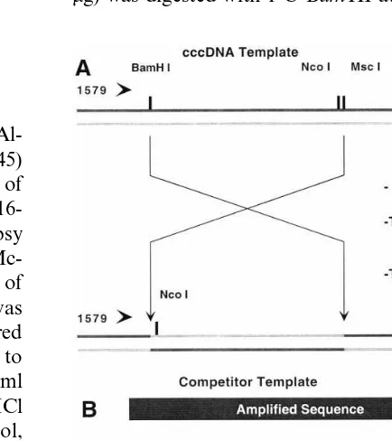
2.2. Construction of pCCCompet
The plasmid pCCCompet was the source of the competitor template used in the PCR assay; its construction is outlined in Fig. 1. The precursor plasmid was pUC19DHBVRI-Sph, in which the 2843 bp EcoRI –SphI restriction fragment of DHBV-16 has been inserted into the correspond-ing sites of pUC19. The precursor plasmid (0.2 mg) was digested with 1 UBamHI at 37°C for 1 h.
The resulting cohesive ends were filled using 2 U of T4 DNA polymerase. The reaction was ex-tracted with phenol, then twice with chloroform and the DNA precipitated with ethanol. The blunt-ended DNA was digested with 1.5 U of
MscI. Nucleic acids were precipitated with iso-propanol then dissolved in 10ml of ligation buffer containing 4 U of T4 ligase and incubated at 25°C for 16 h. An aliquot of the ligation reaction was transformed intoE.coliDH5a(Hanahan, 1983) . Colonies with plasmids containing the BamHI/
MscI fragment were identified by colony hy-bridization with a radiolabeled DHBV probe. Plasmid DNA minipreps from these (Ausubel, 1992) were screened by restriction digestion with
EcoRI andNcoI to identify those containing plas-mids of the desired structure. pCCCompet has the 712 bp sequence between the BamHI and MscI inverted with respect to the wild-type sequence and a 4 bp insertion where the BamHI site was filled. The competitor template for the quantita-tive PCR assays was generated by using 10 ng of pCCCompet in a PCR with primer set 1579/2771. The PCR product was purified using a QIAquick kit (Qiagen Inc., Mississauga, Ont., Canada) and quantitated by the PicoGreen method.
2.3. cccDNA-Specific DHBV PCR
Primers were chosen that converge on the gaps present on both strands of the DHBV genome. Primer 2771 (5%-GAATCTGATTTCCAATA-3%) and primer 1579 (5% -ACGGGTCTACTATTTTA-3%) were used in 50ml reactions containing 20 – 100 ng of duck liver biopsy DNA, varying amounts of competitor template, primers at 250 nM, de-oxynucleotide triphosphates at 250 mM each, 2.5 mM MgCl2, 50 mM spermidine·4 HCl, 1.7 U Taq
polymerase (GibcoBRL) in its recommended buffer. Reactions were assembled on ice then loaded into an MJ Research PTC-100 thermocy-cler heated to 92°C. After 2 min at this tempera-ture the reactions were cycled 30 times through a program of 94°C for 30 s, 53°C for 60 s, and 72°C for 150 s. After a final incubation at 72°C for 5 min, 2.5 U ofNcoI restriction enzyme was added to each reaction and incubated at 37°C for 2 h. The digestion products were separated on a 0.8%
agarose gel by electrophoresis at 70 V for 2 h. Both the gel and the 0.5×TBE (Ausubel, 1992) electrophoresis buffer contained 0.2 mg/ml ethid-ium bromide. An image of the gel’s UV fluores-cence was captured using a video imaging system (UVP, DiaMed Lab Supplies Inc., Mississauga, ON, Canada) and the intensity of the bands was measured using NIH Image 1.62b7 software. The intensity of the upper, 1094 bp, band indicated the amount of competitor product present in the reaction. The lower, 772 bp, band was the product of cccDNA template in the reaction. The intensity of this band was multiplied by 1.41, to correct for its smaller size, before comparison to the intensity of the competitor product. The mass of duck genome was taken to be 2.65 pg/diploid genome (Vendrely, 1958) when calculating the cccDNA copy number per genome. A linear plot of the log10(cccDNA PCR product/competitor
PCR product) versus log10 input competitor
tem-plate was used to estimate the amount of cccDNA in the liver DNA sample.
2.4. Quantitation of cccDNA by Southern transfer
The plasmid used to construct the standard curve, pVg, contains 1 kbp of Drosophila DNA from the 6estigial locus cloned in the pUC19 derivative pT7T319U. Varying amounts of this plasmid, ranging from 4×10⁶to 3.2×10⁷copies,
were added to 20 ml of restriction buffer each containing 1 mg of genomic DNA from a 2 week old, congenitally infected duckling and 5 U of
pD2eco11, which contains a single DHBV genome, linearized at its EcoRI site, cloned into pUC19. This plasmid (10 ng), labeled with [a-32P] dCTP by random priming (GibcoBRL, Life Tech-nologies, Burlington, Ont., Canada) to 5×10⁸
cpm/mg, 106 cpm of this probe were denatured then hybridized to the blot at 65°C for 16 h. After hybridization the blot was rinsed in several changes of 2×SSPE-1% SDS at 25°C then washed twice in 0.2×SSPE-0.1% SDS at 65°C for 30 min each. The amount of probe bound to the pVg and cccDNA bands was measured using a Fuji Phosphoimager system and Image Gauge software.
3. Results
3.1. Selecti6ity for DHBV cccDNA
Hepatocytes infected with DHBV contain sev-eral species of viral replicative intermediates. In addition to cccDNA, they contain some com-pletely replicated, open circular, viral DNA and a large amount of partially replicated DNA (Mason et al., 1982) . With the exception of cccDNA, these forms are covalently linked to protein and will be depleted, though not eliminated by phenol extraction during purification of liver DNA. We designed our PCR primers to selectively amplify the cccDNA form of the genome. While they amplified cccDNA very efficiently we found that an ocDNA template, prepared from extracellular virus (ECV), could also produce a detectable product if present in greater than about 106copies
in our standard PCR reaction. The selectivity of the assay for cccDNA was estimated by compar-ing the efficiency of PCR on the ECV-derived, ocDNA, template and on an ungapped template representative of the cccDNA form. As an un-gapped template we used the competitor con-structed for the cccDNA assay because its product can be distinguished from that of ocDNA template by the position of its NcoI restriction site. A PCR containing 105 molecules of
un-gapped template and a 100-fold excess of un-gapped, ocDNA template nevertheless produced 60 times more product from the ungapped template than
from the gapped, ECV-derived template (results not shown). These results indicate that, though the specificity of the PCR for cccDNA is not absolute, it is sufficiently high that it can form the basis of a quantitative assay for DHBV cccDNA provided that contamination of the sample with ocDNA is not massive.
The left-most lane of Fig. 3A shows the species of DHBV DNA present in the total duck liver DNA samples analyzed in the cccDNA PCR as-say. The most abundant species migrates as ex-pected of intact cccDNA. A less intense upper-most band migrates as expected of ocDNA. This species is present at about 1/5 the level of the cccDNA. There are two distinct sources for DNA migrating with this mobility. ocDNA can be gen-erated by nicking of the cccDNA form during DNA isolation (Newbold et al., 1995). This frac-tion of ocDNA will contribute to the cccDNA pool size measured by our assay. In addition, the gapped DHBV DNA from mature virions will also migrate as ocDNA during agarose gel elec-trophoresis. In light of the results presented above, the small amount of this species present in the DNA sample will not compromise the cc-cDNA assay. A third discrete DHBV species present in the genomic DNA sample migrates at 3.0 kb. This is linear DHBV (linDNA) formed in minor amounts during DHBV replication (Staprans et al., 1991). Like virion-derived ocDNA, small amounts of linDNA should not be amplified efficiently by the cccDNA-selective primers used in the assay and should not interfere with the cccDNA assay.
Our cccDNA extraction method differs from the modified Hirt extraction method commonly used to isolate cccDNA (Wu et al., 1990). We validated our method by extracting cccDNA from equal numbers of primary duck hepatocytes by the two methods and assaying the resulting sam-ples for cccDNA by our competitive PCR assay. Identical amounts of cccDNA were isolated by the two methods (results not shown).
3.2. cccDNA assay competitor
primers as the target template, amplifies with equal efficiency as the target, and produces a PCR product that can be separated from the product of the target template and quantified. As outlined in Fig. 1, we produced such a template by inverting a large segment of the DHBV genome within the region amplified by the PCR primers chosen for the assay. This inversion satisfied two objectives. First, it changed the position of an NcoI restric-tion site permitting the product of the competitor template to be easily distinguished from the product of the wild-type DHBV sequence found in cccDNA (Fig. 1B). Second, the inversion pre-vented the formation of significant amounts of heteroduplex between the products of the two templates. Stable heteroduplexes can form if the competitor and target products differ only slightly in sequence. Such heteroduplexes complicate the separation and quantification of the competitor and target PCR products (Zimmermann and Mannhalter, 1996).
The inversion competitor is almost the same size, has the same base composition, and, except for the regions around the inversion breakpoints, has the same sequence as the cccDNA template albeit on different strands of the duplex. It was hoped, therefore, that it would amplify with the same efficiency as the cccDNA target template. Preliminary experiments suggested that this hope was justified and a test of the PCR assay’s accu-racy by Southern blotting, described below, con-firmed this important point.
3.3. The cccDNA quantitati6e PCR assay
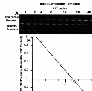
The results of a typical quantitative PCR assay for DHBV cccDNA is shown in Fig. 2. An identi-cal, known, amount of total duck liver DNA from a 2-week old animal was added to a series of PCR reactions each differing in the amount of competi-tor template they were known to contain. After PCR, the products were digested with NcoI re-striction endonuclease which permits discrimina-tion between the products of the two templates in the assay series after agarose gel electrophoresis. The uppermost, 1090 bp, band visible on the stained gel is the product of the competitor tem-plate. As expected, the intensity of this band
increases with increasing amounts of input com-petitor template. The next band, of 772 bp, is the product of the cccDNA template. The intensity of this band decreases with increasing amounts of competitor. The intensity of these bands can be quantitated and, after correcting for the intensity difference due to the difference in the size of the two products, used to estimate the amount of cccDNA present in the duck liver DNA. A simple plot of the two band intensities versus the amount of input competitor template yields an equiva-lence point where the two curves intersect. This can be used to estimate cccDNA copy number. However, a plot of the log of the ratio of the band intensities versus the log of the copies of input competitor template yields a linear plot as in Fig. 2B. Thex-intercept of this plot is used to estimate the equivalence point, the point at which the amounts of input competitor and cccDNA tem-plates are the same. This plot is more robust than the simple plot mentioned above since small tube-to-tube variation in PCR efficiency do not affect the ratio of the two PCR products but may affect their absolute amounts. The copy number esti-mate from the data shown in Fig. 2 is 20.8 copies cccDNA/diploid genome. The assay is quite re-producible. Four repetitions of the assay on the same sample gave a mean of 19.992.2 copies per diploid genome.
3.4. Accuracy of the cccDNA PCR assay
Fig. 2. (A) Quantitative PCR of DHBV cccDNA. 22.2 ng of duck liver DNA (8.4×10³diploid genomes) was added to each of a
series of PCR reactions containing the indicated amounts of competitor template. After PCR the products were digested withNcoI restriction endonuclease and separated and visualized on an agarose gel. The uppermost band is the product of the competitor template which increases in intensity with increasing amounts of its template. The second band, the product of the cccDNA template, decreases in intensity with increasing amounts of competitor template. Duplicate gel loadings were made of PCR reactions containing 0 and 8 – 60×104 copies of competitor template. (B) Determination of cccDNA copy number. The intensity of the
cccDNA and competitor PCR product bands was measured, corrected for intensity differences due to the difference in size between the two products, and the ratio of the intensity of the two bands was calculated for each PCR reaction. The log10of this ratio was
plotted against the log10of the copies of input competitor template. Where the log of the ratio is zero, the amounts of the two PCR
products are the same. In this case the equivalence point is at 1.74×10⁵copies of input competitor template corresponding to a
cccDNA copy number of 20.8 copies per diploid genome.
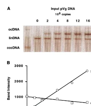
only to increase the size of the plasmid such that it migrates slightly more slowly than linearized DHBV cccDNA during electrophoresis. The blots were hybridized to a radiolabeled probe prepared from a plasmid containing pUC19 and a unit genome of DHBV. Linking these two sequences within the same plasmid ensures that they are labeled with equal efficiency. Probe prepared from this template will hybridize to both DHBV se-quences present in the duck liver DNA sample
restriction digest. Because the hybridization target in this plasmid (2.68 kb) is smaller than in the DHBV (3.02 kb) the hybridization signal mea-sured for these bands was multiplied by 1.12 before comparison to the DHBV signal. A plot of the intensity of the two species versus the amount
of pVg added to each sample is shown in Fig. 3B. The point at which the two curves intersect was used to estimate a cccDNA copy number of 16.2 copies/diploid genome. PCR assays, in quadrupli-cate, of the same sample gave an estimate of 19.992.2 copies per diploid genome. Thus, two
Fig. 3. (A) Quantitation of DHBV cccDNA by Southern blot. A series of reactions were assembled, each contained 0.8mg of duck
liver DNA (3.0×10⁵ diploid genomes). DNA in the leftmost lane was undigested and the positions of ocDNA, linDNA, and
cccDNA are indicated in the left margin. In the other lanes, duck liver DNA was mixed with the indicated amounts of the quantitation standard pVg DNA and linearized withEcoRI. A Southern blot was prepared by standard methods. The blot was hybridized to a radiolabeled probe prepared from a pUC19 plasmid carrying a unit length DHBV genome. The DHBV portion of this probe detected the cccDNA on the blot while the vector portion detected the quantitation standard, pVg. The pattern of hybridization was visualized and quantitated with a phosphoimager. The positions of linearized pVg, linearized cccDNA (and the comigrating linearized ocDNA), and linDNA shortened byEcoRI digestion are shown in the right margin. (B) Determination of cccDNA copy number. The intensity of the bands in the upper panel were measured by phosphoimager analysis; the units of intensity are arbitrary. After correction for the differing hybridization target size in the pVg and cccDNA molecules, the intensities were plotted as a function of the copies of pVg added to the reactions. As expected, the intensity of the cccDNA band was almost constant while that of pVg increased. The equivalence point, marked by the intersection of the two lines, occurs at 5.02×10⁶copies
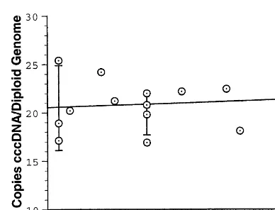
Fig. 4. Range of the cccDNA PCR assay. Quantitative PCR assays were performed using the same duck liver DNA prepa-ration but varying the amounts of DNA/PCR reaction over the range 2.5 – 60 ng. The cccDNA copy number estimates were plotted as a function of the amount of input DNA. The best fit line accommodating these data is shown. The copy number estimate is nearly invariant over the range tested but the reproducibility of the assay is reduced at both low and high levels of input template.
The sensitivity limit of the quantitative assay is about 2×10⁴ cccDNA molecules. This is about
100 times more sensitive than the Southern blot method described above. If the goal is the simple detection, rather than quantitation, of cccDNA the limit is about ten times lower. At present the sensitivity of the assay is limited by the detection of the PCR products by staining with ethidium bromide. Greater sensitivity could perhaps be achieved if dyes, such as ethidium homodimers, with higher affinity for DNA and greater fluores-cent enhancement were used.
4. Discussion
We have devised a selective, quantitative, and specific assay for DHBV cccDNA. The selectivity of the assay is based on the same design, PCR primers that converge on a region of DHBV that is free of gaps only in cccDNA, as developed by Schlicht and his colleagues for qualitative cc-cDNA assays (Ko¨ck and Schlicht, 1993; Ko¨ck et al., 1996). They found that ocDNA templates could be amplified with such primers but at an efficiency only 0.1 – 1% of that on a cccDNA template. They noted that the products formed from each strand of an ocDNA template in a PCR with ‘cccDNA-specific’ primers contain a region of complementarity at their 3% ends which can anneal with one another, be elongated in later cycles, and then be amplified exponentially by the ‘cccDNA specific’ primer pair. Our experiments confirm both the lack of absolute specificity of the primers for cccDNA and the low efficiency of the primers on an ocDNA template. Our results indi-cate that at least a 100-fold excess of ocDNA to cccDNA would be required to interfere seriously with the quantitative assay. Southern blot analysis of our liver DNA preparations (Fig. 3A) shows that ocDNA contamination is much lower than this limit.
In creating a competitive PCR assay one must create a competitor template that is very similar to the target template, so that both templates amplify with identical efficiency, yet differs from the target, so that the PCR products from the two templates can be separated and quantitated. Ini-assays, based on very different principles, gave
similar values for cccDNA copy number. This result increases our confidence in the accuracy of the PCR assay we have developed. It also implies that the competitor template we have constructed amplifies with the same efficiency as the cccDNA template during the PCR reactions, a fundamen-tal requirement for an accurate competitive PCR assay.
3.5. Sensiti6ity of the assay
tially we introduced of a novel restriction site into the competitor template to achieve these ends. This approach, however, was frustrated by the formation of heteroduplexes between products of the competitor and target templates. These het-eroduplexes were resistant to digestion with the differentiating restriction enzyme and prevented accurate quantitation of the two PCR products. To avoid this problem we created a competitor template with a large inversion within the am-plified region. This rearrangement changes the position of a restriction site so that the PCR products of competitor and cccDNA template can be readily distinguished and, at the same time, prevents heteroduplex formation. This competitor structure was key to the success of our assay.
Despite its importance in the replication cycle of the hepadnaviruses, it is remarkable how little is known about the cccDNA pool size within infected hepatocytes. HBV copy number in hu-man liver is very poorly documented. In the livers of woodchucks transiently infected with WHV the cccDNA copy number is 10 – 25 per cell (Kajino et al., 1994); the livers of two woodchucks chroni-cally infected with WHV were found to have a similar cccDNA level of about 20 copies per hepatocyte (Summers et al., 1991; Tencza and Newbold, 1997)
Summers et al. (1991) estimate that primary duck hepatocytes contain 22 copies of cccDNA/ cell 10 days after in vitro infection with DHBV. The method used to make this estimate is not described in their paper, hence, the accuracy of this estimate is difficult to evaluate. The best estimate to date is that of Jilbert et al. who conducted a thorough longitudinal study of tran-sient and chronic DHBV infections in duck liver (Jilbert et al., 1992). One of the parameters they measured was the cccDNA levels in infected hepa-tocytes. They isolated cccDNA by a modified Hirt extraction method and used quantitative Southern blotting to measure the amounts of cccDNA. They found that liver cells from 2-week-old, con-genitally infected ducks contain about 7 – 10 copies of cccDNA/diploid genome. Since they estimated that only 60% of these cells were hepa-tocytes, they concluded that each infected cell contains about 15 – 20 copies of cccDNA. This
value is quite similar to that we obtained using two independent methods, competitive PCR and quantitative Southern blotting. In our estimates we made no attempt to correct the values for the presence of red blood cells and other nonhepato-cytes in our tissue samples. The values we report are averaged over the entire cell population. It is possible that there is considerable cell-to-cell vari-ation in cccDNA copy number. Another potential source of variability would be nonuniform infec-tion of the duck liver. This would be of particular concern in techniques, such as needle biopsy, that sample only a small fraction of the liver. Since in both chronic and acute DHBV infections virtually every hepatocyte contains replicating virus (Jilbert et al., 1992) we do not think this is a serious problem.
We have developed a quantitative assay for DHBV cccDNA that is sensitive, selective, and accurate. This assay will be useful in studying the biology of this crucial replicative intermediate. In particular, it can be used to monitor cccDNA levels during the course of antiviral chemotherapy in this animal model system. This will be essential for determining when treatment might be stopped without incurring a rebound in virus replication. We hope to modify this assay for similar monitor-ing of HBV cccDNA in human liver samples.
Acknowledgements
The authors would like to thank Gary Ritzell for the gift of pVg DNA and Dr Rajan George for the synthesis of the oligodeoxynucleotides used in this work. This work was generously supported by GlaxoWellcome Canada.
References
Ausubel, F.M. (Ed.), 1992. Current Protocols In Molecular Biology. Greene Publishing Associates and Wiley-Inter-science, New York.
Clementi, M., Menzo, S., Bagnarelli, P., Manzin, A., Valenza, A., Varaldo, P.E., 1993. Quantitative PCR and RT-PCR in virology. PCR Methods Appl. 2, 191 – 196.
cessa-tion of treatment with the nucleoside analogue ganciclovir. Antivir. Res. 27, 171 – 178.
Dienstag, J.L., Perrillo, R.P., Schiff, E.R., Bartholomew, M., Vicary, C., Rubin, M., 1995. A preliminary trial of lamivudine for chronic hepatitis B infection. New Engl. J. Med. 333, 1657 – 1661.
Guidotti, L.G., Rochford, R., Chung, J., Shapiro, M., Purcell, R., Chisari, F.V., 1999. Viral clearance without destruction of infected cells during acute HBV infection. Science 284, 825 – 829.
Hanahan, D., 1983. Studies on transformation ofEscherichia coliwith plasmids. J. Mol. Biol. 166, 557 – 580.
Jilbert, A.R., Wu, T.-T., England, J.M., Hall, P.M., Carp, N.Z., O’Connell, A.P., Mason, W.S., 1992. Rapid resolu-tion of duck hepatitis B virus infecresolu-tions occurs after mas-sive hepatocellular involvement. J. Virol. 66, 1377 – 1388. Kajino, K., Jilbert, A.R., Saputelli, J., Aldrich, C.E., Cullen,
J., Mason, W.S., 1994. Woodchuck hepatitis virus infec-tions: very rapid recovery after a prolonged viremia and infection of virtually every hepatocyte. J. Virol. 68, 5792 – 5803.
Ko¨ck, J., Schlicht, H.-J., 1993. Analysis of the earliest steps of hepadnavirus replication: genome repair after infectious entry into hepatocytes does not depend on viral poly-merase activity. J. Virol. 67, 4867 – 4874.
Ko¨ck, J., Theilmann, L., Galle, P., Schlicht, H.-J., 1996. Hepatitis B virus nucleic acids associated with human peripheral blood mononuclear cells do not originate from replicating virus. Hepatology 23, 405 – 413.
Mason, W.S., Aldrich, C., Summers, J., Taylor, J.M., 1982. Asymmetric replication of duck hepatitis B virus DNA in liver cells: free minus-strand DNA. Proc. Natl. Acad. Sci. USA 79, 3997 – 4001.
Miller, R.H., Robinson, W.S., 1984. Hepatitis B virus DNA forms in nuclear and cytoplasmic fractions of infected human liver. Virology 137, 390 – 399.
Moraleda, G., Saputelli, J., Aldrich, C.E., Averett, D., Con-dreay, L., Mason, W.S., 1997. Lack of effect of antiviral therapy in nondividing hepatocyte cultures on the closed circular DNA of woodchuck hepatitis virus. J. Virol. 71, 9392 – 9399.
Newbold, J.E., Xin, H., Tencza, M., Sherman, G., Dean, J., Bowden, S., Locarnini, S., 1995. The covalently closed duplex form of the hepadnavirus genome exists in situ as a heterogeneous population of viral minichromosomes. J. Virol. 69, 3350 – 3357.
Staprans, S., Loeb, D., Ganem, D., 1991. Mutations affecting hepadnavirus plus-strand synthesis dissociate primer cleav-age from translocation and reveal the origin of linear viral DNA. J. Virol. 65, 1255 – 1262.
Summers, J., Smith, P.M., Horwich, A.L., 1990. Hepadnavirus envelope proteins regulate covalently closed circular DNA amplification. J. Virol. 64, 2819 – 2824.
Summers, J., Smith, P.M., Huang, M., Yu, M., 1991. Morpho-genetic and regulatory effects of mutations in the envelope proteins of an avian hepadnavirus. J. Virol. 65, 1310 – 1317. Tencza, M.G., Newbold, J.E., 1997. Heterogeneous response for a mammalian hepadnavirus infection to acyclovir: drug-arrested intermediates of minus-strand viral DNA synthesis are enveloped and secreted from infected cells as virion-like particles. J. Med. Virol. 51, 6 – 16.
Tuttleman, J.S., Pourcel, C., Summers, J., 1986. Formation of the pool of covalently closed circular viral DNA in hepad-navirus-infected cells. Cell 47, 451 – 460.
Vendrely, R., 1958. La notion d’espece a travers quelques donne´es biochemiques re´centes et le cycle L. Ann. Inst. Pasteur (Paris) 94, 143 – 166.
Wu, T.-T., Coates, L., Aldrich, C.E., Summers, J., Mason, W.S., 1990. In hepatocytes infected with duck hepatitis B virus, the template for viral RNA synthesis is amplified by an intracellular pathway. Virology 175, 255 – 261. Zimmermann, K., Mannhalter, J.W., 1996. Technical aspects
of quantitative competitive PCR. Biotechniques 21, 274 – 279.